
The intersection of veterinary medicine and advanced technology has fundamentally altered how we identify, diagnose, and treat common ailments in domestic animals. One of the most persistent challenges in canine dermatology is the identification of dermatophytosis, commonly known as ringworm. While the name suggests a parasitic involvement, ringworm is a fungal infection that presents complex visual data points. In the modern era, the question “what does ringworm in a dog look like” is no longer answered solely by the naked eye of a clinician; it is answered through high-resolution digital imaging, machine learning algorithms, and advanced spectral analysis.
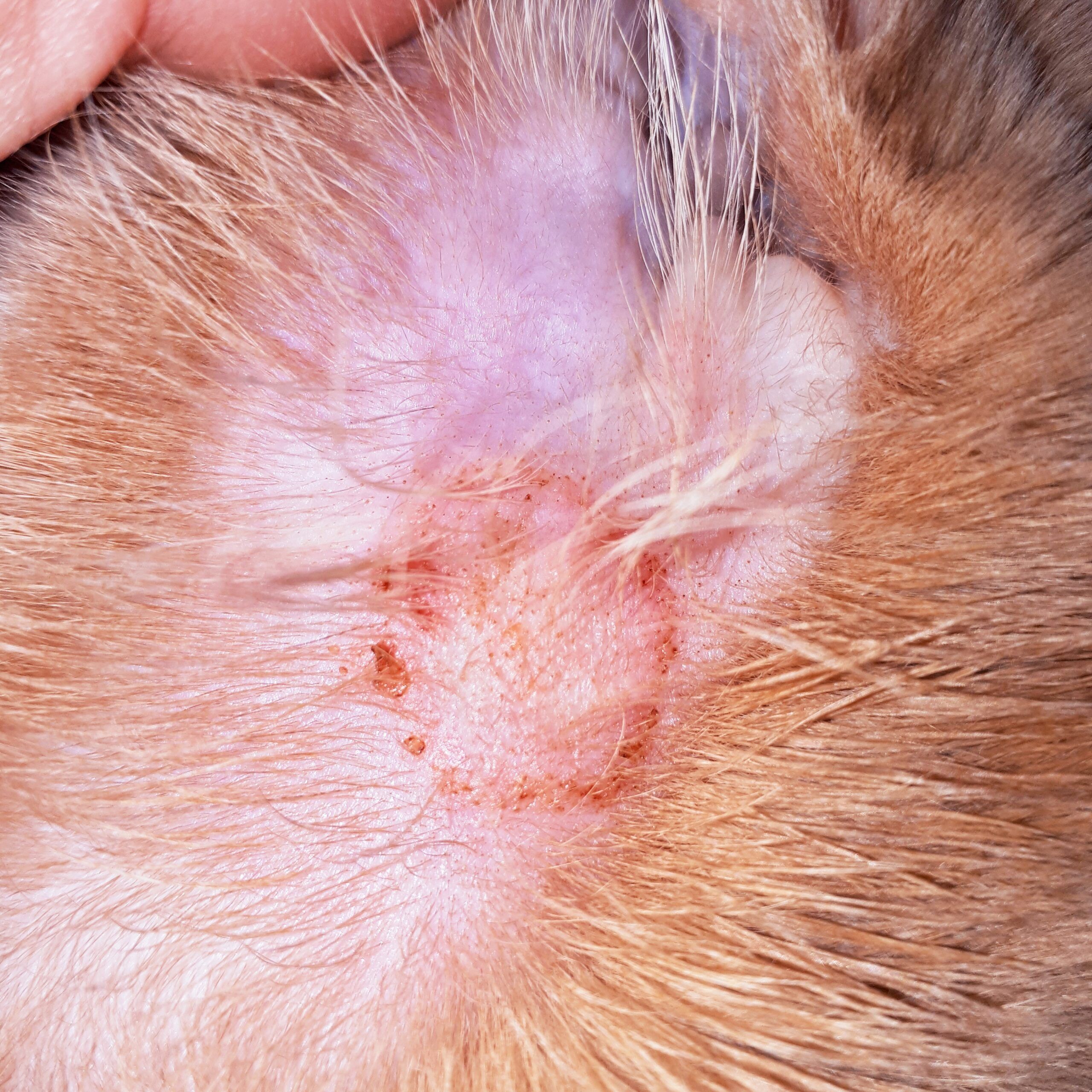
As we transition into an era of “Pet-Tech,” the identification of skin pathologies has moved from subjective observation to objective, data-driven diagnostics. Understanding the visual manifestations of ringworm through the lens of technology allows for earlier intervention, more accurate tracking of contagion vectors, and a significant reduction in zoonotic transmission to human owners.
The Evolution of Veterinary Diagnostics: From Wood’s Lamps to Digital Imaging
Traditionally, the visual identification of ringworm relied on the Wood’s lamp, a tool that utilizes ultraviolet light to induce fluorescence in specific fungal strains. However, the technological landscape has evolved, turning “looking” into a sophisticated process of optical data collection.
Understanding the Optical Signature of Microsporum canis
The primary culprit in canine ringworm, Microsporum canis, possesses a unique chemical property: it produces pteridine, a metabolite that fluoresces a vivid apple-green under specific UV wavelengths (approximately 365 nanometers). Modern diagnostic gadgets have refined this process. High-output LED Wood’s lamps now integrate narrow-band pass filters that eliminate ambient light interference, allowing tech-enabled clinics to see the “glow” of ringworm even in well-lit environments. This optical signature is the first digital “marker” used to differentiate fungal spores from simple dander or medication residue.
Digital Enhancement and High-Resolution Dermatoscopy
Moving beyond simple UV light, the industry has seen an influx of digital dermatoscopes. These devices are essentially high-definition cameras equipped with polarized light sources and magnification lenses (often up to 100x). When a technician asks what ringworm looks like through a dermatoscope, they are looking for “comma hairs” and “corkscrew hairs”—microscopic structural deformities in the hair shaft caused by fungal invasion. These visual markers are often invisible to the human eye but are easily captured by digital sensors, allowing for the creation of a visual baseline that can be tracked over time via cloud-based software.
Machine Learning and Computer Vision in Canine Dermatology
The most significant leap in answering what ringworm looks like in dogs comes from the field of Artificial Intelligence (AI). Computer vision—the ability of a machine to interpret and understand the visual world—is now being applied to dermatological databases to provide instantaneous diagnostic support.
Training Neural Networks for Pattern Recognition
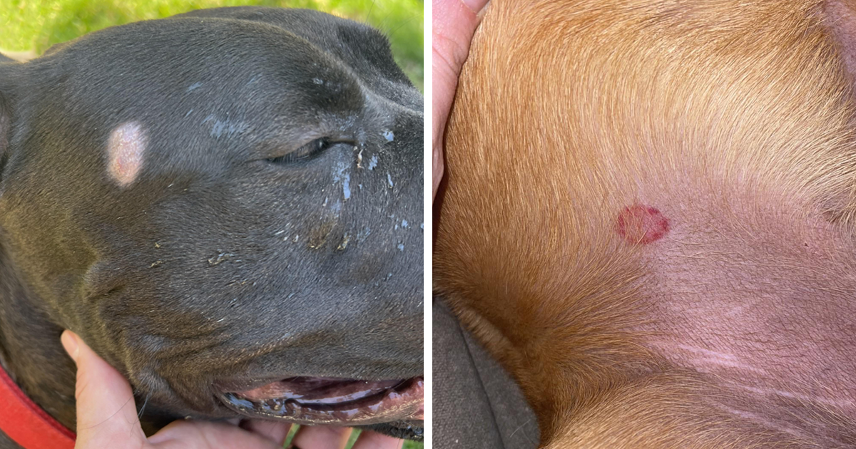
To a computer, a ringworm lesion is a collection of pixel intensities, edge gradients, and color distributions. Developers are currently training Convolutional Neural Networks (CNNs) on thousands of verified images of canine skin diseases. By processing these datasets, the AI learns to identify the classic “annular” (ring-like) pattern of ringworm: a red, inflammatory periphery with a central area of alopecia (hair loss) and scaling. The software scans for specific geometric irregularities that distinguish a fungal infection from a bacterial “hot spot” or an allergic reaction.
The Role of Big Data in Differentiating Pathologies
One of the difficulties in manual diagnosis is that ringworm is a “great mimicker.” It can look like folliculitis, demodicosis (mange), or autoimmune disorders. Technology solves this through the aggregation of big data. By cross-referencing a digital image of a dog’s skin with a global database of confirmed cases, AI tools can provide a “probability score.” For instance, an algorithm might determine there is an 88% statistical likelihood that a lesion is dermatophytosis based on its border morphology and the degree of follicular crusting, providing a level of precision that transcends traditional visual inspection.

Telemedicine and Remote Diagnostic Tools for Pet Owners
The accessibility of technology means that the first step in identifying “what ringworm looks like” often happens in the owner’s home rather than a sterile clinic. The rise of telehealth platforms has necessitated the development of consumer-grade visual assessment tools.
AI-Powered Mobile Apps: A Preliminary Screening Tool
Several emerging startups are focusing on “at-home triage” apps. Using the high-resolution cameras found on modern smartphones, these apps guide the user to take a clear, well-lit photo of the suspicious area. The app’s internal logic then analyzes the image for the hallmark signs of ringworm: the circular red rash, the presence of “cigarette ash” scaling, and localized hair breakage. While these tools do not replace a fungal culture (the gold standard of testing), they serve as a critical tech-layer in the diagnostic funnel, prompting owners to seek professional help before the infection spreads.
Integration with Electronic Health Records (EHR)
Once an image is captured via a smartphone or a clinic’s digital dermatoscope, it is no longer just a picture; it is a data point in a dog’s Electronic Health Record (EHR). Modern veterinary software suites now allow for “image longitudinal tracking.” By overlaying images taken on Day 1, Day 7, and Day 21, the software can calculate the rate of lesion shrinkage or the progress of hair regrowth. This visual analytics approach ensures that the treatment protocol—often a combination of systemic antifungals and topical tech-washes—is working effectively.
Security, Ethics, and the Future of Veterinary Bio-Data
As we rely more on technology to visualize and diagnose canine ringworm, we encounter new challenges regarding data security and the ethical use of AI in veterinary medicine.
Data Privacy in Genomic Sequencing and Imaging
The “visual” aspect of ringworm is now being paired with genomic sequencing. Technology allows us to look at the DNA of the fungi to determine its exact strain and origin. However, this creates a vast repository of biological data. Ensuring that these digital assets—both the images of the pet and the genetic data of the pathogen—are stored securely in encrypted cloud environments is a burgeoning sub-sector of veterinary IT. Protecting “pet privacy” and owner metadata is becoming as crucial as the diagnosis itself.
Ensuring Accuracy and Preventing Algorithmic Bias
A significant concern in the tech space is the potential for algorithmic bias. If an AI is trained primarily on images of short-haired dogs with light skin (like Beagles or Labradors), it may struggle to identify what ringworm looks like on a dark-skinned, long-haired breed like a Briard or a Black Russian Terrier. The tech industry is currently working toward “inclusive datasets” to ensure that diagnostic software is accurate across all canine phenotypes. This involves using infrared imaging and multi-spectral sensors that can “see” through thick coats or identify inflammation on hyper-pigmented skin where a red ring might not be visible to the human eye.
The Future: Wearable Sensors and Real-Time Monitoring
Looking forward, the question of what ringworm looks like will move from static images to real-time bio-feedback. We are seeing the development of smart collars equipped with multi-spectral sensors that monitor the skin’s surface. These wearables can detect changes in skin temperature (indicating inflammation) or alterations in the skin’s microbiome through chemical sensors.
In this future scenario, the “look” of ringworm is identified by a shift in a data dashboard before a single hair falls out. This proactive technological stance represents the pinnacle of modern veterinary care—where software, hardware, and biological insight converge to keep both our pets and our homes safe from infection.
By leveraging these technological advancements, we transform the subjective observation of a “red spot” into a sophisticated diagnostic workflow. The integration of AI, high-definition imaging, and secure data management ensures that the visual identification of canine ringworm is faster, more accurate, and more integrated into the broader ecosystem of digital health than ever before. For the modern pet owner and the tech-forward veterinarian, “what it looks like” is just the beginning of what the data can tell us.
aViewFromTheCave is a participant in the Amazon Services LLC Associates Program, an affiliate advertising program designed to provide a means for sites to earn advertising fees by advertising and linking to Amazon.com. Amazon, the Amazon logo, AmazonSupply, and the AmazonSupply logo are trademarks of Amazon.com, Inc. or its affiliates. As an Amazon Associate we earn affiliate commissions from qualifying purchases.