
In the realm of modern diagnostics, few technologies command as much respect and utility as Magnetic Resonance Imaging (MRI). Far from a simple X-ray, MRI represents a pinnacle of technological innovation, offering an unparalleled view into the human body’s intricate architecture without the use of ionizing radiation. When people ask, “What does MRI?”, they are often seeking to understand not just what the acronym stands for (Magnetic Resonance Imaging), but how this sophisticated piece of medical hardware functions, what it reveals, and why it has become an indispensable tool across numerous medical specialties. This article delves deep into the technological marvel that is MRI, exploring its fundamental principles, unique advantages, diverse applications, and the exciting future it portends in diagnostic medicine.
The Core Science: How MRI Works
At its heart, an MRI scanner is a powerful radio transmitter and receiver, employing a combination of magnetic fields and radio waves to generate detailed images of organs and soft tissues within the body. Unlike X-rays or CT scans that utilize ionizing radiation, MRI is based on the principles of nuclear magnetic resonance, a phenomenon observed at the atomic level. The process is a fascinating interplay of physics and computer science, orchestrated to reveal the subtle differences in tissue composition that can signify health or disease.
Harnessing Magnetic Fields
The journey of an MRI scan begins with a superconducting magnet – the most crucial and often the largest component of the scanner. This magnet creates an incredibly strong and uniform static magnetic field, typically measured in Teslas (T). Clinical MRI scanners range from 1.5T to 3T, with research machines reaching much higher field strengths. When a patient enters this magnetic field, the vast majority of water molecules in their body (which are abundant) respond. Each water molecule contains hydrogen atoms, and each hydrogen atom possesses a single proton. These protons act like tiny bar magnets, normally spinning and orienting randomly. The powerful magnetic field of the MRI scanner aligns these protons, causing them to point in the same direction, much like compass needles aligning with the Earth’s magnetic field. This alignment is a foundational step, creating a net magnetization within the patient’s body that can be manipulated and measured.
Radiofrequency Pulses and Signal Detection
Once the protons are aligned, the MRI scanner introduces a second type of energy: radiofrequency (RF) pulses. These short bursts of electromagnetic energy are specifically tuned to the resonant frequency of hydrogen protons in the strong magnetic field. When the RF pulse is applied, it briefly “knocks” the aligned protons out of alignment, tipping them at a specific angle relative to the main magnetic field. Imagine pushing a swinging pendulum to momentarily alter its natural rhythm. When the RF pulse is switched off, the protons relax and realign with the main magnetic field. As they relax, they release the absorbed energy in the form of a faint radio signal. It is this signal that the MRI scanner’s antenna coils detect. The strength and timing of these emitted signals vary depending on the type of tissue the protons reside in – whether it’s fat, water, bone, or muscle. This fundamental difference in relaxation properties is what allows MRI to distinguish between various tissues.
Gradient Coils and Spatial Encoding
Capturing a simple radio signal from the entire body wouldn’t provide diagnostic images; spatial information is crucial. This is where gradient coils come into play. These are smaller, rapidly switching electromagnets positioned within the main magnet. By altering the magnetic field strength systematically across different parts of the body, gradient coils create slight variations in the magnetic field. This causes protons at different locations to resonate at slightly different frequencies when the RF pulse is applied, and consequently, emit signals at different frequencies as they relax. This precise manipulation allows the MRI system to “encode” the spatial origin of the emitted signals. By rapidly switching these gradients on and off, the scanner can effectively map out the body in slices, allowing for the creation of incredibly detailed two-dimensional cross-sections and ultimately, three-dimensional reconstructions. The rapid switching of these gradient coils is also the primary source of the loud knocking, thumping, and buzzing sounds that patients hear during an MRI scan.
Computer Processing and Image Reconstruction
The raw data collected by the receiver coils is a complex array of radio signals varying in frequency, phase, and amplitude. This intricate data is fed into a powerful computer system. Sophisticated algorithms and a mathematical technique called the Fourier Transform are employed to process and translate these signals into actual images. The computer reconstructs these signals into pixels, forming a detailed grayscale image. Different tissues appear with varying degrees of brightness (or signal intensity) depending on their hydrogen content and their specific relaxation times (T1 and T2 relaxation). Radiologists, experts in interpreting these images, can then discern anatomical structures, identify abnormalities, and make diagnoses based on these highly detailed visual representations.
Beyond X-rays and CT Scans: The Unique Advantages of MRI
While other imaging modalities like X-rays, ultrasound, and CT scans play vital roles in diagnostics, MRI offers distinct advantages that make it indispensable for certain conditions. Its unique operating principles grant it capabilities that set it apart.
Superior Soft Tissue Contrast
One of MRI’s most celebrated strengths is its exceptional ability to differentiate between various soft tissues. Unlike X-rays, which are best for bone, or CT scans that excel at visualizing both bone and some soft tissues, MRI shines when it comes to organs, muscles, tendons, ligaments, cartilage, and particularly the brain and spinal cord. It can reveal subtle lesions, inflammations, tumors, and structural abnormalities in soft tissues with clarity that other methods simply cannot match. This superior contrast resolution is paramount for diagnosing neurological disorders, musculoskeletal injuries, and certain types of cancer.
Non-Invasive and Radiation-Free
A significant advantage of MRI is its non-invasive nature and the absence of ionizing radiation. This makes it a safer option, especially for patients requiring multiple scans, pregnant women (with careful consideration), or children. While some MRI scans may involve the injection of a contrast agent (typically gadolinium-based) to enhance visibility of certain structures, the procedure itself does not expose the patient to harmful radiation. This contrasts sharply with X-rays and CT scans, which utilize radiation and thus carry cumulative risks over time.
Versatility in Imaging Planes
MRI scanners possess the remarkable ability to produce images in any desired orientation – axial (cross-sectional), sagittal (side-to-side), or coronal (front-to-back), as well as oblique planes. This multi-planar imaging capability allows clinicians to view anatomical structures from various perspectives, providing a comprehensive understanding of complex pathologies and their relationship to surrounding tissues. This flexibility is particularly valuable in planning surgeries or precisely localizing lesions.
Functional and Advanced Imaging Capabilities
Beyond basic anatomical imaging, MRI technology has evolved to offer a suite of advanced techniques that provide insights into physiological processes and tissue function.
-
Functional MRI (fMRI): Measures brain activity by detecting changes in blood flow (BOLD response), crucial for understanding cognitive functions and mapping eloquent brain areas.
-
Diffusion Tensor Imaging (DTI): Visualizes the white matter tracts (nerve fibers) in the brain, aiding in the study of neurological disorders and brain connectivity.
-
Magnetic Resonance Angiography (MRA): Focuses on blood vessels, providing detailed images of arteries and veins without invasive catheterization.
-
Magnetic Resonance Spectroscopy (MRS): Detects biochemical changes in tissues, useful for characterizing tumors or metabolic disorders.
These advanced applications push MRI beyond mere anatomical visualization, offering a window into the dynamic processes occurring within the body.
Key Applications Across Medical Specialties
The technological prowess of MRI has made it an indispensable diagnostic tool across virtually all medical disciplines. Its ability to provide detailed, radiation-free images of soft tissues has revolutionized the diagnosis and management of countless conditions.
Neurological Imaging
MRI is the gold standard for imaging the brain and spinal cord. It is extensively used to diagnose:
-
Stroke: Identifying areas of reduced blood flow or hemorrhage.
-
Brain Tumors: Characterizing size, location, and invasiveness.
-
Multiple Sclerosis (MS): Detecting plaques and lesions in the brain and spinal cord.
-
Epilepsy: Locating structural abnormalities that may trigger seizures.
-
Degenerative Diseases: Such as Alzheimer’s and Parkinson’s, by revealing changes in brain volume or structure.
-
Spinal Cord Injuries and Disorders: Pinpointing nerve compression, herniated discs, and spinal cord lesions.
Musculoskeletal System

For orthopedic specialists and rheumatologists, MRI is invaluable for assessing joint injuries and conditions:
-
Ligament and Tendon Tears: Such as ACL tears in the knee or rotator cuff tears in the shoulder.
-
Cartilage Damage: Detecting meniscal tears or articular cartilage degeneration.
-
Bone Marrow Abnormalities: Identifying infections, tumors, or stress fractures that might not be visible on X-rays.
-
Soft Tissue Masses: Characterizing cysts, tumors, or inflammatory processes in muscles and fascia.
Cardiovascular Imaging
Cardiac MRI (CMR) has emerged as a powerful tool for evaluating heart structure and function:
-
Heart Muscle Damage: Assessing scar tissue after a heart attack or detecting inflammation (myocarditis).
-
Congenital Heart Defects: Providing detailed anatomical views in pediatric and adult patients.
-
Heart Valve Function: Evaluating the severity of valve stenosis or regurgitation.
-
Aortic Aneurysms and Dissections: Visualizing the large blood vessels originating from the heart.
Abdominal and Pelvic Imaging
MRI provides excellent visualization of internal organs in the abdomen and pelvis:
-
Liver: Detecting tumors, cirrhosis, or other liver diseases.
-
Kidneys: Assessing masses, cysts, or congenital abnormalities.
-
Pancreas and Biliary System: Identifying pancreatitis, tumors, or gallstones.
-
Uterus and Ovaries: Diagnosing fibroids, endometriosis, or ovarian cysts.
-
Prostate: Crucial for detecting and staging prostate cancer, especially with multi-parametric MRI (mpMRI).
Oncology
MRI plays a critical role in the diagnosis, staging, and monitoring of various cancers:
-
Tumor Detection: Identifying primary tumors and metastases in soft tissues.
-
Staging: Determining the extent of cancer spread.
-
Treatment Planning: Guiding biopsies and radiation therapy.
-
Response Monitoring: Assessing the effectiveness of chemotherapy or radiation.
-
Breast MRI: Used for high-risk screening and further evaluation of suspicious lesions.
The User Experience: What to Expect During an MRI Scan
For patients, understanding the MRI process can alleviate anxiety. While the technology is complex, the patient’s role is primarily to remain still and communicate.
Preparation and Safety Protocols
Prior to an MRI, patients undergo a thorough screening process. Because of the extremely powerful magnetic field, any ferromagnetic objects (iron-containing metals) are strictly prohibited in the scanner room. This includes certain types of implants (pacemakers, cochlear implants, some aneurysm clips), metal fragments, jewelry, and even some tattoo inks. Patients must remove all metal objects and change into a gown. In some cases, a contrast agent containing gadolinium may be injected intravenously to enhance the visibility of certain tissues or blood vessels; patients are typically screened for kidney function before receiving contrast.
The Scanner Environment
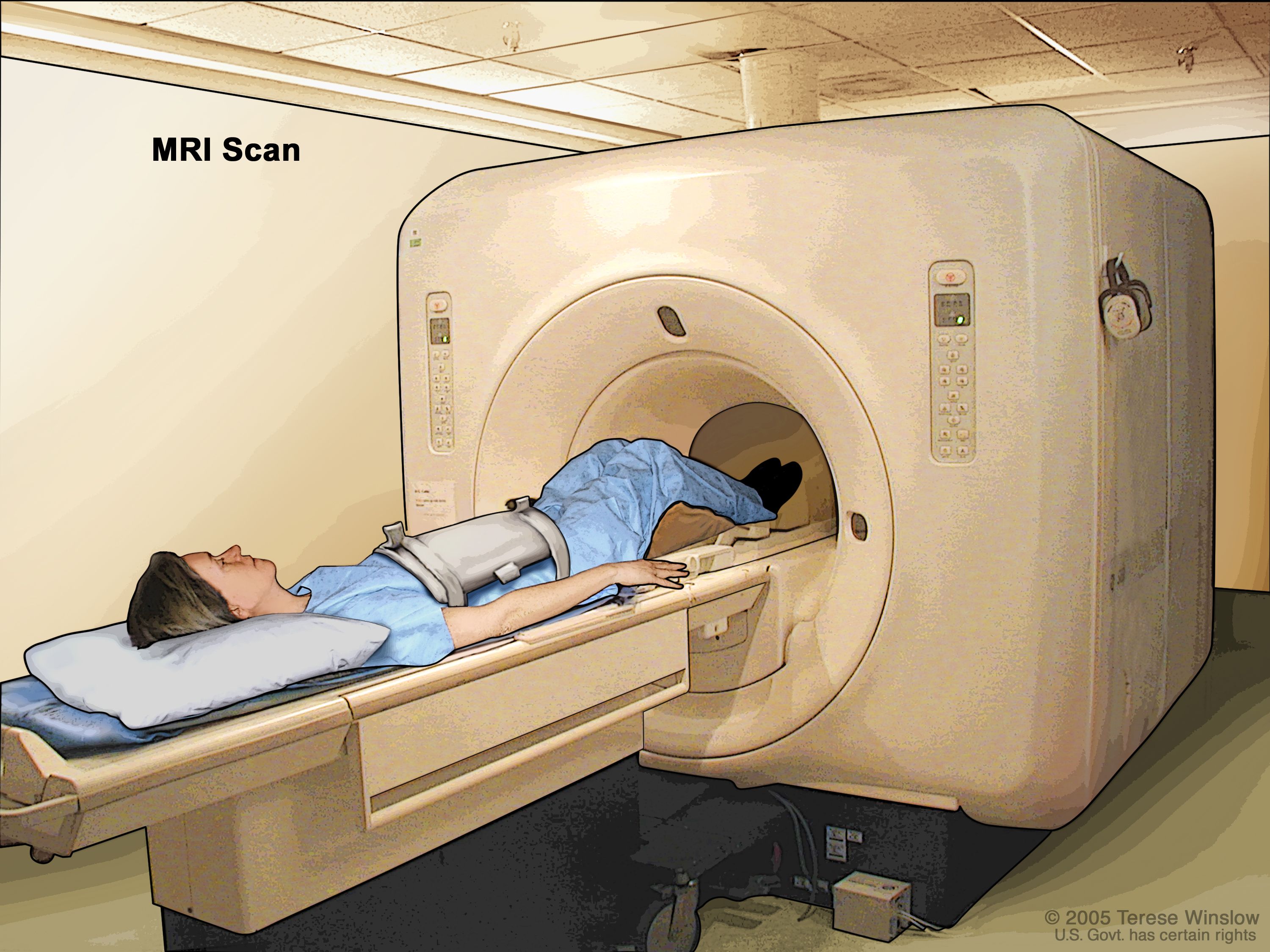
An MRI scanner typically consists of a large, tube-shaped magnet. Patients lie on a movable table that slides into the center of this tunnel. While some “open MRI” machines exist to reduce claustrophobia, traditional closed-bore scanners provide the highest field strength and image quality. The most notable aspect of the scan itself is the noise. The rapid switching of the gradient coils generates loud knocking, buzzing, and thumping sounds. Patients are provided with earplugs or headphones (often playing music) to mitigate the noise and allow for communication with the technologist.
Scan Duration and Patient Comfort
MRI scans can range from 15 minutes to over an hour, depending on the area being imaged and the complexity of the study. Remaining absolutely still during this time is crucial for clear images. Any movement can cause motion artifacts, blurring the images and potentially necessitating repeat sequences. Technologists are trained to guide patients through the process, providing breathing instructions and offering reassurance. For patients with claustrophobia or anxiety, options like open MRI, sedation, or pre-scan relaxation techniques can be discussed with their physician.
The Evolution of MRI Technology: Trends and Future Outlook
The field of MRI is not static; it’s a dynamic area of technological innovation, constantly pushing the boundaries of what’s possible in diagnostic imaging.
Higher Field Strengths and Image Resolution
The trend towards higher magnetic field strengths continues, with 3T becoming standard in many clinical settings and ultra-high field systems (7T and beyond) emerging for specialized research and clinical applications. These higher fields offer improved signal-to-noise ratio, leading to increased spatial resolution and clearer, more detailed images, particularly beneficial for visualizing minute structures in the brain and joints.
Faster Scan Times and Enhanced Patient Experience
Developers are relentlessly working on techniques to reduce scan times. Faster imaging sequences, parallel imaging, and compressed sensing algorithms are significantly cutting down the duration patients need to spend in the scanner. This not only improves patient comfort and reduces motion artifacts but also increases patient throughput, making MRI more accessible. Advances in coil technology and quieter gradient systems are also contributing to a more patient-friendly experience.
AI and Machine Learning Integration
Artificial intelligence (AI) and machine learning (ML) are poised to revolutionize MRI at multiple levels. AI algorithms can reconstruct images faster and with higher quality from less raw data, allowing for shorter scans. They can also assist radiologists in image analysis, automating tedious tasks like organ segmentation, detecting subtle abnormalities, and even aiding in diagnosis by identifying patterns indicative of disease. Predictive analytics based on MRI data could also play a role in personalized medicine.
Open MRI and Specialized Scanners
While traditional closed-bore MRI offers superior image quality, the development of more open-architecture MRI systems continues. These are particularly valuable for claustrophobic patients, bariatric patients, and those requiring interventional procedures under MRI guidance. Specialized scanners, like dedicated extremity MRI units, are also becoming more common, offering high-resolution imaging for specific body parts with a smaller footprint and lower cost.

The Future of Diagnostic Imaging
The future of MRI is bright, promising even greater diagnostic precision, increased patient comfort, and broader accessibility. As technology advances, MRI will likely integrate more seamlessly with other diagnostic tools and patient data, forming a comprehensive picture for personalized medicine. From refining our understanding of neurological diseases to enabling earlier and more accurate cancer detection, MRI continues to be a cornerstone of modern healthcare technology, offering insights that were once unimaginable. Its journey from a complex physics experiment to an everyday clinical tool is a testament to human ingenuity and the relentless pursuit of better health outcomes.
aViewFromTheCave is a participant in the Amazon Services LLC Associates Program, an affiliate advertising program designed to provide a means for sites to earn advertising fees by advertising and linking to Amazon.com. Amazon, the Amazon logo, AmazonSupply, and the AmazonSupply logo are trademarks of Amazon.com, Inc. or its affiliates. As an Amazon Associate we earn affiliate commissions from qualifying purchases.