In the modern clinical environment, the term “contracted gallbladder” is more than a physiological observation; it is a data point generated by high-sophistication diagnostic hardware and interpreted by complex software algorithms. For medical professionals and HealthTech developers alike, understanding what a contracted gallbladder means through the lens of technology is essential for improving patient outcomes and streamlining diagnostic workflows. From the physics of piezoelectric transducers to the integration of deep-learning AI, the journey from a sonic pulse to a digital diagnosis represents the cutting edge of 21st-century medical technology.
The Physics of Sound: Ultrasound Technology and Gallbladder Volume Analysis
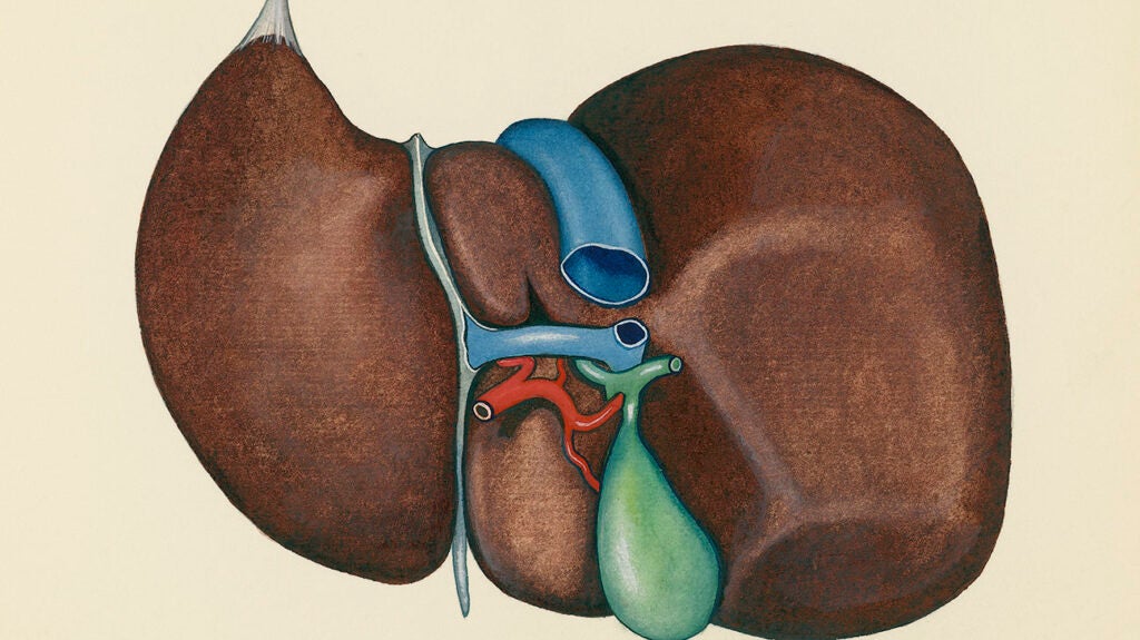
At its core, identifying a contracted gallbladder—a state where the organ appears shrunken or collapsed—relies heavily on the evolution of ultrasound technology. While CT and MRI scans play a role, ultrasound remains the primary technological tool due to its non-invasive nature and real-time imaging capabilities.
High-Frequency Transducers and Signal Resolution
The ability to distinguish between a healthy, distended gallbladder and a pathologically contracted one depends on the axial and lateral resolution of the ultrasound probe. Modern transducers utilize advanced piezoelectric crystals that can operate at varying frequencies. High-frequency probes (7–15 MHz) allow for superior detail of the gallbladder wall, which is critical when the organ is contracted. In a tech-driven diagnostic suite, the signal-to-noise ratio is optimized by software-defined imaging, allowing the technician to see whether the contraction is “postprandial” (natural contraction after a meal) or indicative of chronic cholecystitis.
Digital Signal Processing (DSP) and Speckle Reduction
Raw ultrasound data is inherently “noisy.” To determine if a gallbladder is truly contracted or simply obscured by bowel gas, modern ultrasound machines employ sophisticated Digital Signal Processing. Speckle reduction imaging (SRI) uses software algorithms to analyze the texture of the liver and gallbladder tissues, smoothing out graininess while preserving the sharp edges of the gallbladder wall. This technological layer ensures that the “contracted” status is not a false positive caused by poor image quality or artifacts.
The Rise of AI in Radiology: Automating the Detection of a Contracted Gallbladder
The interpretation of a contracted gallbladder is shifting from a purely human visual task to a collaborative effort between radiologists and Artificial Intelligence (AI). Computer-Aided Diagnosis (CAD) systems are now being integrated directly into imaging software to provide objective measurements that human eyes might overlook.
Deep Learning and Pattern Recognition
Artificial Intelligence models, particularly Convolutional Neural Networks (CNNs), are trained on massive datasets containing millions of gallbladder images. These models can identify a “contracted gallbladder” by comparing the current scan against an atlas of known pathologies. The AI looks for specific geometric markers: the thickness of the wall, the volume of the lumen, and the presence of echogenic shadows (calculi). By automating the segmentation of the gallbladder, these AI tools provide a volumetric analysis that is far more precise than a manual 2D measurement.
Reducing Inter-Operator Variability
One of the greatest challenges in ultrasound technology is its dependence on the skill of the operator. A technician might angle the probe in a way that makes a healthy gallbladder look contracted. AI-driven “Smart Scanning” assistants provide real-time feedback to the technologist, ensuring the probe is positioned optimally for a true volumetric reading. If the software detects a contracted gallbladder, it can automatically prompt the technician to capture additional planes of view, ensuring that the data sent to the physician is comprehensive and accurate.

Interconnectivity and the Digital Health Ecosystem
A diagnostic finding like a contracted gallbladder does not exist in a vacuum. In the modern tech-forward hospital, this finding is a digital packet that moves through a complex ecosystem of Picture Archiving and Communication Systems (PACS) and Electronic Health Records (EHR).
PACS Integration and DICOM Standards
The Digital Imaging and Communications in Medicine (DICOM) standard is the backbone of medical imaging tech. When a contracted gallbladder is identified, the high-resolution images are uploaded to a PACS server. Modern PACS utilize cloud-based architecture, allowing a specialist in another city to review the high-fidelity data instantly. This connectivity is vital; if a patient presents with a contracted gallbladder in a rural clinic, the “tech stack” allows for immediate consultation with a hepatobiliary surgeon at a major medical center.
Cloud Computing and Longitudinal Data Analysis
One of the most powerful aspects of modern medical software is the ability to track changes over time. Cloud-based EHRs allow clinicians to compare a current scan showing a contracted gallbladder with scans from five years prior. Predictive analytics software can analyze these longitudinal trends to determine if the contraction is part of a progressive inflammatory disease. This move from “snapshot” diagnostics to “trend” diagnostics is a hallmark of the Big Data revolution in healthcare.
Future Frontiers: Augmented Reality and Predictive Analytics in Biliary Tech
As we look toward the future, the technology used to interpret gallbladder states is moving beyond 2D screens and into the realms of immersive data visualization and predictive modeling.
3D Reconstruction and Augmented Reality (AR)
While traditional ultrasound provides a “slice” of the gallbladder, new software can synthesize these slices into a 3D volumetric model. Surgeons are beginning to use Augmented Reality (AR) headsets to overlay these 3D models onto a patient’s body during pre-operative planning. If a gallbladder is chronically contracted and scarred, the AR system can highlight the “danger zones” around the bile ducts, using data-driven insights to guide the surgeon’s hand and reduce the risk of complications.
Wearable Ultrasound and Continuous Monitoring
The next frontier in HealthTech is the transition from clinical-grade hardware to wearable devices. Researchers are currently developing “patch” ultrasound sensors that can be worn by the patient. For individuals with recurring biliary issues, such a device could theoretically monitor gallbladder contraction cycles in real-time, sending data to a smartphone app. This would allow doctors to see exactly how the gallbladder responds to different stimuli throughout the day, providing a level of granular data that was previously impossible to obtain.

Conclusion: The Digital Transformation of Clinical Insight
What does a contracted gallbladder mean in the age of technology? It means a complex interplay of sonic physics, algorithmic interpretation, and cloud-based data management. We have moved past the era where a diagnosis was a simple “yes” or “no” based on a blurry film. Today, a contracted gallbladder is a rich source of digital information that, when processed through the right tech stack, leads to faster interventions, safer surgeries, and a deeper understanding of human physiology. As AI continues to refine its predictive capabilities and imaging hardware reaches even higher resolutions, the “contracted gallbladder” will remain a key focus of the ongoing synergy between medicine and technology.
aViewFromTheCave is a participant in the Amazon Services LLC Associates Program, an affiliate advertising program designed to provide a means for sites to earn advertising fees by advertising and linking to Amazon.com. Amazon, the Amazon logo, AmazonSupply, and the AmazonSupply logo are trademarks of Amazon.com, Inc. or its affiliates. As an Amazon Associate we earn affiliate commissions from qualifying purchases.