
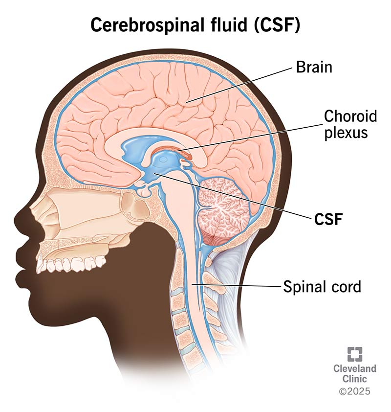
The seemingly simple question, “What does cerebral fluid look like?” unravels a complex tapestry of biological inquiry that modern technology is uniquely positioned to explore. While traditionally answered through direct observation—a clear, colorless fluid resembling water—today’s technological landscape allows us to delve far deeper. We no longer just perceive its macroscopic appearance; instead, we dissect its microscopic structure, chemical composition, dynamic flow, and subtle pathological variations with unprecedented precision. This article navigates how cutting-edge technology, from advanced imaging and AI-driven analytics to computational modeling, is revolutionizing our understanding of cerebrospinal fluid (CSF), transforming a basic biological query into a sophisticated diagnostic and research frontier.

The Digital Lens: Visualizing CSF with Next-Gen Imaging
For decades, the initial assessment of CSF involved a visual inspection, followed by basic lab tests. Today, technology provides a digital lens, offering extraordinary resolution and insight into what CSF “looks like” at various scales, revealing nuances invisible to the naked eye. This digital transformation is not just about seeing better; it’s about seeing differently, enabling earlier detection and more precise characterization of neurological conditions.
High-Resolution Microscopy and Spectroscopy: Beyond the Naked Eye
Traditional microscopy allows for cellular analysis, identifying red or white blood cells that might indicate hemorrhage or infection. However, modern high-resolution microscopy, often integrated with advanced digital imaging systems, pushes these boundaries. Techniques such as phase-contrast microscopy, differential interference contrast (DIC), and fluorescence microscopy can highlight subtle morphological changes in cells or even detect microorganisms with greater clarity and contrast, helping clinicians “see” infectious agents or abnormal cellular structures more distinctly.
Beyond visual morphology, spectroscopic techniques offer a chemical fingerprint of CSF. Raman spectroscopy, Fourier-transform infrared (FTIR) spectroscopy, and nuclear magnetic resonance (NMR) spectroscopy can analyze the molecular composition of CSF without extensive sample preparation. These technologies detect minute changes in protein profiles, metabolite concentrations, or the presence of abnormal molecules. For instance, subtle alterations in protein secondary structures, indicative of neurodegenerative diseases like Alzheimer’s or Parkinson’s, can be identified long before macroscopic changes manifest. This provides a chemical “look” at CSF, far more detailed than any visual inspection could offer. These spectral insights are crucial for early diagnosis and monitoring disease progression, turning invisible chemical changes into quantifiable data points.
AI-Enhanced Image Analysis: Pattern Recognition and Anomaly Detection
The sheer volume and complexity of data generated by high-resolution imaging and microscopy can overwhelm human analysis. This is where Artificial Intelligence (AI) and machine learning (ML) become indispensable. AI algorithms are trained on vast datasets of CSF images—both healthy and diseased—to learn intricate patterns, textures, and subtle anomalies that might escape the human eye.
For example, in cytological analysis of CSF, AI can automate the counting and classification of cells, identifying abnormal cell shapes or clusters suggestive of malignancy with higher speed and consistency than manual methods. In analyzing spectroscopic data, AI can rapidly process complex spectra to identify specific biomarkers or signatures associated with various neurological disorders, often with superior accuracy. By acting as a tireless, ultra-observant assistant, AI significantly enhances our ability to “see” and interpret the true appearance and characteristics of CSF, flagging deviations that warrant further investigation. This technology is not just augmenting human capabilities; it’s redefining the limits of what we can discern from biological samples.
Decoding the Unseen: AI and Machine Learning in CSF Diagnostics
The true power of AI in understanding what CSF looks like lies not just in visual interpretation but in its ability to decode the unseen—the complex interplay of biomarkers and genetic information. Machine learning models can process multi-modal data from CSF, integrating imaging, spectroscopic, proteomic, and genomic insights to build a comprehensive picture of a patient’s neurological health.
Predictive Analytics for Neurological Conditions
The ability to predict disease onset or progression is a holy grail in medicine, and AI-driven predictive analytics applied to CSF data is bringing this closer to reality. Machine learning algorithms can analyze patterns in CSF biomarker concentrations, genetic markers, and even subtle changes in fluid dynamics to identify individuals at high risk for neurodegenerative diseases (e.g., predicting the conversion from mild cognitive impairment to Alzheimer’s disease), inflammatory conditions, or certain types of stroke.
By correlating these CSF “looks” (data points) with clinical outcomes over time, AI models learn to recognize early warning signs that are often too subtle for human clinicians to synthesize from disparate data points. For instance, a combination of amyloid-beta, tau protein levels, and specific inflammatory markers, when analyzed by a sophisticated algorithm, can offer a more robust prediction than any single marker alone. This allows for proactive interventions and personalized treatment strategies, fundamentally changing how we approach neurological care.
Automating Biomarker Identification and Quantification
The process of identifying and quantifying biomarkers in CSF traditionally involves labor-intensive, often manual, laboratory techniques. Automation and robotics, powered by AI and ML, are transforming this landscape. High-throughput screening platforms can process hundreds of CSF samples rapidly, automatically isolating, purifying, and quantifying a wide array of biomarkers using techniques like mass spectrometry or advanced immunoassay systems.
AI algorithms then take over, analyzing the vast amounts of quantitative data generated, identifying relevant biomarkers, correcting for potential errors, and flagging results that fall outside normal ranges. This not only significantly increases efficiency and reduces human error but also enables the discovery of novel biomarkers that might have been missed in less comprehensive manual analyses. The automated, AI-driven quantification provides an objective and detailed “look” at the molecular profile of CSF, which is critical for both routine diagnostics and groundbreaking research.
Data Flow and Digital Twins: Simulating CSF Dynamics
Beyond its static appearance or molecular composition, CSF is a dynamic fluid, constantly circulating and interacting with brain tissue. Understanding “what it looks like” also involves comprehending its flow patterns and pressure dynamics. Here, advanced computational technologies offer an invaluable window into these complex processes.
Computational Fluid Dynamics (CFD) in Brain Health
Computational Fluid Dynamics (CFD) is a branch of fluid mechanics that uses numerical methods and algorithms to solve and analyze problems that involve fluid flows. Applied to CSF, CFD creates highly detailed, patient-specific 3D models of the brain’s ventricles and subarachnoid spaces based on MRI or CT scans. These digital twins allow researchers and clinicians to simulate the flow of CSF, understand pressure gradients, and predict how pathologies like hydrocephalus (excess CSF accumulation), cysts, or tumors might impede or alter its circulation.
By visualizing these simulated flow patterns, clinicians can “see” what abnormal CSF dynamics look like, helping them plan surgical interventions, predict the efficacy of shunts, or understand the impact of various conditions on intracranial pressure. This technology moves beyond a snapshot of CSF appearance to a dynamic, predictive understanding of its behavior within the complex intracranial environment.
Virtual Reality and Augmented Reality for Medical Training and Visualization
The complex anatomy of the brain and the intricate pathways of CSF can be challenging to visualize and understand, even for experienced medical professionals. Virtual Reality (VR) and Augmented Reality (AR) technologies are emerging as powerful tools to enhance this understanding. By leveraging patient-specific imaging data, VR platforms can generate immersive 3D models of the brain and its CSF spaces, allowing medical students and surgeons to “walk through” the anatomy, examine fluid pathways, and simulate surgical procedures in a risk-free environment.
AR applications, on the other hand, can overlay digital CSF flow models onto a physical patient during surgery or diagnostic procedures, providing real-time insights into underlying dynamics. This interactive visualization transforms how we perceive and learn about CSF, making its complex “look” and behavior tangible and explorable in ways previously impossible. It facilitates better surgical planning, enhances educational experiences, and ultimately improves patient outcomes.
The Future of CSF Analysis: Integration and Innovation
The trajectory of technological advancement in CSF analysis points towards even greater integration, non-invasiveness, and predictive power. The future will likely see a convergence of various tech modalities to create holistic, real-time pictures of neurological health.
Wearable Sensors and Non-Invasive Monitoring (Future Implications)
While direct CSF sampling is invasive, future technologies might offer non-invasive or minimally invasive ways to infer CSF characteristics. Research is underway on wearable sensors that could indirectly monitor intracranial pressure fluctuations, changes in brain fluid dynamics (e.g., through advanced acoustic or impedance measurements), or even detect specific biomarkers in peripheral fluids that correlate strongly with CSF changes. For instance, advanced optical sensors could potentially detect minute changes in tissue properties that reflect CSF volume or pressure. While still in early development, these technologies aim to provide continuous, real-time “looks” at factors influencing CSF health, enabling proactive monitoring outside of clinical settings.
Ethical AI and Data Security in Medical Diagnostics
As AI takes on a more central role in interpreting sensitive CSF data, the ethical considerations and data security implications become paramount. Developing robust, unbiased AI models requires diverse and representative datasets, ensuring that diagnostic tools are equitable across different patient populations. Furthermore, protecting the privacy and security of highly sensitive patient neurological data is crucial. Blockchain technology, secure multi-party computation, and advanced encryption techniques are being explored to ensure data integrity and confidentiality in the context of AI-driven medical diagnostics. The future of CSF analysis hinges not just on technological capability, but also on responsible and ethical implementation.

Conclusion
The question “What does cerebral fluid look like?” has evolved dramatically with the advent of sophisticated technology. No longer content with a macroscopic glance, we now employ advanced imaging, AI-driven analytics, and computational modeling to dissect its every facet—from microscopic cellular changes and molecular compositions to dynamic flow patterns. Technology has provided a multi-dimensional lens, transforming CSF from a simple biological fluid into a rich source of diagnostic and prognostic information. As these technological frontiers continue to expand, our ability to understand, predict, and intervene in neurological conditions will only grow more precise, paving the way for a new era of brain health management powered by innovation.
aViewFromTheCave is a participant in the Amazon Services LLC Associates Program, an affiliate advertising program designed to provide a means for sites to earn advertising fees by advertising and linking to Amazon.com. Amazon, the Amazon logo, AmazonSupply, and the AmazonSupply logo are trademarks of Amazon.com, Inc. or its affiliates. As an Amazon Associate we earn affiliate commissions from qualifying purchases.