
In the realm of orthopedic medicine, the question “what does a torn meniscus look like?” has evolved from a matter of physical palpation and surgical exploration to a sophisticated exercise in high-definition digital visualization. For decades, the meniscus—the C-shaped piece of tough, rubbery cartilage that acts as a shock absorber between your shinbone and thighbone—was a “black box” of sorts. Today, however, breakthroughs in medical technology, signal processing, and artificial intelligence have transformed how we visualize, diagnose, and treat this common yet debilitating injury.
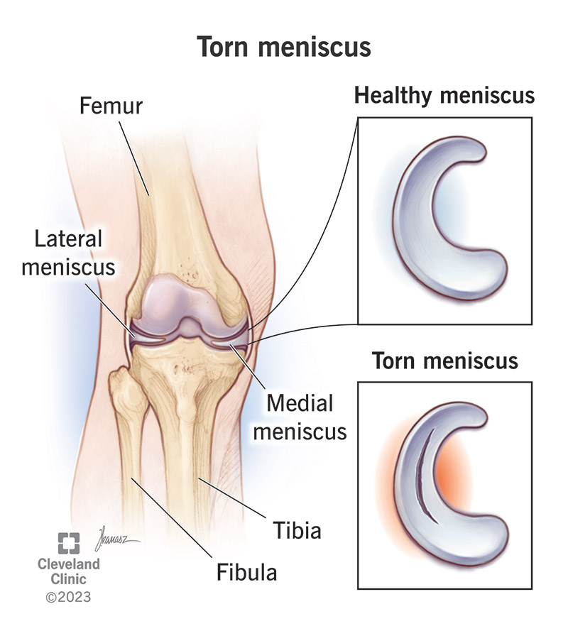
To understand what a torn meniscus “looks” like in the modern era, one must look past the biological tissue and into the advanced tech stack that powers contemporary radiology and digital health.
The Digital Lens: How MRI Technology Visualizes Soft Tissue Damage
When a clinician seeks to visualize a meniscal tear, the gold standard is Magnetic Resonance Imaging (MRI). Unlike X-rays, which are excellent for bone density but fail to capture soft tissue, MRI utilizes powerful magnets and radio waves to create detailed cross-sectional images. In the context of technology, “what a tear looks like” is defined by signal intensity and pixel variance.
The Physics of the Image: T1 and T2 Weighted Sequences
In a digital MRI output, a healthy meniscus appears as a solid black, wedge-shaped structure. This is because fibrocartilage has low water content and does not emit a strong signal, resulting in a dark “void” on the screen. A tear, conversely, allows joint fluid (which is high in protons) to penetrate the structure.
From a technological standpoint, radiologists look for “hyperintensity”—bright white lines or spots—cutting through the black wedge. Tech-driven imaging protocols, such as T2-weighted fat-saturated sequences, allow the software to suppress the signal from surrounding fat, making the “bright” fluid within the “dark” tear stand out with high contrast.
High-Resolution Precision: From 1.5T to 7T Scanning
The “look” of a tear is also a function of Tesla (T) strength—the measurement of magnetic field power. Traditional 1.5T scanners provided a grainy view that sometimes led to “false positives” or missed small horizontal cleavages. The shift toward 3T and the experimental 7T scanners represents a massive leap in hardware capability. These high-field magnets provide a superior signal-to-noise ratio, allowing for thinner “slices” of the knee to be imaged. This prevents “partial volume averaging,” a digital artifact where the computer blurs a tear with healthy tissue because the image slice is too thick.
The Role of Artificial Intelligence in Identifying Knee Pathology
The most significant recent trend in medical technology isn’t the camera itself, but the software interpreting the data. As the volume of diagnostic images grows globally, Artificial Intelligence (AI) and Machine Learning (ML) have become essential tools in defining what a torn meniscus looks like to a computer.
Computer Vision and Pattern Recognition in Radiology
AI models, specifically Convolutional Neural Networks (CNNs), are now trained on millions of labeled MRI slices. To an AI, a torn meniscus looks like a statistical deviation in pixel arrangement. While a human eye might miss a subtle “parrot-beak” tear or a hidden “root tear,” AI algorithms can analyze the geometric integrity of the meniscal borders with mathematical precision. These tools can highlight areas of concern, effectively acting as a digital “highlighter” for the radiologist, ensuring that subtle fraying—which might look like a mere shadow to a tired human eye—is flagged for review.

Reducing Human Error: AI as a Second Pair of Eyes
In the tech-driven diagnostic workflow, “looking” at a tear is no longer a solitary task. AI tools provide “Computer-Aided Detection” (CADe). These systems are particularly adept at identifying “discoid menisci”—a structural abnormality that is more prone to tearing. By comparing a patient’s unique anatomy against a vast database of pathological “looks,” the software can predict the likelihood of a tear with an accuracy rate that often rivals or exceeds senior consultants. This technology reduces the “diagnostic lag,” ensuring that what a tear “looks like” is determined in seconds rather than days.
Beyond the Static Image: Dynamic Imaging and 3D Modeling
A torn meniscus does not just “look” like a static line on a screen; it “looks” like a functional failure during movement. The tech industry is moving away from static 2D slices toward dynamic, multi-dimensional representations of knee health.
Cinematographic MRI and Functional Assessment
Newer “Cine-MRI” technology allows clinicians to see the meniscus in motion. Because some tears (like “bucket-handle” tears) only displace when the knee is flexed or extended, a static MRI might miss the true extent of the damage. Digital frame-by-frame analysis allows surgeons to see how the cartilage “flaps” or “locks” during joint rotation. This represents a shift from seeing a tear as an image to seeing it as a mechanical event.
Digital Twins: Simulating Surgical Outcomes
One of the most exciting trends in “MedTech” is the creation of a “Digital Twin.” Using the data from an MRI, software can generate a 3D volumetric model of a patient’s knee. Surgeons can then “look” at the tear from any angle in a virtual environment. This tech allows for preoperative planning where a surgeon can virtually “trim” the meniscus or simulate a repair to see how it affects the stress distribution across the joint. In this context, a torn meniscus looks like a 3D structural vulnerability that can be stress-tested in a digital sandbox before a single incision is made.
The Future of Wearable Tech in Meniscal Health Monitoring
The question of what a torn meniscus looks like is also being answered by data points outside the radiology suite. Wearable technology is providing a “biometric look” at internal injuries through the analysis of gait and mechanical load.
Smart Braces and Biometric Feedback
Modern “Smart Braces” equipped with integrated sensors and accelerometers can detect the subtle changes in a patient’s stride caused by a meniscal tear. To this technology, a tear looks like an “asymmetry profile.” If a patient is subconsciously offloading weight or if the knee is exhibiting “micro-instability” during the terminal extension phase of a step, the wearable sensors log this as a specific data signature. This “digital phenotype” of an injury allows for early intervention before a minor fray becomes a major rupture.
Remote Patient Monitoring and Data-Driven Recovery
Following a diagnosis or surgery, the “look” of the meniscus is tracked through recovery data. Telehealth platforms and remote monitoring tools use computer vision via a smartphone camera to track a patient’s range of motion. If the software detects a “catch” or a limited degree of flexion, it correlates that visual data with the known physical characteristics of a meniscal tear. This creates a continuous loop of visual and digital data, ensuring that the “look” of a healing meniscus is trending toward the “look” of a healthy one.
Conclusion: The Convergence of Tech and Orthopedic Care
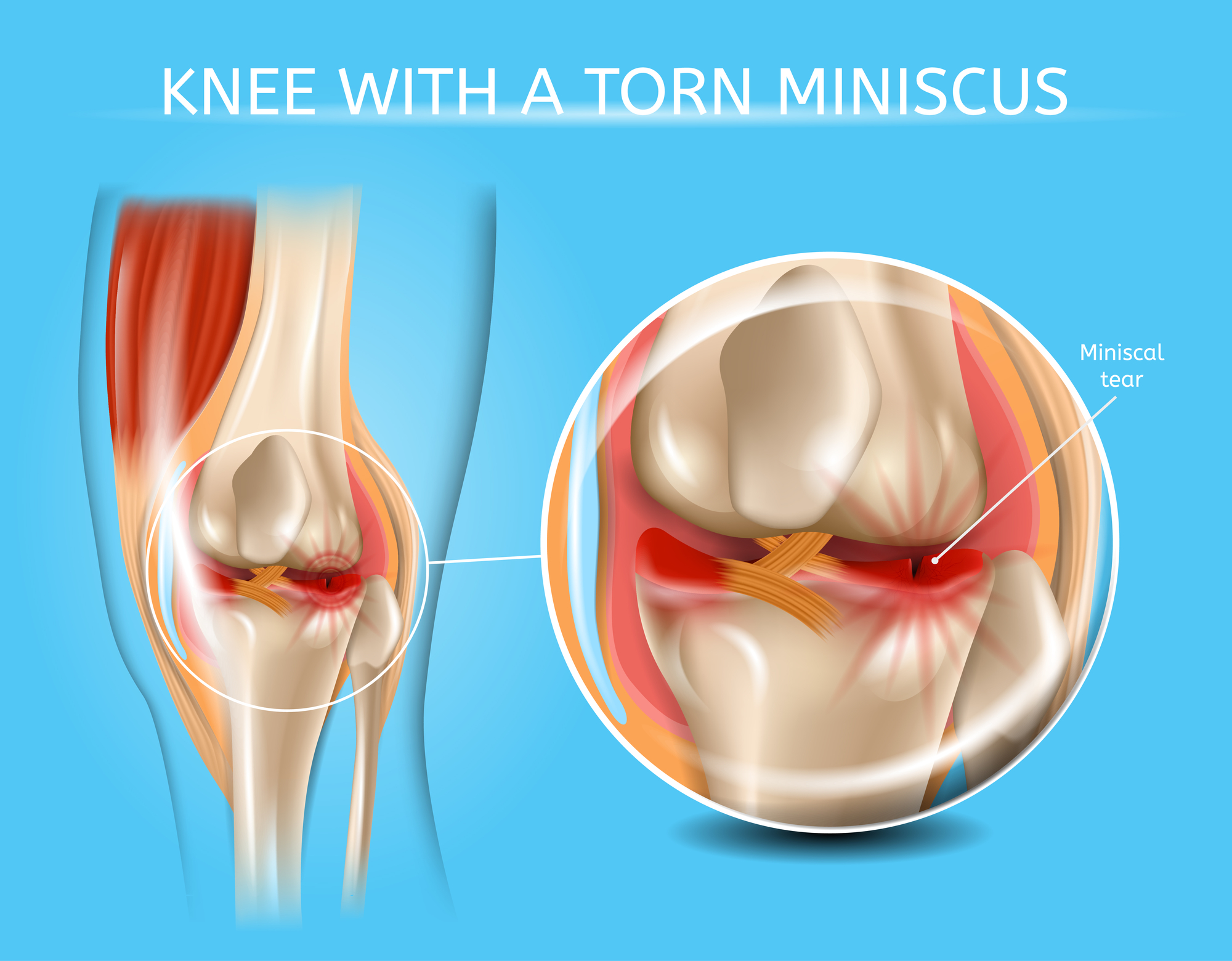
Ultimately, what a torn meniscus looks like depends entirely on the technology used to observe it. To a 3T MRI, it looks like a hyperintense signal disrupting a dark fibrocartilage wedge. To an AI algorithm, it looks like a pixel anomaly that deviates from a trained norm. To a 3D modeling program, it looks like a mechanical flaw in a complex structural system.
As we look toward the future, the integration of these technologies—AI, high-field imaging, 3D modeling, and wearables—is creating a holistic, 360-degree view of musculoskeletal health. We are no longer limited to “looking” at a tear through a grainy lens. Instead, we are analyzing it through a sophisticated tech stack that promises higher diagnostic accuracy, personalized surgical interventions, and more effective recovery protocols. The digital transformation of orthopedics has ensured that when we ask what an injury looks like, the answer is more detailed, accurate, and actionable than ever before.
aViewFromTheCave is a participant in the Amazon Services LLC Associates Program, an affiliate advertising program designed to provide a means for sites to earn advertising fees by advertising and linking to Amazon.com. Amazon, the Amazon logo, AmazonSupply, and the AmazonSupply logo are trademarks of Amazon.com, Inc. or its affiliates. As an Amazon Associate we earn affiliate commissions from qualifying purchases.