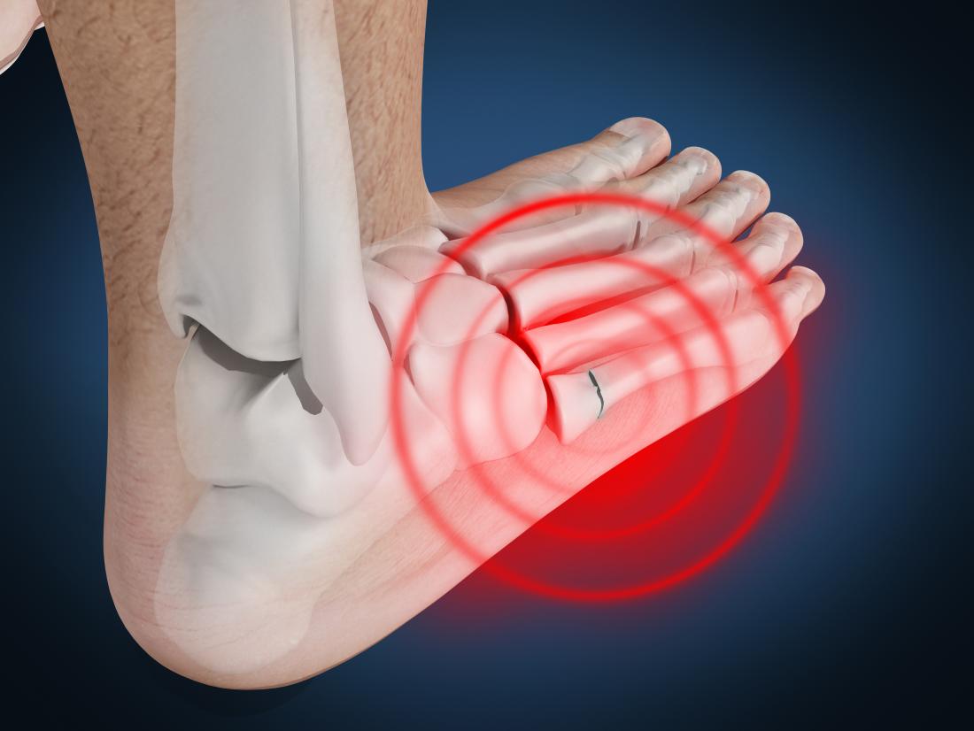
The human foot is a marvel of biomechanical engineering, a complex structure of 26 bones, 33 joints, and over 100 muscles, tendons, and ligaments working in concert to provide support, mobility, and balance. When this intricate system is subjected to excessive force, whether from a direct impact, a fall, or repetitive stress, one or more of these bones can crack or break. This injury, known as a fractured foot, can range from a hairline crack to a complete separation of bone fragments. While the physical symptoms – pain, swelling, bruising – are readily apparent, understanding the visual representation and diagnostic nuances of a fractured foot increasingly relies on advanced technological tools. This article delves into what a fractured foot looks like, not just from a clinical observation standpoint, but through the lens of cutting-edge technology that allows us to visualize, diagnose, and ultimately, heal these complex injuries.

Visualizing the Invisible: Advanced Imaging Technologies
The initial assessment of a suspected foot fracture often begins with a physical examination. However, to confirm a diagnosis, pinpoint the exact location and severity of the fracture, and rule out other soft tissue injuries, medical professionals rely on sophisticated imaging techniques. These technologies have revolutionized the way we “see” inside the body, transforming guesswork into precise data-driven diagnosis.
The Foundation: Radiography (X-rays)
Standard X-rays remain the cornerstone of initial fracture diagnosis. These machines utilize electromagnetic radiation to pass through the body, with denser tissues like bone absorbing more radiation than softer tissues. The transmitted radiation then exposes a detector (film or digital sensor), creating an image where bones appear white and soft tissues appear in shades of gray.
What to Look For on an X-ray:
- Displacement: A clear break in the bone’s continuity. If the bone fragments are no longer aligned, this is considered displaced.
- Angulation: The bone fragments are bent at an unnatural angle.
- Shortening: The fractured bone is shorter than its intact counterpart.
- Comminution: The bone is broken into multiple pieces.
- Avulsion Fractures: A small piece of bone is pulled away by a ligament or tendon.
- Stress Fractures: Often appear as subtle thickening of the bone cortex or a faint line of lucency (darkness) within the bone. Early stress fractures might not be visible on initial X-rays and may require follow-up imaging.
The clarity and detail of modern digital X-ray systems far surpass older film-based methods. High-resolution detectors capture finer details, allowing radiologists to identify even subtle hairline fractures that might have been missed previously. Furthermore, digital images can be manipulated – enhanced, magnified, and contrasted – to optimize visualization, aiding in the accurate identification of fracture lines.
Beyond the Basics: Advanced Imaging Modalities
While X-rays provide a crucial initial overview, certain complex fractures or situations requiring a more in-depth look necessitate advanced imaging techniques. These technologies offer greater detail, can visualize soft tissues, and provide three-dimensional perspectives.
Computed Tomography (CT) Scans: The Three-Dimensional View
CT scans employ a series of X-ray beams taken from multiple angles around the body. A computer then processes these images to create cross-sectional “slices” or tomograms of the bone. This provides an exceptionally detailed, three-dimensional view of the fracture.
When CT Scans are Indispensable:
- Intra-articular Fractures: Fractures that extend into a joint surface. CT scans are vital for assessing the degree of joint involvement, detecting small fragments, and planning surgical repair.
- Complex Fractures: Particularly those involving multiple bone fragments or significant displacement, where traditional X-rays may not offer sufficient clarity.
- Pre-Surgical Planning: CT provides surgeons with precise anatomical information, allowing them to visualize the fracture in detail, plan the surgical approach, and anticipate potential challenges.
- Post-Surgical Assessment: CT can be used to evaluate the healing of bone after surgery and the placement of hardware.
The visual output of a CT scan is typically a series of axial, coronal, and sagittal images, offering a comprehensive understanding of the fracture’s geometry and spatial relationships. Advanced visualization software can even render these slices into interactive 3D models, allowing surgeons to “hold” and manipulate the patient’s anatomy on a screen.
Magnetic Resonance Imaging (MRI): Illuminating Soft Tissues and Subtle Fractures
MRI uses strong magnetic fields and radio waves to generate detailed images of both bone and soft tissues. Unlike X-rays or CT, MRI does not involve ionizing radiation. It is particularly adept at visualizing cartilage, ligaments, tendons, and bone marrow.
The Role of MRI in Fracture Diagnosis:
- Occult Fractures: Fractures that are not visible on initial X-rays but are suspected due to persistent pain and tenderness. MRI can detect edema (swelling) within the bone marrow, which is often an early sign of a stress fracture.
- Associated Soft Tissue Injuries: Fractures are often accompanied by ligamentous tears, tendon injuries, or cartilage damage. MRI is the gold standard for diagnosing these concomitant injuries, which can significantly impact treatment and recovery.
- Osteomyelitis: In cases of suspected infection of the bone, MRI can help identify the extent of the infection and any associated soft tissue involvement.
- Avascular Necrosis: A condition where bone tissue dies due to a lack of blood supply, which can sometimes occur after a fracture. MRI is excellent at detecting early signs of avascular necrosis.
On MRI, fractures often appear as areas of high signal intensity (bright spots) within the bone on T2-weighted or STIR sequences, indicating edema. This allows for the detection of fractures that might be completely invisible on conventional X-rays.
Ultrasound: A Dynamic and Accessible Tool
While less common for initial fracture diagnosis compared to X-rays or CT, ultrasound technology is emerging as a valuable tool, particularly for certain types of fractures and in specific clinical settings. Ultrasound uses high-frequency sound waves to create images. It is particularly useful for visualizing superficial structures and assessing soft tissues.
Potential Applications of Ultrasound:
- Distal Tibia and Fibula Fractures: Studies have shown ultrasound can be effective in detecting certain fractures in these locations, especially in pediatric populations.
- Assessing Periosteal Reaction: Ultrasound can identify subtle signs of bone healing or periosteal (the membrane covering the bone) reaction, which can be indicative of a fracture.
- Dynamic Assessment: Ultrasound allows for real-time imaging, enabling the assessment of joint stability and soft tissue integrity during movement, which can be helpful in identifying ligamentous injuries associated with fractures.
- Point-of-Care Diagnosis: Its portability and lack of radiation make ultrasound an attractive option for rapid assessment in emergency settings or remote locations.
Digital Reconstruction and 3D Modeling: Enhancing Surgical Planning and Education
The advent of advanced imaging has paved the way for sophisticated digital reconstruction and 3D modeling techniques, offering unparalleled insights into fractured foot anatomy and profoundly impacting surgical planning, patient education, and medical training.
From Slices to Surfaces: The Power of Reconstruction Software
Raw imaging data from CT and MRI scans are processed by specialized software to create highly detailed, multi-dimensional representations of the fractured foot. This involves reconstructing the 2D slices into a cohesive 3D model.
Key Benefits of Digital Reconstruction:
- Comprehensive Anatomical Visualization: Surgeons can view the fracture from any angle, isolating specific bones and joints. This allows for a much deeper understanding of the fracture pattern, displacement, and comminution than was previously possible.
- Virtual Surgical Planning: Before an operation, surgeons can use these 3D models to simulate different surgical approaches, test the fit of implants, and determine the optimal trajectory for screws and plates. This virtual rehearsal significantly reduces operative time and improves accuracy.
- Patient-Specific Implants: In complex cases, 3D models can be used to design and manufacture patient-specific implants and surgical guides, ensuring a precise fit and improved surgical outcomes.
3D Printing: Bringing Digital Models to Life
The integration of 3D printing technology with digital imaging has created a powerful synergy. Anatomical models of the fractured foot, derived from patient scans, can now be physically printed.
The Impact of 3D Printed Models:
- Enhanced Surgical Dexterity: Surgeons can physically hold and examine a tangible replica of the fractured foot, allowing them to palpate the fracture lines and practice complex maneuvers on the model before operating on the patient. This tactile experience is invaluable for complex reconstructions.
- Improved Patient Communication: A 3D printed model can be a powerful tool for explaining the injury and the proposed surgical plan to patients. Seeing and touching a replica of their own anatomy can demystify the condition and foster greater patient understanding and buy-in.
- Medical Education and Training: 3D printed anatomical models provide realistic training tools for medical students and residents, offering hands-on experience with complex fractures without the risks associated with cadaveric dissection or operating on live patients.
Artificial Intelligence and Machine Learning: The Future of Fracture Detection
The integration of Artificial Intelligence (AI) and Machine Learning (ML) into medical imaging is rapidly transforming the field, promising to enhance the accuracy, efficiency, and accessibility of fracture diagnosis. AI algorithms can be trained on vast datasets of medical images to identify patterns and anomalies that may be subtle or easily overlooked by the human eye.
AI-Powered Image Analysis: Augmenting Radiologist Capabilities
AI algorithms are being developed to assist radiologists in detecting and characterizing fractures from various imaging modalities.
How AI is Making a Difference:
- Automated Fracture Detection: AI systems can scan X-rays, CT scans, and even MRI images to flag potential fracture sites, drawing the radiologist’s attention to areas of concern. This can act as a “second pair of eyes,” reducing the risk of missed diagnoses.
- Quantitative Analysis: AI can precisely measure fracture displacement, angulation, and fragment size, providing objective data to support diagnostic decisions and treatment planning.
- Triage and Prioritization: In busy radiology departments, AI can help prioritize urgent cases by identifying critical findings such as unstable fractures or those requiring immediate intervention.
- Predictive Analytics: Future applications of AI may involve predicting fracture healing rates or the likelihood of complications based on imaging characteristics and patient data.
Machine Learning for Enhanced Visualization and Reconstruction
Beyond simple detection, ML is being employed to refine image processing and reconstruction techniques.
Advancing Image Quality and Interpretation:
- Noise Reduction and Image Enhancement: ML algorithms can effectively reduce image noise and enhance contrast in medical scans, leading to clearer and more interpretable images, especially in cases where image quality might be compromised.
- Iterative Reconstruction: In CT imaging, ML is being used to improve iterative reconstruction techniques, allowing for reduced radiation dose while maintaining or even improving image quality.
- Automated Segmentation: ML models can automatically segment different anatomical structures within an image, simplifying the process of creating 3D models and isolating specific areas of interest.
While still in its nascent stages for fracture diagnosis, the potential of AI and ML in this domain is immense. As these technologies mature and are integrated into clinical workflows, they will undoubtedly play a crucial role in visualizing, understanding, and ultimately treating fractured feet with greater precision and efficiency. The “look” of a fractured foot, as seen through the sophisticated lenses of modern technology, is becoming increasingly detailed, objective, and predictive, heralding a new era in orthopedic care.
aViewFromTheCave is a participant in the Amazon Services LLC Associates Program, an affiliate advertising program designed to provide a means for sites to earn advertising fees by advertising and linking to Amazon.com. Amazon, the Amazon logo, AmazonSupply, and the AmazonSupply logo are trademarks of Amazon.com, Inc. or its affiliates. As an Amazon Associate we earn affiliate commissions from qualifying purchases.