
Managing a medical condition like diabetes requires a heightened level of vigilance, particularly regarding foot health. For those leveraging health-tech ecosystems—such as wearable sensors, integrated glucose monitoring apps, and AI-driven diagnostic platforms—understanding the visual markers of complications is critical. When digital health monitoring systems flag anomalies, recognizing the physical appearance of a diabetic foot ulcer can be the difference between a minor intervention and a long-term clinical challenge.
The Digital Anatomy of a Diabetic Foot Ulcer
In the landscape of modern digital health, an ulcer is not merely a wound; it is a clinical data point that manifests through specific physical characteristics. Because diabetic neuropathy often mutes pain receptors, patients frequently rely on visual inspection or AI-assisted image analysis to detect these developments.

The Initial Visual Markers: Erythema and Callus Formation
Before an ulcer fully opens, the skin often exhibits warning signs that sophisticated digital health trackers aim to identify via thermal imaging or high-resolution photography. The process typically begins with localized erythema—redness that indicates inflammation. In many cases, this is preceded by a thickened, hyperkeratotic area known as a callus.
A callus in a diabetic patient is not a benign skin condition. It acts as a pressure point. When you observe a yellowish, hardened, or translucent patch of skin on the sole of the foot or over a bony prominence, it serves as a digital indicator of mechanical stress. If left unmanaged, the pressure causes tissue breakdown beneath the callus, often resulting in a sub-callous hemorrhage, which presents as a dark, purple, or black spot visible through the thickened skin.
The Appearance of the Open Ulcer
Once the skin barrier is breached, the ulcer assumes a distinct appearance. Clinically, a typical diabetic foot ulcer is often circular or irregular in shape, with a “punched-out” appearance. The wound bed itself may vary in color depending on the stage of healing or infection:
- Granulation tissue: A healthy, healing ulcer will display a beefy, red, granular base. This is the physiological goal for treatment protocols.
- Slough: If the wound appears covered in a yellowish or tan, stringy substance, it indicates necrotic tissue or debris that requires professional debridement.
- Eschar: A black, leathery, or dry coating suggests necrotic tissue that has hardened. This is a high-risk visual indicator that often requires clinical intervention.
The Role of Smart Monitoring and AI Diagnostics
Modern software solutions have revolutionized how we categorize and track these visual markers. Through deep learning algorithms, smartphone applications are now capable of analyzing images of the foot to assess wound severity and track healing trajectories.
Leveraging Machine Learning for Wound Classification
AI-powered diagnostic tools are trained on thousands of clinical images to distinguish between normal skin changes and pathologically significant ulcers. These tools look for the “Periwound” area—the skin immediately surrounding the ulcer.
If the surrounding skin appears shiny, tightly stretched, or shows a clear border of edema (swelling), the software interprets this as a potential sign of cellulitis or deep tissue involvement. By digitizing the observation process, patients can upload images to secure portals, allowing healthcare providers to receive quantitative data on the wound’s dimensions, depth, and color composition, rather than relying solely on subjective descriptions.
Integrating IoT and Wearable Sensors
The tech-driven approach to ulcer prevention often involves the use of smart insoles and IoT-enabled socks. These devices measure plantar pressure distribution and skin temperature. When a specific area of the foot shows a persistent temperature elevation—a sign of the inflammatory process that precedes an ulcer—the software triggers an alert.
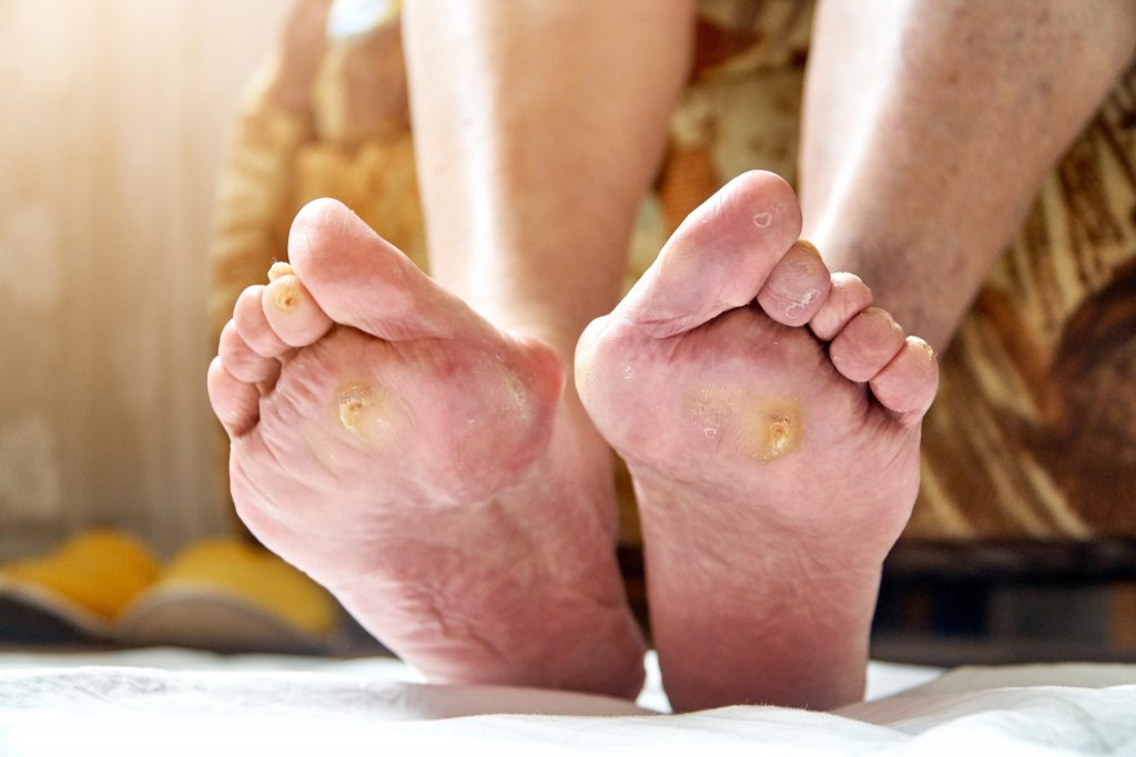
When a user receives such an alert, they are prompted to visually inspect the area for the markers described above. This symbiosis between hardware sensors and visual self-assessment creates a proactive defense loop, preventing the ulcer from reaching advanced, more difficult-to-treat stages.
Warning Signs: When Digital Documentation is Not Enough
While software applications are excellent for monitoring, they are not a substitute for clinical diagnostics. Understanding when a visual change necessitates immediate professional attention is a pillar of responsible health management.
Indicators of Infection: The Digital Health Alert
Infection in a diabetic foot ulcer is a clinical emergency. Technology can assist in detection, but the user must recognize the following visual cues:
- Purulent Exudate: If the ulcer produces a thick, cloudy, or foul-smelling discharge, the software’s alert system will typically classify this as a high-risk event.
- Spreading Erythema: If the redness around the ulcer begins to track upward (a phenomenon often called “streaking”), it suggests a rapid spread of infection into the lymphatic system.
- Crepitus and Gas: In advanced digital imaging, particularly those that support multi-spectral analysis, pockets of gas or air can sometimes be detected, though this is usually reserved for advanced clinical settings.
If a patient captures an image of a wound that suddenly shows these characteristics, modern health apps are designed to bypass routine scheduling and provide direct tele-consultation links to wound care specialists.
The Importance of Consistent Visual Logging
Consistent tracking is the core of the digital health philosophy. A single image is a snapshot; a gallery of images is a trend report. By maintaining a longitudinal record of the foot’s appearance through an encrypted health app, patients provide their medical team with an objective visual history.
This is particularly important for differentiating between stagnant, chronic wounds and those that are responding to modern therapies like negative pressure wound therapy (NPWT) or bioengineered skin substitutes. If the color of the wound bed shifts from yellow to red, the patient knows the treatment is working. If it shifts from red to black or grey, the digital record serves as evidence that the current care plan requires immediate revision.
Technological Best Practices for Foot Self-Examination
To maintain the accuracy of your digital health data, the physical examination must be conducted under optimal conditions. Technology is only as good as the input it receives.
Environmental Optimization for Image Capture
When performing a self-check or using an app to scan your feet, ensure the environment mimics clinical lighting. High-intensity, angled lighting is often used by dermatologists to highlight the depth of an ulcer or the elevation of a callus. Avoid shadows that could hide the extent of the wound.
Use high-resolution camera settings and, if possible, a fixed distance to ensure that successive photos are comparable for the AI algorithms that track surface area reduction. This consistency allows the software to generate accurate growth or regression charts, providing a clear “look” at the health of the foot over time.

The Feedback Loop between Patient and Platform
The ultimate goal of using technology for diabetic foot management is to normalize the observation process. By integrating daily, systematic checks into your digital routine—much like checking your glucose levels—the anxiety associated with foot health is replaced by a data-driven strategy.
A diabetic foot ulcer looks different at every stage of its progression, from a simple pressure-induced callus to a deep, infected wound. By combining the clinical knowledge of these visual markers with the diagnostic power of today’s software and sensor technology, individuals can proactively manage their health, minimize the risk of severe complications, and ensure that their feet remain a foundation for their active, digital-forward lifestyle. The visual recognition of these markers, bolstered by the precision of modern applications, represents the new frontier in diabetic care—moving away from reactive emergency medicine and toward a future of predictive, persistent, and informed personal health management.
aViewFromTheCave is a participant in the Amazon Services LLC Associates Program, an affiliate advertising program designed to provide a means for sites to earn advertising fees by advertising and linking to Amazon.com. Amazon, the Amazon logo, AmazonSupply, and the AmazonSupply logo are trademarks of Amazon.com, Inc. or its affiliates. As an Amazon Associate we earn affiliate commissions from qualifying purchases.