
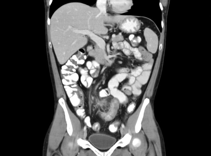
In the rapidly evolving landscape of health technology, the Computed Tomography (CT) scan has transitioned from a basic diagnostic tool to a sophisticated nexus of high-speed hardware and artificial intelligence. When we ask, “What does a CT scan show in the abdomen?” we are no longer just looking at static X-ray shadows. Instead, we are interrogating a massive dataset processed by complex algorithms to visualize the intricate architecture of the human torso. For tech enthusiasts, developers, and digital health strategists, the modern abdominal CT scan represents one of the most successful integrations of Big Data, computer vision, and edge computing in the professional world today.

The Evolution of Compute in Medical Imaging: Hardware and Software Synergy
At its core, a CT scan is a feat of engineering that marries physics with high-performance computing. To understand what the scan reveals within the abdomen, one must first understand the “tech stack” that makes it possible. Unlike traditional X-rays, which provide a 2D projection, the CT scan utilizes a rotating gantry that captures thousands of “slices” of the body.
Multi-Detector Row CT (MDCT) Technology
The hardware advancement known as Multi-Detector Row CT (MDCT) has revolutionized the speed and resolution of abdominal imaging. In the past, capturing a clear image of the moving gut, a pulsating aorta, or a breathing diaphragm was a challenge due to motion artifacts. Modern scanners now utilize 64, 128, or even 640 slices. This hardware allows for sub-millimeter isotropic resolution, meaning the digital data is equally detailed in all three dimensions. From a technical perspective, this creates a volumetric dataset rather than a series of pictures, allowing software to “re-slice” the abdomen in any plane—axial, sagittal, or coronal—without loss of quality.
The Role of Edge Computing in Real-Time Reconstruction
The raw data generated during an abdominal scan is enormous. To convert raw X-ray attenuation data into viewable images, the system must perform “filtered back-projection” or “iterative reconstruction.” This requires immense processing power. Modern CT suites utilize specialized GPUs and edge computing clusters located directly within the hospital’s infrastructure to process these algorithms in near real-time. This ensures that while the patient is still on the table, the software is already rendering a 3D map of their vascular system and organ structures, identifying potential anomalies with high-speed mathematical precision.
AI and Machine Learning: Enhancing Abdominal Diagnostics
The most significant trend in current medical technology is the integration of Artificial Intelligence (AI) and Machine Learning (ML) into the radiology workflow. When a CT scan looks at the abdomen, the software is increasingly doing the “heavy lifting” of initial identification, acting as a sophisticated layer of computer vision.
Automated Lesion Detection and Segmentation
One of the primary functions of AI in abdominal CT imaging is automated segmentation. In a dense scan containing the liver, spleen, kidneys, pancreas, and bowel, manually outlining each organ is time-consuming. Modern AI tools use deep learning models—trained on millions of previous scans—to automatically segment organs and identify lesions. For instance, if the scan identifies a focal point in the liver, the software can analyze the “texture” of the pixels (radiomics) to determine if the growth matches the digital signature of a benign cyst or a malignant tumor. This tech allows for a level of granular analysis that exceeds the human eye’s ability to distinguish subtle grayscale variations.

Predictive Analytics and Opportunistic Screening
Beyond simply showing what is currently in the abdomen, tech-driven CT analysis is moving toward predictive diagnostics. Using ML algorithms, a standard abdominal scan can be used for “opportunistic screening.” For example, a scan taken to investigate stomach pain can be processed by an AI tool to measure bone density in the spine (identifying osteoporosis) or to quantify visceral fat and muscle mass (sarcopenia). This turns a single diagnostic event into a multi-variable data report, providing a comprehensive view of the patient’s biological “tech specs” and long-term health trajectory.
Digital Security and Interoperability in Radiologic Data
As the resolution of abdominal CT scans increases, so does the digital footprint of the data. Managing this information requires a robust tech infrastructure centered on security and seamless interoperability. An abdominal CT scan is not just a file; it is a complex container of metadata and high-resolution imagery.
Protecting DICOM Files and Patient Privacy
The standard format for medical imaging is DICOM (Digital Imaging and Communications in Medicine). From a digital security standpoint, protecting these files is paramount. Because an abdominal CT contains unique anatomical markers (like a digital fingerprint of the spine or vascular tree), it is considered highly sensitive Personal Health Information (PHI). Cybersecurity in this niche involves end-to-end encryption and strict access controls. Furthermore, the rise of “adversarial AI”—where malicious actors could theoretically inject fake lesions into a digital scan—has led to the development of digital watermarking and blockchain-based verification systems to ensure the integrity of the scan data from the gantry to the doctor’s workstation.
Cloud-Based Image Sharing and Collaborative Diagnostics
The era of carrying a physical CD of your CT scan is ending. The current tech trend leans toward vendor-neutral archives (VNA) and cloud-based Picture Archiving and Communication Systems (PACS). These platforms allow high-resolution abdominal datasets to be streamed to specialists anywhere in the world. This interoperability is powered by high-speed fiber networks and sophisticated compression algorithms that allow for “lossless” viewing, ensuring that a surgeon in a different time zone sees the exact same pixel-perfect detail of a patient’s renal artery as the technician who performed the scan.
Future Trends: From 3D Rendering to Digital Twins
The future of what a CT scan shows in the abdomen lies in the synthesis of radiology with virtual reality (VR) and generative technology. We are moving away from 2D gray-scale images toward immersive, data-rich environments.
Cinematic Rendering and Virtual Dissection
Cinematic rendering is a relatively new post-processing technique that uses global illumination algorithms—the same technology used in high-end Hollywood CGI and video games—to create photorealistic 3D visualizations of CT data. When applied to the abdomen, this tech allows surgeons to perform a “virtual dissection.” They can rotate a hyper-realistic model of a patient’s gallbladder or colon, seeing exactly how blood vessels wrap around an organ before they ever make an incision. This minimizes surgical risk and represents a pinnacle of how consumer-grade graphics tech is being repurposed for life-saving medical applications.
The Integration of GenAI in Radiology Reporting
Generative AI (GenAI) is also beginning to impact how CT results are communicated. Natural Language Processing (NLP) models can now take the complex, structured data from an abdominal scan and translate it into a readable report for the patient or a summary for the primary care physician. Moreover, GenAI is being used to synthesize “synthetic data” to train new models, helping researchers understand rare abdominal conditions without compromising real patient privacy.
As we look toward the next decade, the abdominal CT scan will continue to evolve. It will become faster, safer (using lower radiation doses optimized by AI), and more integrated into the broader “Digital Twin” concept—where every scan feeds into a persistent digital model of an individual’s health. What the CT scan shows in the abdomen is no longer just a picture of our insides; it is a high-resolution, AI-augmented, and highly secure digital map of our biological reality.
aViewFromTheCave is a participant in the Amazon Services LLC Associates Program, an affiliate advertising program designed to provide a means for sites to earn advertising fees by advertising and linking to Amazon.com. Amazon, the Amazon logo, AmazonSupply, and the AmazonSupply logo are trademarks of Amazon.com, Inc. or its affiliates. As an Amazon Associate we earn affiliate commissions from qualifying purchases.