
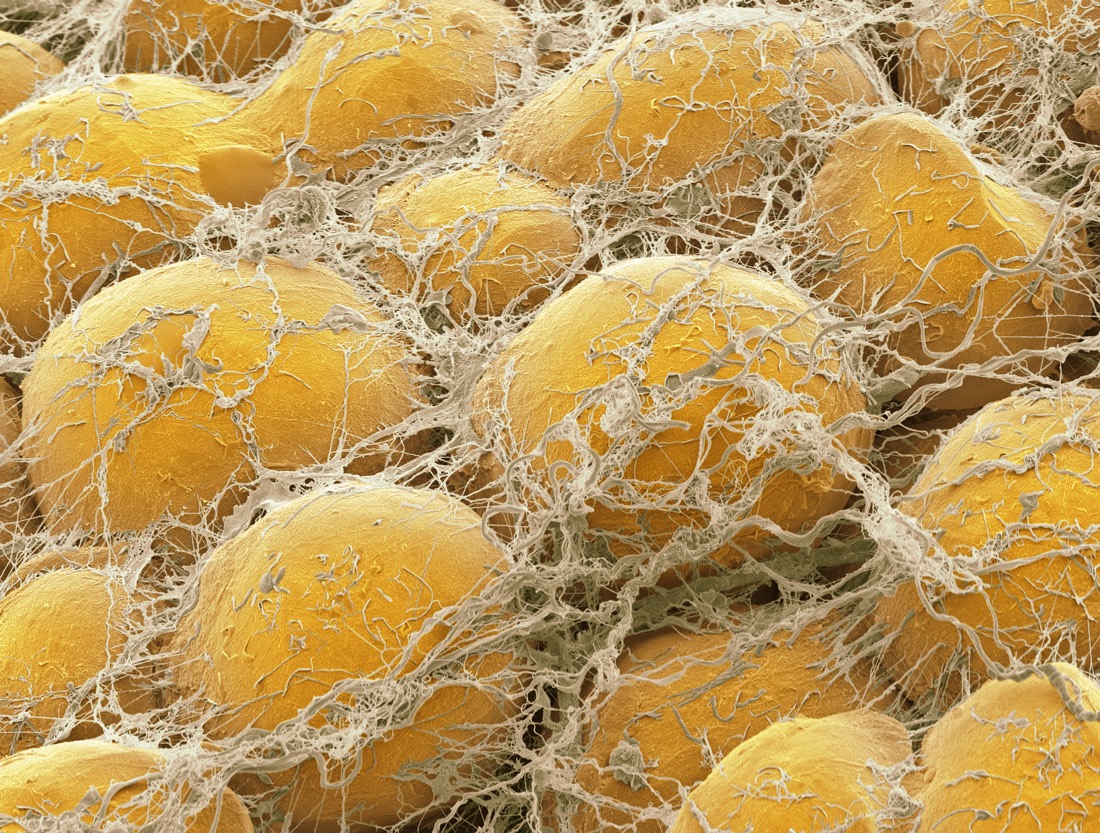
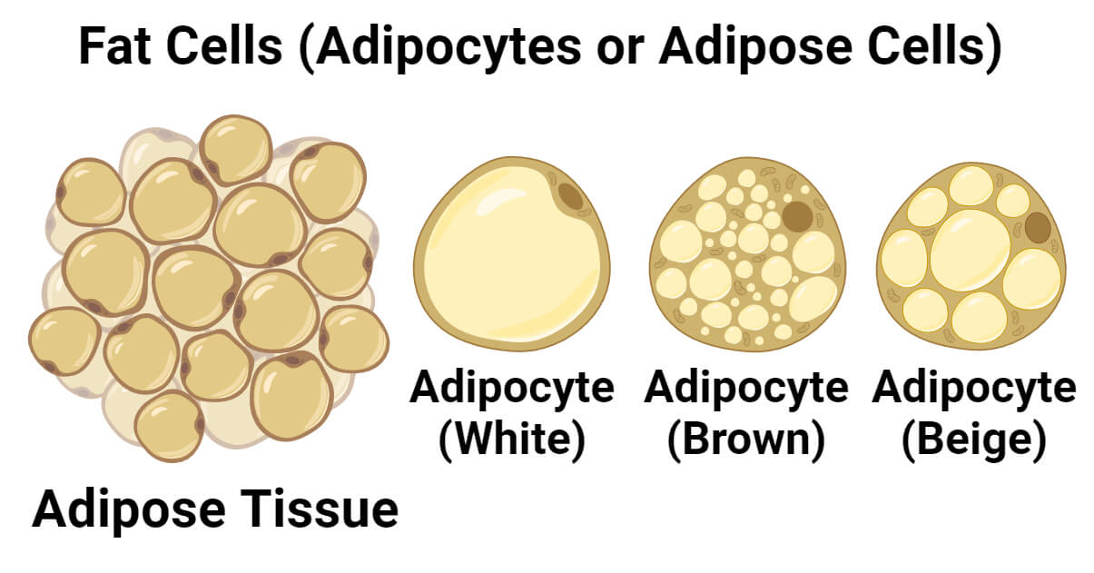
In the traditional biological sense, fat cells—or adipocytes—are often simplified in textbooks as yellow, bulbous droplets. However, in the realm of modern technology, the question of “what do fat cells look like” has shifted from basic biology to a complex intersection of high-resolution imaging, artificial intelligence, and digital health modeling. Today, we don’t just look at fat cells through a glass lens; we reconstruct them through billions of data points, 3D rendering software, and machine learning algorithms.
Understanding the visual architecture of adipose tissue is no longer just the domain of doctors. It is a frontier for software developers, hardware engineers, and data scientists who are building the next generation of diagnostic tools. By leveraging advanced tech, we can now see the microscopic nuances of these cells in ways that were impossible a decade ago, revealing a dynamic, communicative organ rather than a static storage unit.

The Evolution of Cellular Imaging: From Optical Lenses to Electron Microscopy
The history of visualizing fat cells is a roadmap of technological progress. In the early days of microscopy, fat cells appeared as translucent, structural voids because the chemicals used to prepare slides often dissolved the lipids within. Today’s technology allows us to see the “invisible” with startling clarity.
From Optical Microscopy to Cryo-Electron Imaging
Traditional light microscopy provided a two-dimensional, often distorted view of fat cells. The breakthrough came with the integration of Cryo-Electron Microscopy (Cryo-EM) and Scanning Electron Microscopy (SEM). These technologies allow researchers to freeze cells in their natural state, preventing the structural collapse that previously hindered visualization. Through SEM, a fat cell looks like a shimmering, spherical orb, often clustered together like a high-tech honeycomb. The resolution is so high that we can see the delicate network of capillaries and connective tissues that feed these cells, providing a digital map of human metabolism.
The Role of Fluorescence and Digital Contrast Enhancement
Modern software has revolutionized the “look” of fat cells through digital staining. Using fluorescent proteins and high-speed digital cameras, tech-enabled microscopes can highlight specific components of a fat cell. For example, the large lipid droplet can be rendered in neon blue, while the mitochondria—the cell’s power plants—glow in vibrant green. This digital contrast isn’t just for aesthetics; it allows computer vision algorithms to quantify the health of the cell automatically. By converting light signals into binary data, software can measure the exact volume and surface area of a cell, providing precise metrics for metabolic research.
3D Modeling and AI: Seeing the Invisible Through Data
While a single image tells a story, 3D modeling and Artificial Intelligence (AI) provide the full narrative. The question of what fat cells look like is now answered through volumetric rendering—creating a digital twin of adipose tissue that can be manipulated in virtual space.
Algorithmic Reconstruction of Adipose Tissue
Computed Tomography (CT) and Magnetic Resonance Imaging (MRI) generate “slices” of the body, but it is the post-processing software that defines the visual output. Advanced algorithms take these 2D slices and reconstruct them into 3D models. These models allow us to see the difference between subcutaneous fat (the layer just under the skin) and visceral fat (the dangerous fat surrounding internal organs). In these digital environments, fat cells are rendered as complex geometric structures. Software like OsiriX or specialized AI plugins can “segment” the fat, stripping away muscle and bone on the screen to show the sheer density and distribution of the adipocytes in high-definition 3D.
AI-Driven Diagnostic Tools for Metabolic Health
AI tools are now being trained to identify “white” vs. “brown” fat cells with higher accuracy than the human eye. Brown fat cells are smaller, iron-rich, and metabolically active. To an AI, these cells look different in terms of pixel density and heat signatures. Machine learning models, such as convolutional neural networks (CNNs), analyze thousands of images to detect early signs of cellular dysfunction. If a fat cell begins to expand beyond its healthy capacity—a state known as hypertrophy—AI software can flag this as a precursor to metabolic syndrome. In this context, the “look” of a fat cell is a data point that triggers a preventative healthcare intervention.
Wearable Tech and Personal Body Composition Apps
The visualization of fat cells has moved from the laboratory to the palm of our hands. Consumer-grade technology now allows individuals to visualize their body composition through sophisticated sensors and mobile applications.
Digital Twins and Bio-Impedance Analysis
Smart scales and high-end wearables use Bio-electrical Impedance Analysis (BIA) to estimate body fat percentage. While these devices don’t “see” a cell in the literal sense, the accompanying apps use the data to create a visual representation of the user’s internal health. This is often referred to as a “Digital Twin.” By inputting biometric data into an app, users can see a 3D avatar of themselves that reflects their adiposity levels. These apps translate invisible electrical resistance into a visual medium, showing users where their fat cells are most concentrated and how their lifestyle choices are impacting cellular volume over time.
The Future of Real-Time Cellular Monitoring
We are approaching an era where “what fat cells look like” could be a real-time feed on a smartwatch. Emerging tech in the field of “lab-on-a-chip” and near-infrared spectroscopy (NIRS) aims to monitor lipid levels through the skin. Imagine a dashboard that provides a graphical representation of lipid storage and mobilization. This tech would turn the abstract concept of “fat” into a manageable digital asset, allowing for precision nutrition and exercise regimens based on the actual visual and chemical state of the user’s adipose tissue.
Security and Ethics in Biological Data Imaging
As we gain the ability to visualize fat cells with such precision, we encounter a new frontier of digital security. Biological data is the most personal form of information, and the high-resolution images of our cellular structures are increasingly becoming part of our permanent digital footprint.
Protecting Biometric Visual Data
A 3D scan of an individual’s fat distribution is as unique as a fingerprint. As hospitals and tech companies store these high-resolution “cellular portraits,” the risk of data breaches grows. Digital security in the medical tech space must evolve to protect visual biological data. Encryption protocols (such as AES-256) are now being applied to DICOM (Digital Imaging and Communications in Medicine) files to ensure that the visualization of a patient’s internal health remains private. The “look” of your cells could, in theory, be used by insurance companies to predict future health costs, making the security of these images a top priority for developers.
Blockchain in Medical Imaging Storage
To combat the risks of centralized data storage, some health-tech startups are looking toward blockchain technology. By decentralizing the storage of 3D cellular models, patients can retain “ownership” of their biological images. Using Smart Contracts, a patient could grant temporary access to a specialist to view their fat-cell distribution data without the image ever residing on a vulnerable central server. This intersection of fintech and medtech ensures that as we get better at seeing what is inside us, we also get better at protecting that vision from unauthorized eyes.
Conclusion: The Digital Transformation of the Adipocyte
The question “what do fat cells look like” no longer has a single answer. To a scientist using an electron microscope, they are complex, shimmering orbs of energy storage. To a software engineer, they are a series of volumetric data points. To a consumer, they are a graph on a health app or a 3D avatar on a smartphone screen.
Technology has transformed the fat cell from a misunderstood “filler” tissue into a high-definition map of human health. As AI continues to refine our diagnostic capabilities and 3D rendering becomes even more lifelike, our ability to visualize, monitor, and manage our cellular health will only increase. We are moving toward a future where the digital and the biological are inextricably linked, providing us with a window into our own bodies that is as clear, detailed, and actionable as the latest 4K display.
aViewFromTheCave is a participant in the Amazon Services LLC Associates Program, an affiliate advertising program designed to provide a means for sites to earn advertising fees by advertising and linking to Amazon.com. Amazon, the Amazon logo, AmazonSupply, and the AmazonSupply logo are trademarks of Amazon.com, Inc. or its affiliates. As an Amazon Associate we earn affiliate commissions from qualifying purchases.