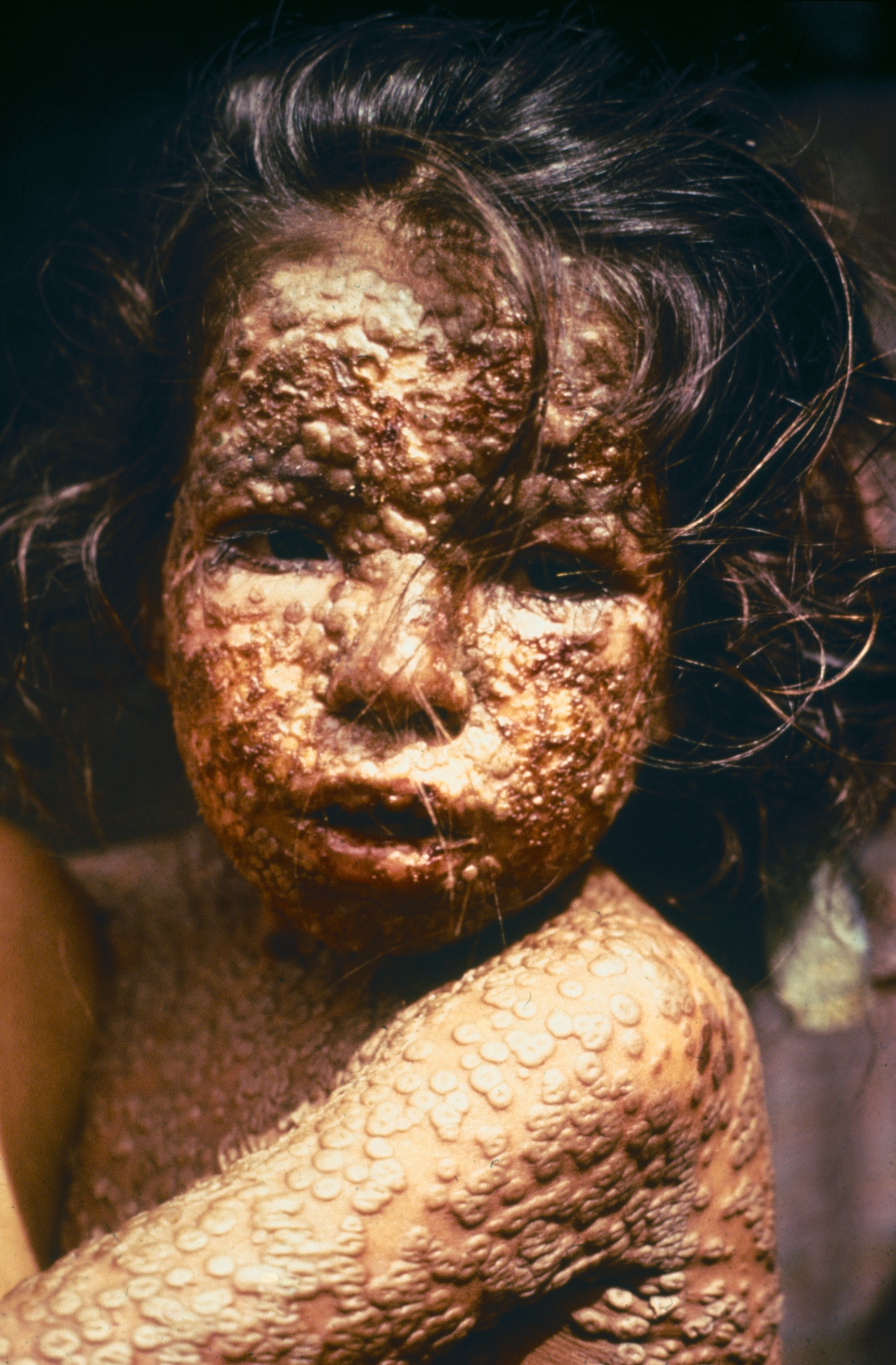
Smallpox remains the only human infectious disease to be completely eradicated through global vaccination efforts, officially declared gone by the World Health Organization (WHO) in 1980. For the modern generation, the question “what did smallpox look like?” is no longer a matter of clinical observation, but a challenge of digital reconstruction and historical preservation. Because the disease is no longer present in the wild, our understanding of its visual manifestation—the distinct, deep-seated pustules and the harrowing progression of the rash—now relies almost exclusively on the intersection of medical science and cutting-edge technology.
From AI-driven image restoration to high-fidelity virtual reality simulations, technology is bridging the gap between a forgotten terror and modern medical preparedness. This article explores the sophisticated tech stacks and methodologies currently used to visualize, archive, and simulate the appearance of smallpox in a world that has, thankfully, forgotten its face.
Digital Pathology and the High-Resolution Archiving of Variola
The physical evidence of smallpox is largely confined to a few high-security laboratories in the United States and Russia. However, for the global scientific community to study the disease without risking a catastrophic leak, technology has stepped in to create “digital twins” of historical samples.
The Transition from Physical Slides to Digital Twin Collections
Historically, medical students and researchers relied on glass slides and wax models to understand the morphology of the Variola virus. Today, Whole Slide Imaging (WSI) technology allows pathologists to convert these fragile historical specimens into ultra-high-resolution digital files. These digital twins can be magnified thousands of times without loss of clarity, allowing researchers to see the cellular impact of the virus in ways that were impossible during the height of the epidemic. By using cloud-based repositories, international health organizations can share these visual data points instantly, ensuring that the “look” of the disease is codified in a format that will never degrade.
Leveraging Scanning Electron Microscopy (SEM) for Nanoscale Visualization
To truly understand what smallpox “looked” like, we must look beyond the human eye’s capabilities. Scanning Electron Microscopy (SEM) and Cryo-electron microscopy (Cryo-EM) have been instrumental in visualizing the structural biology of the orthopoxvirus family. Through advanced rendering software, the raw data from these microscopes is transformed into 3D models. These tech-driven visualizations reveal the complex, brick-shaped structure of the virus, providing a blueprint that is essential for developing modern antivirals and synthetic vaccines.
AI-Driven Reconstruction: Restoring the “Face” of a Forgotten Plague
Because smallpox was eradicated before the advent of high-definition digital photography, much of our visual record consists of grainy, black-and-white photos or hand-drawn medical illustrations from the 19th and early 20th centuries. Artificial Intelligence is now being used to “upcycle” this historical data.
Generative AI and Historical Medical Imaging
Generative Adversarial Networks (GANs) are currently being utilized to restore and colorize archival footage of smallpox patients. By training AI models on the color palettes of modern, related viruses (such as Mpox or cowpox) and applying them to historical black-and-white imagery, researchers can create medically accurate color reconstructions. This technology provides a more visceral and accurate understanding of the “erythematous macules” and “umbilicated pustules” that characterized the disease, allowing contemporary clinicians to recognize potential symptoms that they have never seen in person.
Predictive Modeling: Simulating the Progression of the Variola Virus
AI isn’t just for looking at the past; it’s used for predictive visualization. Using machine learning algorithms, researchers can simulate how smallpox might manifest in different skin tones and demographics—visual data that was often missing or poorly documented in colonial-era medical records. These AI models synthesize dermatological data to create a “visual progression map,” showing the evolution of the rash from day one to day twenty-one. This tech is vital for biosecurity, providing a visual benchmark for early detection software.
Virtual Reality (VR) and Immersive Medical Education
In the absence of real-world cases, medical universities are turning to immersive technologies to train the next generation of healthcare providers. If a smallpox-like pathogen were ever to re-emerge, the first line of defense would be the ability of a triage nurse or GP to visually identify it.
Creating Risk-Free Diagnostic Environments for Modern Clinicians
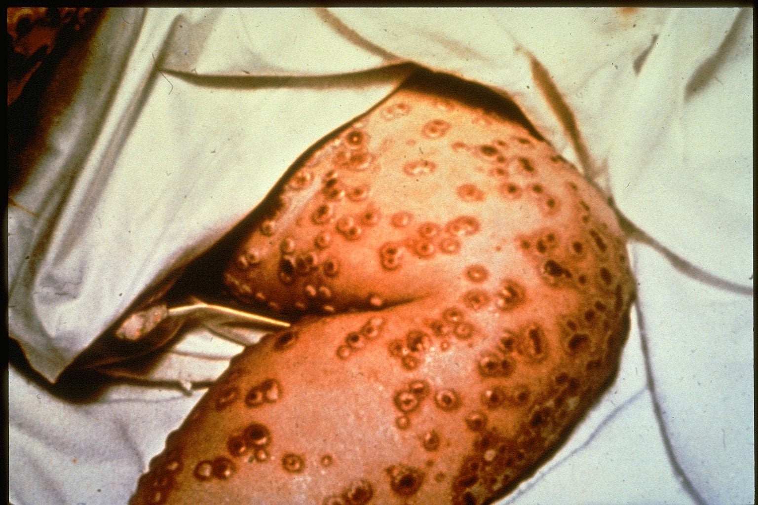
Virtual Reality (VR) platforms are now capable of simulating a clinical environment where a student can interact with a “digital patient” exhibiting smallpox symptoms. These simulations use high-fidelity textures and haptic feedback to mimic the appearance and physical distribution of the rash. By putting on a VR headset, a doctor can “walk around” the patient, observing the centrifugal distribution of the pustules—a key diagnostic feature where the rash is more dense on the face and limbs than on the trunk. This immersive tech provides a level of “visual muscle memory” that a textbook simply cannot offer.
The Ethical Framework of Digital Disease Simulation
As we use technology to recreate the visual horrors of smallpox, a new niche of “Digital Bioethics” has emerged. Tech developers must balance the need for clinical accuracy with the psychological impact of these realistic simulations. Advanced software allows for the “modular” adjustment of severity, enabling educators to show various strains of the disease (Variola Major vs. Variola Minor) in a controlled, digital setting. This ensures that the visual history of the disease is used for education rather than sensationalism, maintaining a professional standard in medical tech.
Data Visualization and the Mapping of the “Great Destroyer”
Understanding what smallpox looked like also involves looking at it from a macro perspective—how it looked as it moved through a population. Modern data visualization tools are being used to map historical outbreaks, turning ancient parish records and quarantine logs into dynamic digital maps.
Geospatial Analysis: Reconstructing the Visual Spread of Historical Outbreaks
Using Geographic Information Systems (GIS), historians and epidemiologists can visualize the “shape” of an epidemic. By inputting historical data into GIS software, we can see heat maps of how smallpox moved through cities like London or Boston in the 1700s. These visualizations provide insights into the “visual footprint” of the disease—showing how density, trade routes, and early inoculation efforts altered its path. Seeing the disease move across a map helps modern tech-driven health organizations model potential future outbreaks of other high-consequence pathogens.
Infographic Tech: Turning Biological Data into Accessible Visual Knowledge
In the digital age, the “look” of a disease is often distilled into data visualizations for public consumption. Using tools like D3.js or advanced Adobe Suite workflows, medical illustrators create 3D cross-sections of skin layers affected by Variola. These tech-driven illustrations explain the “why” behind the “look”—showing how the virus replicates in the dermis to create the characteristic deep-seated lesions. This level of technical clarity is essential for public health communication, ensuring that if a public health emergency were to occur, the information is both visually clear and scientifically grounded.
The Future of Visualizing Eradicated Pathogens
As we move further away from the 1977 last natural case of smallpox, the role of technology in maintaining our visual “memory” of the disease becomes even more critical. We are entering an era where Synthetic Biology and Digital Epidemiology will be the primary ways we interact with the history of the Variola virus.
The integration of AI, VR, and high-resolution digital pathology ensures that the answer to “what did smallpox look like” is preserved with perfect fidelity. By using these technological tools, we are not just looking at a ghost of the past; we are building a digital fortress of knowledge. This “tech-first” approach to medical history ensures that even though the virus has been defeated, our visual and diagnostic awareness remains sharp, protecting future generations through the power of digital preservation and advanced visualization.
aViewFromTheCave is a participant in the Amazon Services LLC Associates Program, an affiliate advertising program designed to provide a means for sites to earn advertising fees by advertising and linking to Amazon.com. Amazon, the Amazon logo, AmazonSupply, and the AmazonSupply logo are trademarks of Amazon.com, Inc. or its affiliates. As an Amazon Associate we earn affiliate commissions from qualifying purchases.