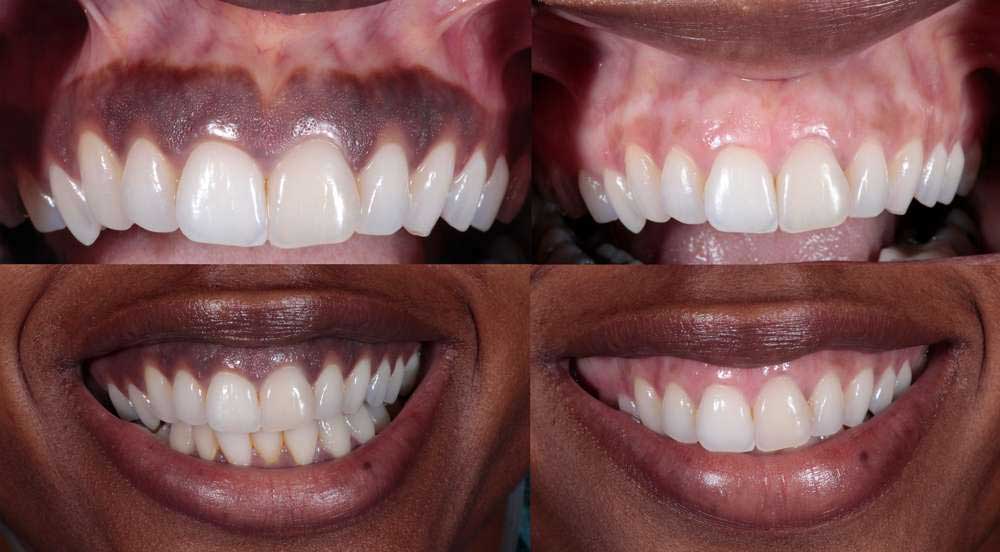
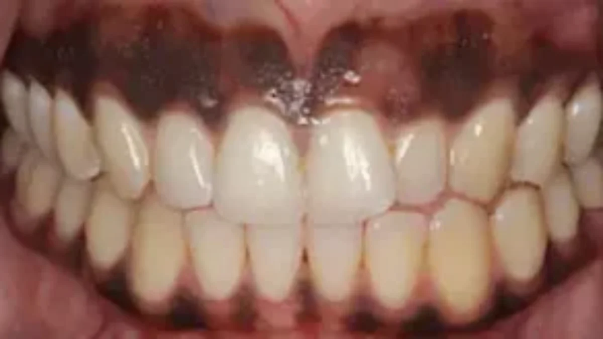
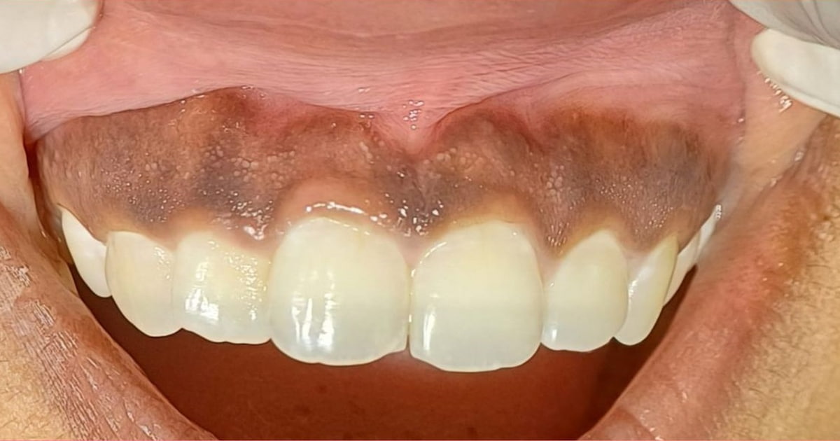
The appearance of dark gums, medically known as gingival hyperpigmentation, has long been a subject of clinical interest and cosmetic concern. While the causes range from physiological factors like melanin production to external influences such as systemic medications or “smoker’s melanosis,” the modern approach to addressing this condition has shifted entirely into the realm of high-tech intervention.
In the contemporary dental landscape, understanding what causes gums to be dark is no longer just about identifying a biological trait; it is about leveraging advanced software, laser physics, and artificial intelligence to provide precise, minimally invasive solutions. This article explores the technological frontier of periodontal aesthetics, focusing on the tools and trends redefining how we treat oral discoloration.
Digital Diagnostics: How AI and Imaging Identify Gum Discoloration
Before a treatment plan can be established, clinicians must accurately diagnose the etiology of the dark pigmentation. The integration of digital imaging and machine learning has revolutionized this initial phase, moving beyond simple visual inspection to data-driven analysis.
Computer-Aided Diagnosis (CAD) in Periodontics
Artificial Intelligence (AI) is now being trained to recognize patterns in gingival tissue that the human eye might miss. Computer-aided diagnosis systems use deep learning algorithms to analyze thousands of intraoral images, categorizing the intensity and distribution of melanin or metallic stains (such as amalgam tattoos). By feeding a “dark gum” image into these AI models, the software can differentiate between benign physiological pigmentation and potential indicators of systemic issues, such as Addison’s disease or Peutz-Jeghers syndrome. This technological triage ensures that aesthetic treatments are only performed when the underlying cause is confirmed to be non-pathological.
High-Resolution Intraoral Scanning and Color Analysis
The rise of the 3D intraoral scanner has replaced traditional impressions, but its utility extends to colorimetry. Modern scanners, equipped with high-definition optical sensors, can capture the specific “RGB” values of gum tissue. This allows for a baseline digital map of the patient’s mouth. By using specialized software, periodontists can quantify the “darkness” of the gums on a numerical scale, providing a more objective measure for treatment progress than subjective “before and after” photos. This level of precision is vital for patients seeking a specific aesthetic outcome, as the tech allows for a predictable simulation of post-treatment results.
Laser Technology: The Gold Standard for Melanin Ablation
Once the cause of dark gums is identified as melanin-related, the conversation shifts to removal. Gone are the days of painful surgical “scraping” with scalpels. The modern standard is laser depigmentation, a field driven by rapid advancements in laser physics and pulse technology.
Er:YAG vs. Diode Lasers: The Physics of Depigmentation
Two primary technologies dominate the market for treating dark gums: the Er:YAG (Erbium-doped Yttrium Aluminum Garnet) laser and the Diode laser. Each functions on a different principle of “selective photothermolysis.”
The Diode laser (typically 810–980 nm) is highly absorbed by melanin and hemoglobin. When the laser light hits the dark areas of the gums, the energy is converted into heat, vaporizing the pigmented cells while leaving the underlying connective tissue relatively untouched. Conversely, the Er:YAG laser targets water molecules within the tissue, causing a “micro-explosion” that peels away the pigmented layer with incredible coolness and precision. The technological trend is leaning toward “dual-wavelength” devices that allow clinicians to switch modes depending on the depth and intensity of the pigmentation.
Precision and Pulse Control in Soft Tissue Surgery
Modern laser units are now equipped with “SuperPulse” technology. In the past, constant heat from a laser could cause collateral thermal damage to the surrounding healthy tissue, leading to a painful recovery. Today’s software-controlled lasers deliver energy in billionths of a second. This allows the tissue to cool down between pulses, a concept known as “Thermal Relaxation Time.” For the patient, this means the procedure often requires no local anesthesia and results in almost zero bleeding, as the laser simultaneously cauterizes the area. This is a prime example of how hardware engineering has solved a biological challenge.
Telehealth and Remote Monitoring in Post-Operative Care
The treatment of dark gums does not end when the laser is turned off. The stability of the results—ensuring the dark pigment does not return—depends heavily on post-operative monitoring. This is where digital health tools and mobile applications have become indispensable.
Mobile Health (mHealth) Apps for Healing Visualization
Post-procedure, patients are often required to track their healing process. New dental apps allow patients to take daily “selfies” of their gums using a standardized calibration tool (usually a small sticker placed on the tooth). The app’s software then compares the daily images to track the “re-epithelialization” of the tissue. If the software detects signs of infection or premature repigmentation, it alerts the clinician immediately. This remote monitoring tech reduces the need for frequent in-person follow-ups, making the treatment more accessible for a global demographic.
The Role of Teledentistry in Aesthetic Consultation
The initial inquiry into “what causes gums to be dark” often begins with a Google search and ends in a teledentistry portal. Cloud-based platforms now allow patients to upload high-resolution images for a virtual “smile analysis.” Specialized software can then overlay a digital filter to show the patient what their gums would look like after a 20% or 50% reduction in melanin. This “virtual try-on” tech is a powerful tool in cosmetic dentistry, bridging the gap between digital curiosity and clinical reality.
Future Trends: Bio-Printing and Regenerative Tech
As we look toward the next decade, the tech addressing dark gums is moving from “removal” to “regeneration.” While current methods focus on taking away the dark pigment, future innovations focus on the cellular level.
Tissue Engineering for Uniform Gingival Tone
In cases where dark gums are caused by deep-seated issues or tissue thinness (where the dark color of the underlying root shows through), “gum grafting” is often required. The high-tech evolution here is 3D bio-printing. Researchers are currently developing bio-printed scaffolds infused with the patient’s own growth factors. Instead of harvesting tissue from the roof of the mouth, a 3D printer can create a customized, collagen-based “mesh” that is surgically placed to thicken the gums. This tech ensures a uniform, healthy pink tone by physically shielding the dark underlying structures.
Machine Learning and Predictive Outcomes
The holy grail of periodontal tech is the ability to predict “melanocyte migration.” A common issue with treating dark gums is that the pigment can eventually return, as melanin-producing cells migrate back into the treated area. Current research is utilizing machine learning to analyze the genetic and lifestyle data of patients (such as smoking habits or UV exposure) to predict the likelihood of recurrence. Future software modules will likely provide a “Recurrence Probability Score,” allowing clinicians to adjust the intensity of the laser treatment or the frequency of maintenance visits based on a personalized algorithmic profile.
The Intersection of Aesthetics and Innovation
The question of “what causes gums to be dark” has evolved from a simple biological query into a catalyst for technological advancement. We have moved from a baseline understanding of melanin to a sophisticated ecosystem of AI diagnostics, precision laser ablation, and remote monitoring.
For the modern consumer and clinician, the focus is no longer just on the “why,” but on the “how”—how technology can provide a safer, faster, and more predictable path to aesthetic health. As laser pulse control becomes even more refined and bio-printing moves from the lab to the clinic, the ability to manage gingival hyperpigmentation will stand as a testament to the power of tech in modern medicine. The dark tint of the gums is merely a digital data point in a much larger, tech-driven journey toward the perfect smile.
aViewFromTheCave is a participant in the Amazon Services LLC Associates Program, an affiliate advertising program designed to provide a means for sites to earn advertising fees by advertising and linking to Amazon.com. Amazon, the Amazon logo, AmazonSupply, and the AmazonSupply logo are trademarks of Amazon.com, Inc. or its affiliates. As an Amazon Associate we earn affiliate commissions from qualifying purchases.