
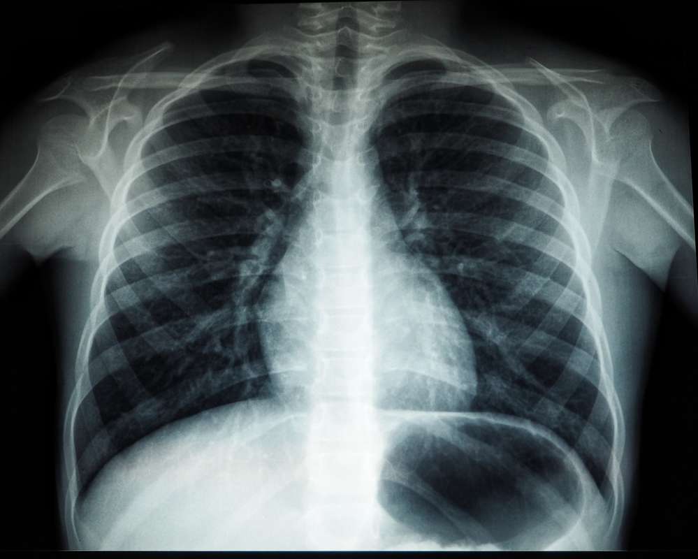
The chest X-ray, or chest radiograph, stands as one of the oldest and most frequently performed diagnostic imaging procedures in medicine. For over a century, this foundational technology has provided clinicians with invaluable insights into the thoracic cavity, offering a non-invasive glimpse into the lungs, heart, blood vessels, airways, and bones of the chest. However, to interpret “what can chest X-rays reveal” through the lens of modern technology is to embark on a journey far beyond the static film images of the past. Today, the reveal is not just about a shadow on a plate, but about the intricate data captured, processed, and analyzed by sophisticated digital systems, augmented by artificial intelligence, and seamlessly integrated into a broader technological healthcare ecosystem. This exploration delves into the technological capabilities and advancements that empower chest X-rays to reveal more than ever before, touching upon digital transformation, AI integration, advanced imaging techniques, and the future trajectory of this indispensable diagnostic tool.

The Digital Transformation of Radiography
The most profound shift in the history of chest X-rays, perhaps since Roentgen’s initial discovery, has been the transition from analog film-based radiography to fully digital systems. This technological leap has revolutionized how images are acquired, processed, stored, and interpreted, significantly enhancing the diagnostic potential of the humble X-ray.
From Film to Pixels: Enhancing Image Quality and Accessibility
The move away from chemical processing and darkrooms began with Computed Radiography (CR), which utilized phosphor plates to capture X-ray energy, later scanned by a laser to convert latent images into digital data. This was a crucial intermediate step. The true revolution, however, came with Direct Radiography (DR) systems. DR systems employ flat-panel detectors, often made with amorphous silicon or selenium, that directly convert X-ray photons into electrical signals, which are then digitized. This process offers several distinct advantages:
-
Superior Image Quality: Digital detectors boast a wider dynamic range, meaning they can capture a broader spectrum of X-ray intensities. This translates into images with better contrast resolution and detail, allowing for the visualization of subtle pathologies that might be missed on film. Post-processing capabilities further enhance image clarity, enabling technologists and radiologists to adjust brightness, contrast, and apply filters to optimize visualization for specific clinical questions.
-
Reduced Radiation Dose: The increased sensitivity of digital detectors often means that diagnostic quality images can be obtained with a lower dose of radiation compared to traditional film or even CR systems, improving patient safety without compromising diagnostic accuracy.
-
Instant Image Acquisition and Review: Unlike film, which required chemical processing time, digital images appear on a monitor almost instantly after exposure. This drastically reduces patient waiting times, improves workflow efficiency in busy radiology departments, and allows for immediate quality control checks.
-
Elimination of Physical Waste: Digital radiography eliminates the need for hazardous chemicals and the physical storage of film, contributing to a greener and more efficient healthcare environment.
The Power of PACS: Streamlining Workflow and Collaboration
The true utility of digital X-rays is unlocked by Picture Archiving and Communication Systems (PACS). PACS is a medical imaging technology that provides economical storage and convenient access to images from multiple modalities (not just X-rays, but also CT, MRI, etc.). It’s a digital ecosystem that fundamentally alters how medical images are managed:
-
Centralized Digital Archive: Instead of shelves of physical films, images are stored digitally on secure servers. This means images can be retrieved quickly, eliminating the risk of lost or misplaced films. Historical images are instantly accessible for comparison, crucial for tracking disease progression or treatment response.
-
Ubiquitous Access: Radiologists, clinicians, and specialists can access images from virtually anywhere within a hospital network, or even remotely via secure connections (tele-radiology). This facilitates faster consultations, collaborative diagnostic discussions, and more timely patient care, especially in critical situations or for geographically dispersed expertise.
-
Enhanced Workflow Efficiency: PACS integrates with other hospital information systems (HIS) and electronic health records (EHR), allowing patient demographics, clinical notes, and imaging reports to be linked seamlessly. This reduces administrative burdens, minimizes errors, and ensures that all relevant patient information is available at the point of care.
-
Advanced Viewing Tools: PACS workstations come equipped with sophisticated software tools that allow radiologists to manipulate images in various ways – zooming, panning, measuring structures, adjusting window/level settings – all to extract maximum diagnostic information.
AI and Machine Learning: Augmenting Diagnostic Capabilities
Perhaps the most exciting frontier in what chest X-rays can reveal is the integration of Artificial Intelligence (AI) and Machine Learning (ML). AI algorithms are no longer just theoretical concepts; they are actively being deployed to enhance the speed, accuracy, and consistency of chest X-ray interpretation.
Automating Anomaly Detection: Early Warning Systems for Clinicians
AI algorithms, particularly deep learning models trained on vast datasets of annotated chest X-rays, are proving remarkably adept at identifying specific pathologies. These systems can act as “second readers” or “triage assistants” for radiologists:
-
Spotting Subtle Lesions: AI can be trained to recognize patterns indicative of conditions like small pulmonary nodules (which could be early signs of cancer), subtle pneumothoraces (collapsed lung), or signs of pneumonia. These findings can sometimes be overlooked by the human eye, especially in high-volume settings where radiologists are experiencing fatigue.
-
Prioritization and Triage: AI can rapidly analyze incoming X-rays and flag studies with critical findings, pushing them to the top of the radiologist’s worklist. This ensures that life-threatening conditions are addressed with immediate urgency, improving patient outcomes.
-
Consistency and Reduced Variability: Human interpretation, while invaluable, can sometimes vary between radiologists or even for the same radiologist under different circumstances. AI provides a consistent, objective analysis, reducing inter-observer variability and maintaining a high standard of detection.
-
Quantification: Beyond simple detection, AI can quantify findings, such as the size and density of a lesion, or the extent of an abnormality, providing objective metrics that can be tracked over time.
Quantitative Analysis and Predictive Modeling: Beyond Visual Inspection
The power of AI extends beyond merely identifying visual anomalies. Machine learning models can extract quantitative data from X-ray images that are difficult or impossible for humans to discern, opening new avenues for understanding disease.
-
Measuring Disease Progression: AI can precisely measure subtle changes in lung texture or cardiac size over time, providing objective data on how a disease is progressing or responding to treatment. For conditions like interstitial lung disease, this granular data can inform clinical decisions.
-
Predictive Analytics: By correlating imaging features with clinical outcomes and other patient data, AI models are being developed to predict future health risks. For example, specific patterns on a chest X-ray might be linked to an increased risk of cardiovascular events or respiratory failure, allowing for proactive interventions.
-
Biomarker Discovery: AI can identify novel imaging biomarkers – features within the X-ray that correlate with specific biological processes or genetic predispositions. This could lead to a deeper understanding of diseases and personalized treatment approaches.
-
Assessment of Severity: In conditions like COVID-19, AI has been used to assess the severity of lung involvement, helping clinicians make decisions about patient management and resource allocation.
Advanced Imaging Techniques and Their Technological Underpinnings
While the fundamental principle of X-ray generation remains, continuous technological innovation has led to specialized techniques that extend the diagnostic reach of chest radiography.
Dual-Energy X-Rays: Separating Tissue Types for Clearer Diagnoses
Dual-energy X-ray imaging is a sophisticated technique that utilizes two different X-ray energy levels (high and low kilovoltage) during a single exposure or in rapid succession. The technological magic lies in the post-processing of these two images:
-
Material Decomposition: Because different materials (like bone and soft tissue) attenuate X-rays differently depending on the energy level, advanced algorithms can use the data from both exposures to “decompose” the image into separate material-specific images. This results in two primary sets of images: a bone-subtracted image (where bones are removed to better visualize soft tissues like lung parenchyma and vessels) and a soft-tissue-subtracted image (emphasizing bone structures).
-
Enhanced Visualization: For radiologists, this means being able to “see through” ribs to better detect subtle lung nodules or infiltrates that might otherwise be obscured by bone. Conversely, the bone-only image can highlight fractures or calcifications more clearly. This significantly enhances diagnostic confidence, especially in complex cases.
Mobile and Portable X-Ray Units: Bringing Imaging to the Patient
The advent of compact, lightweight, and powerful digital X-ray technology has revolutionized patient care by making diagnostic imaging accessible outside the traditional radiology department.
-
Point-of-Care Diagnostics: Portable X-ray units are indispensable in intensive care units (ICUs), emergency rooms, operating theaters, and even at patients’ bedsides. This eliminates the need to transport critically ill or immobile patients to the radiology suite, reducing risks and delays.
-
Advanced Features: Modern portable units incorporate high-frequency generators, digital flat-panel detectors, and sophisticated battery management systems that allow for multiple exposures on a single charge. Wireless connectivity ensures that images are immediately transmitted to PACS for review, maintaining workflow efficiency even in remote settings.
-
Remote Clinics and Field Operations: The robustness and portability of these units make them ideal for use in mobile clinics, nursing homes, and even disaster zones, extending diagnostic capabilities to underserved populations.
Challenges and Ethical Considerations in AI-Powered Radiology
While the technological advancements in chest X-rays, particularly with AI, promise immense benefits, they also introduce a new set of challenges and ethical considerations that must be meticulously addressed.
Data Bias and Algorithm Transparency: Ensuring Equitable Healthcare
AI models are only as good as the data they are trained on. If the training datasets are not representative of the diverse patient population, the algorithms can develop biases:
-
Racial and Ethnic Bias: If an AI model is predominantly trained on images from one ethnic group, its performance might degrade when applied to patients from other groups, leading to diagnostic disparities.
-
Socioeconomic Bias: Datasets might inadvertently reflect biases in healthcare access, potentially leading to models that perform poorly for underserved populations.
-
“Black Box” Problem: Many deep learning models are “black boxes,” meaning their decision-making process is opaque. Radiologists need “explainable AI” (XAI) that can provide reasons or confidence scores for its findings, building trust and allowing for critical human oversight.
-
Regulatory Oversight: Developing robust regulatory frameworks for AI in medicine is crucial to ensure that these technologies are safe, effective, and ethically deployed.
The Human Element: Radiologist as the Ultimate Interpreter
Despite the remarkable capabilities of AI, it is imperative to remember that AI is an assistive tool, not a replacement for human expertise.
-
Clinical Context: AI lacks the ability to integrate clinical history, physical examination findings, and other laboratory results – the holistic patient context that a human radiologist always considers when interpreting an image.
-
Unusual Cases and Novel Pathologies: AI models are trained on known patterns. They may struggle with highly unusual presentations of disease or entirely novel pathologies that they haven’t encountered in their training data.
-
Legal and Ethical Responsibility: Ultimately, the radiologist bears the legal and ethical responsibility for the diagnosis. AI insights must be critically evaluated and integrated into the overall clinical picture.
-
Evolving Role of the Radiologist: AI will likely shift the radiologist’s role from primary image interpreter to a highly skilled orchestrator of AI tools, focusing on complex cases, quality control, and communicating findings within the broader clinical context.
The Future Landscape of Chest X-Ray Technology
Looking ahead, the evolution of chest X-ray technology will continue to be driven by innovation in digital systems, AI, and connectivity, further enhancing what these fundamental images can reveal.
Integration with Electronic Health Records and Predictive Analytics
The future will see even deeper integration of X-ray data and AI insights directly into comprehensive Electronic Health Records (EHRs). This means:
-
Holistic Patient View: Imaging findings will be instantly contextualized with all other patient data, allowing for more informed clinical decisions.
-
Proactive Care Pathways: AI-driven predictive analytics, powered by X-ray data, will help identify patients at risk for certain conditions earlier, enabling preventive measures or early interventions.
-
Personalized Medicine: X-ray features, combined with genetic and other biomarker data, will contribute to a more personalized approach to disease management.
Miniaturization and Point-of-Care Diagnostics
The trend towards smaller, more powerful, and more intelligent X-ray devices will continue.
-
Handheld X-Ray Devices: Research is progressing on truly handheld X-ray devices that could offer rapid screening or diagnostic capabilities in even more remote or resource-limited settings.
-
Enhanced Connectivity: Future devices will feature even more robust wireless capabilities, enabling seamless data transfer and remote diagnostics from virtually anywhere, powered by 5G and future network technologies.
-
Robotic and Automated Imaging: Robotic systems could assist in positioning patients and acquiring images with greater precision and consistency, especially in challenging environments.

Conclusion
From its humble beginnings as a shadowy image on a photographic plate, the chest X-ray has embarked on an incredible technological journey. Today, “what can chest X-rays reveal” encompasses not just the anatomical structures visible to the eye, but a wealth of digital data, processed by intelligent algorithms, and integrated into a sophisticated healthcare ecosystem. The digital transformation has brought unparalleled clarity and accessibility, while the advent of AI is augmenting diagnostic capabilities to detect the subtle, quantify the unmeasurable, and even predict the unforeseen. As technology continues to advance, the chest X-ray remains a cornerstone of diagnostic medicine, perpetually evolving to reveal deeper, more nuanced insights into human health, solidifying its place at the forefront of medical imaging innovation.
aViewFromTheCave is a participant in the Amazon Services LLC Associates Program, an affiliate advertising program designed to provide a means for sites to earn advertising fees by advertising and linking to Amazon.com. Amazon, the Amazon logo, AmazonSupply, and the AmazonSupply logo are trademarks of Amazon.com, Inc. or its affiliates. As an Amazon Associate we earn affiliate commissions from qualifying purchases.