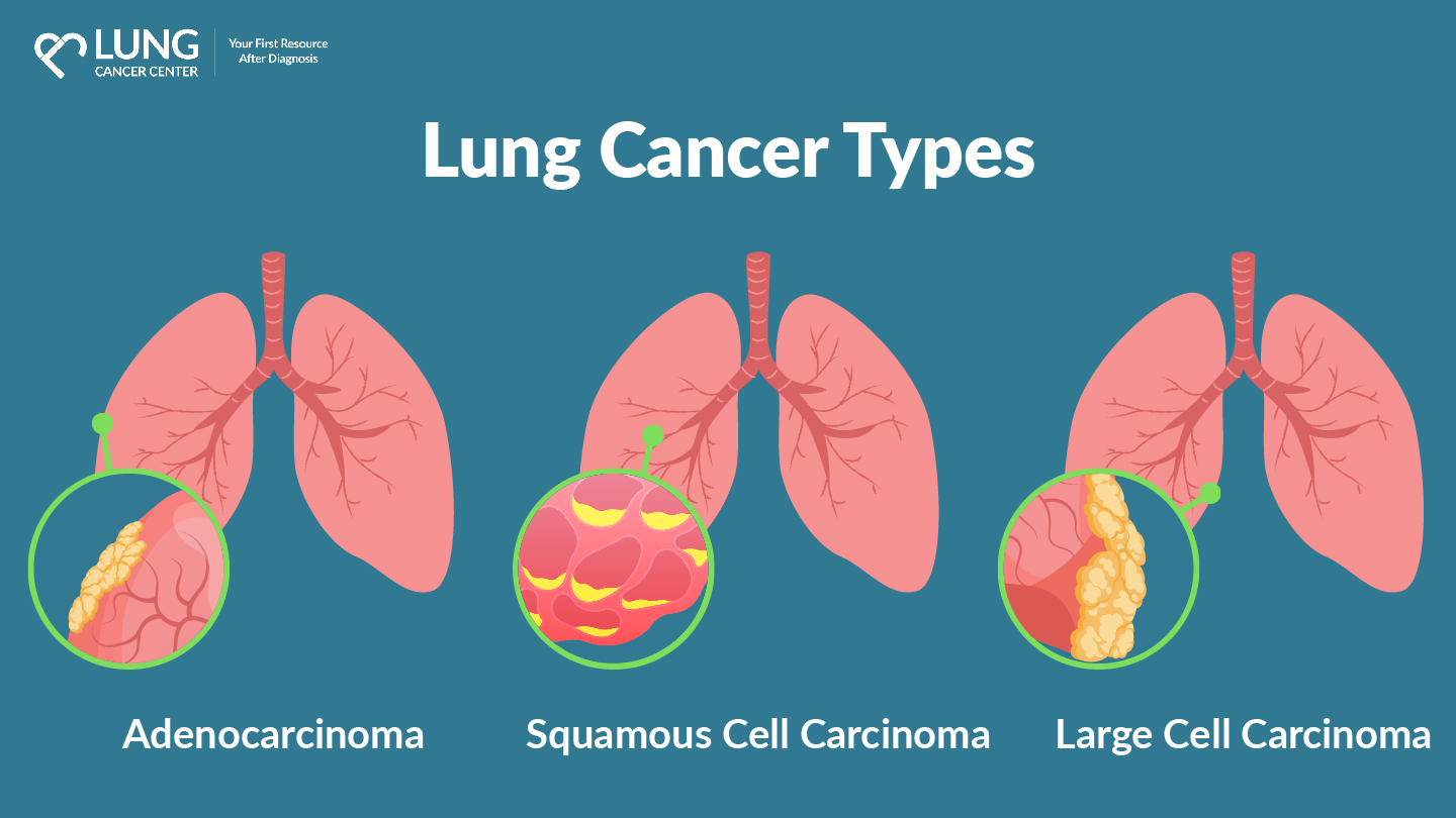
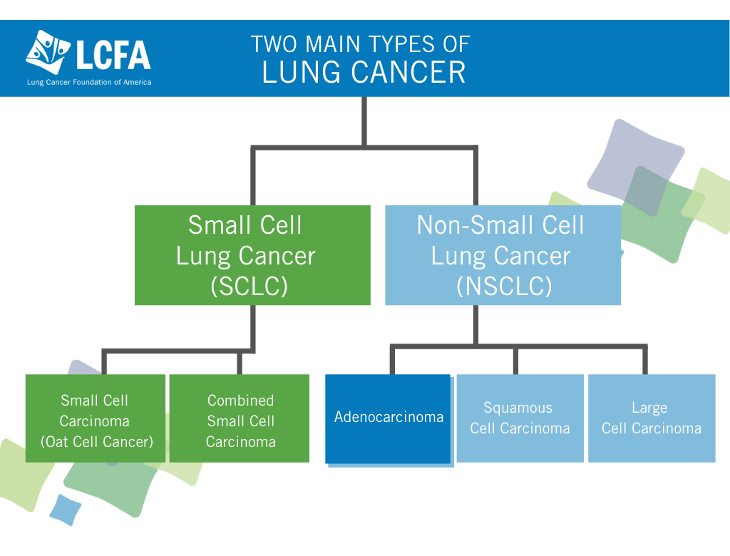
The intersection of healthcare and technology has ushered in a new era of “precision medicine,” where the classification of complex diseases is no longer a matter of manual observation but a result of high-compute data analysis. Historically, identifying the nuances of lung cancer was a laborious process involving physical biopsies and human interpretation of cellular structures. Today, the landscape is dominated by diagnostic technology, software algorithms, and genomic sequencing tools. To understand the five primary types of lung cancer—Adenocarcinoma, Squamous Cell Carcinoma, Large Cell Carcinoma, Small Cell Lung Cancer (SCLC), and Bronchial Carcinoid tumors—one must look through the lens of the technological innovations driving their detection and treatment.
By leveraging Artificial Intelligence (AI), Next-Generation Sequencing (NGS), and advanced imaging software, the tech industry is providing oncologists with the digital tools necessary to differentiate these malignancies with unprecedented accuracy.
The Digital Transformation of Diagnostic Imaging
The first line of defense in identifying the various types of lung cancer is medical imaging. However, the modern “tech-heavy” approach goes far beyond the traditional X-ray. Digital imaging has evolved into a sophisticated ecosystem where software plays a more critical role than the hardware itself.
AI-Powered Computer-Aided Detection (CAD)
Artificial Intelligence has revolutionized the way radiologists interpret Computed Tomography (CT) scans. Computer-Aided Detection (CAD) systems utilize deep learning algorithms trained on millions of clinical images. These AI tools can detect pulmonary nodules as small as a few millimeters, often invisible to the human eye. When differentiating between Non-Small Cell Lung Cancer (NSCLC) and Small Cell Lung Cancer (SCLC), CAD software analyzes texture, density, and growth patterns. For instance, the software can flag the irregular, “spiculated” margins often associated with Adenocarcinoma, allowing for earlier digital classification before a physical biopsy is even performed.
Radiomics and Predictive Analytics
Radiomics is an emerging field in digital health that extracts thousands of quantitative features from medical images using data-characterization algorithms. These features, which include spatial distribution and pixel intensity variations, are processed through machine learning models to create a “digital twin” of the tumor. By applying radiomic workflows, tech platforms can predict the histological subtype of a tumor. This is particularly useful in distinguishing Squamous Cell Carcinoma, which typically forms in the central airways, from Adenocarcinoma, which occurs in the outer regions. The technology essentially turns a standard image into a searchable database of biological information.
Genomic Sequencing: Decoding the 5 Major Classifications
While imaging provides a macroscopic view, the true classification of lung cancer types now happens at the molecular level through bioinformatics and high-throughput sequencing technology. This is where the “Tech” in biotechnology takes center stage.
Next-Generation Sequencing (NGS) Tools
Next-Generation Sequencing (NGS) is a massive leap forward from traditional DNA sequencing. It allows for the simultaneous sequencing of millions of DNA fragments. In the context of the five types of lung cancer, NGS hardware (such as those developed by Illumina or Thermo Fisher) allows technicians to identify specific genetic mutations like EGFR, ALK, and KRAS.
For example, Adenocarcinoma is frequently driven by specific genetic mutations that NGS tools can pinpoint in a matter of hours. By utilizing cloud-based bioinformatics platforms, labs can compare a patient’s genetic profile against global databases to determine if a tumor is a Large Cell Carcinoma or a more aggressive Small Cell Lung Cancer (SCLC).
Identifying Driver Mutations with Bioinformatics
The software used to analyze genomic data is as vital as the sequencing hardware. Bioinformatics pipelines use complex algorithms to filter out genetic “noise” and identify the “driver mutations” that cause a tumor to grow. This digital identification is crucial for distinguishing Bronchial Carcinoid tumors, which are rare and slow-growing, from the more common and faster-growing NSCLC subtypes. The ability to categorize these five types through software-driven genetic analysis has moved lung cancer from a broad diagnosis to a highly specific digital profile.
The Role of Machine Learning in Digital Pathology
Pathology—the study of tissue samples—has transitioned from the physical microscope to the digital workstation. Digital pathology involves scanning traditional glass slides into high-resolution digital images, which are then analyzed by specialized software.
Deep Learning Models for Pathological Slices
Machine learning models, specifically Convolutional Neural Networks (CNNs), are now trained to recognize the cellular patterns of the different lung cancer types. When a tissue sample is scanned, the AI can differentiate between the small, “oat-shaped” cells characteristic of Small Cell Lung Cancer and the larger, flatter cells of Squamous Cell Carcinoma. These deep learning tools serve as a “second pair of eyes” for pathologists, reducing human error and increasing the speed of diagnosis.
Furthermore, these tools can quantify “PD-L1 expression,” a protein marker that determines whether a patient is a candidate for immunotherapy. The integration of image-recognition software into the pathology workflow ensures that the classification of the five types is standardized across different medical institutions.
Natural Language Processing (NLP) in Clinical Data Integration
Beyond image analysis, Natural Language Processing (NLP) is used to scan through thousands of unstructured clinical notes and pathology reports. This tech-driven approach allows researchers to aggregate data on how the five different types of lung cancer respond to various treatments. By using NLP to “read” electronic health records (EHRs), healthcare tech companies can build predictive models that help clinicians understand the progression of Large Cell Carcinoma versus Adenocarcinoma, leading to more tailored technological interventions.
Liquid Biopsies and Non-Invasive Surveillance Tech
One of the most significant technological breakthroughs in the last decade is the development of the liquid biopsy. This technology allows for the detection of cancer types through a simple blood draw, utilizing microfluidic chips and high-sensitivity sensors.
Circulating Tumor DNA (ctDNA) Monitoring
When cancer cells die, they release fragments of DNA into the bloodstream, known as circulating tumor DNA (ctDNA). Advanced digital sensors and sequencing software can capture these fragments. This technology is particularly effective in monitoring the five types of lung cancer for potential mutations or resistance to treatment. For instance, if a patient with Squamous Cell Carcinoma develops a new mutation, liquid biopsy tech can detect this change months before it would appear on a traditional CT scan.
Microfluidic Chip Technology
The hardware used in liquid biopsies often involves microfluidic chips—tiny devices that manipulate minute amounts of fluids. These chips are designed to “catch” rare circulating tumor cells (CTCs). The software integrated with these chips uses fluorescence-based imaging to categorize the cells based on their size and surface markers. This allows for a real-time digital “snapshot” of the cancer’s type and stage, providing a non-invasive alternative to traditional surgical biopsies.
Telemedicine and the Digital Surveillance Ecosystem
Once the type of lung cancer is identified, the technological focus shifts to surveillance and management. The modern oncology tech stack includes wearable devices, remote monitoring apps, and digital therapeutics.
Wearable Tech and IoT for Post-Treatment Surveillance
Internet of Things (IoT) devices, such as smartwatches and specialized medical wearables, are being used to monitor the physiological data of patients undergoing treatment for different types of lung cancer. Patients with Small Cell Lung Cancer, which often requires aggressive chemotherapy and radiation, can be monitored for respiratory distress or heart rate variability in real-time. This data is transmitted to a cloud platform where algorithms flag any anomalies, allowing for immediate intervention.

Digital Therapeutics (DTx) and Patient Empowerment
Digital Therapeutics (DTx) are software-based interventions used to treat or manage medical conditions. For lung cancer patients, DTx apps provide personalized symptom management and psychological support. Depending on whether a patient is dealing with the specific side effects of treatments for Adenocarcinoma or Carcinoid tumors, the software adjusts its recommendations. This level of personalized tech ensures that the management of the disease is as precise as the initial digital diagnosis.
In conclusion, the classification of the five types of lung cancer is no longer strictly a biological endeavor; it is a technological one. From the AI that scans a lung for nodules to the genomic software that identifies a driver mutation, technology is the backbone of modern oncology. As machine learning continues to evolve and sequencing hardware becomes faster and more accessible, our ability to identify, categorize, and treat these five types will only become more precise, moving us closer to a future where digital tools can outpace the progression of the disease itself.
aViewFromTheCave is a participant in the Amazon Services LLC Associates Program, an affiliate advertising program designed to provide a means for sites to earn advertising fees by advertising and linking to Amazon.com. Amazon, the Amazon logo, AmazonSupply, and the AmazonSupply logo are trademarks of Amazon.com, Inc. or its affiliates. As an Amazon Associate we earn affiliate commissions from qualifying purchases.