
The intricate world of human biology often harbors microscopic marvels that, while unseen by the naked eye, play colossal roles in our health and well-being. Among these unsung heroes are podocytes, specialized cells residing within the glomerulus of the kidney. While their primary function in filtration is fundamental, the exploration of podocytes is increasingly intersecting with cutting-edge technological advancements, from diagnostic tools to novel therapeutic approaches. Understanding podocytes, therefore, is not just a biological pursuit but also a gateway to appreciating how technology is revolutionizing nephrology and the treatment of kidney diseases.
The Cellular Architecture of Kidney Filtration
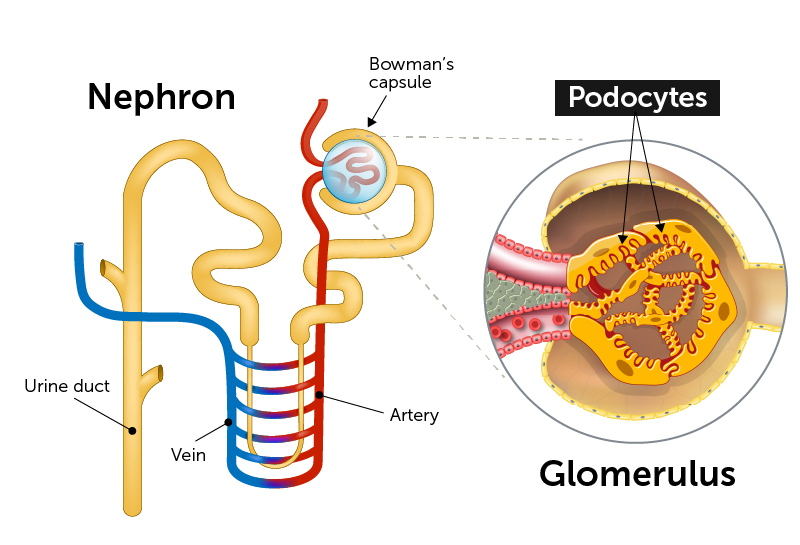
The kidney is a complex filtration system, a marvel of biological engineering designed to remove waste products from the blood and maintain fluid balance. At the heart of this filtration lies the glomerulus, a network of tiny blood vessels where the initial stage of urine formation occurs. This intricate structure is meticulously designed to allow small molecules like water and waste products to pass through while retaining larger molecules such as proteins and blood cells. Podocytes are critical components of this sophisticated filtering unit, acting as the final barrier that prevents essential proteins from entering the urine.

The Glomerular Filtration Barrier: A Multi-Layered Defense
The glomerular filtration barrier is a tripartite structure, each layer contributing uniquely to its selective permeability. The inner layer is the fenestrated endothelium of the glomerular capillaries. These capillaries possess numerous pores, or fenestrations, that allow free passage of water and small solutes. However, these fenestrations are too small for larger components of blood to pass through.
The middle layer is the glomerular basement membrane (GBM), a complex extracellular matrix rich in collagen, laminin, and other glycoproteins. The GBM acts as a physical sieve, possessing a negative charge that repels negatively charged molecules, further preventing proteins like albumin, which also carry a negative charge, from crossing. This charge-based repulsion is a crucial aspect of selective filtration.
The outermost layer of the filtration barrier is formed by the podocytes themselves. These are highly differentiated cells with unique cellular extensions. They are not just passive structural elements but active participants in the filtration process. Their distinctive morphology and function are what make them particularly fascinating from a scientific and technological perspective.
Podocyte Morphology: The Foot Processes and Slits
Podocytes are characterized by their elaborate cellular structure. They are large cells that embrace the outer aspect of the glomerular capillaries. From their main cell body, podocytes extend primary processes, which then branch into a dense network of secondary foot processes, also known as pedicels. These pedicels interdigitate with the foot processes of neighboring podocytes, creating narrow gaps called filtration slits.
These filtration slits are bridged by a specialized protein structure called the slit diaphragm. This diaphragm is a dynamic protein complex, with key components including nephrin, podocin, and FAT1. The slit diaphragm acts as a fine-tuning mechanism for filtration, controlling the passage of molecules based on size and charge. Its precise structure and function are areas of intense research, particularly as disruptions to the slit diaphragm are implicated in various kidney diseases. The intricate arrangement of podocyte foot processes and the slit diaphragm is what ultimately determines the efficiency and selectivity of the glomerular filtration process.
Podocytes and Kidney Disease: A Technological Lens
The health and integrity of podocytes are paramount for normal kidney function. When podocytes are damaged or lost, the glomerular filtration barrier is compromised, leading to proteinuria (protein in the urine) and potentially progressing to chronic kidney disease (CKD) and end-stage renal disease (ESRD). The identification and study of podocyte injury have been significantly advanced by technological innovations.
Advanced Imaging Techniques for Podocyte Visualization
Historically, studying the delicate structure of podocytes and their interactions within the glomerulus posed significant challenges. However, advancements in microscopy have revolutionized our ability to visualize these cells. Techniques like transmission electron microscopy (TEM) were crucial in first revealing the intricate foot process architecture and the slit diaphragm. While powerful, TEM requires sample fixation and can be time-consuming.
More recently, super-resolution microscopy techniques have offered unprecedented detail. These methods can overcome the diffraction limit of light, allowing researchers to visualize cellular structures with nanoscale resolution. This has been instrumental in dissecting the molecular organization of the slit diaphragm and understanding how specific proteins interact. Furthermore, live-cell imaging, often employing fluorescent protein markers, allows researchers to observe podocyte behavior in real-time, including their responses to stress, injury, and therapeutic interventions. These dynamic observations are critical for understanding disease pathogenesis and for evaluating the efficacy of potential treatments.
Biomarker Discovery and Diagnostic Technologies
The early detection of podocyte injury is crucial for intervening and preventing kidney disease progression. Traditionally, proteinuria has been the primary indicator of glomerular damage. However, detecting specific markers of podocyte injury can provide more precise and earlier insights into the health of these critical cells. Technological advancements have enabled the discovery and measurement of novel podocyte-specific biomarkers.
For instance, the measurement of soluble forms of slit diaphragm proteins, such as soluble nephrin, in urine or blood can indicate podocyte damage. Technologies like enzyme-linked immunosorbent assays (ELISAs) and advanced mass spectrometry have made the reliable quantification of these biomarkers possible. These diagnostic tools offer the potential for non-invasive monitoring of podocyte health, enabling earlier diagnosis and more personalized treatment strategies. Furthermore, the development of microfluidic devices, often referred to as “lab-on-a-chip” technology, is paving the way for rapid, point-of-care diagnostics, allowing for quicker assessment of kidney function and podocyte status.
Therapeutic Innovations Driven by Podocyte Research
Understanding the molecular mechanisms underlying podocyte injury has opened new avenues for therapeutic development. Technology plays a pivotal role in translating this biological knowledge into effective treatments.
Gene Therapy and Gene Editing for Podocyte Health
The genetic basis of some kidney diseases that affect podocytes is becoming increasingly clear. With the advent of gene therapy and gene editing technologies like CRISPR-Cas9, there is burgeoning hope for treating these inherited conditions. For diseases caused by mutations in genes essential for podocyte function, such as nephrin or podocin, gene editing offers the potential to correct the faulty gene directly within the podocytes or their precursors.
While still largely in the experimental stages for many kidney diseases, the precision and efficiency of these gene-editing tools are rapidly improving. The challenge lies in the safe and effective delivery of these genetic payloads to the target cells within the kidney. Research is actively exploring various delivery methods, including viral vectors and nanoparticle-based systems, to target podocytes specifically and minimize off-target effects. Success in this area could revolutionize the treatment of a range of genetic kidney disorders.
Drug Discovery and Development Platforms
The development of new drugs to protect or repair podocytes is a significant focus in nephrology. Technology is transforming the drug discovery process. High-throughput screening (HTS) platforms allow researchers to test thousands of potential drug compounds against podocyte cell models or organoids in a rapid and efficient manner. These platforms can identify compounds that mitigate podocyte injury, promote podocyte regeneration, or improve the function of the filtration barrier.
Furthermore, the use of artificial intelligence (AI) and machine learning (ML) in drug discovery is accelerating the identification of promising therapeutic targets and the design of novel drug molecules. AI algorithms can analyze vast datasets of genomic, proteomic, and clinical information to predict drug efficacy and toxicity, significantly streamlining the preclinical stages of drug development. Organ-on-a-chip technology, which incorporates engineered microfluidic devices with living human cells to mimic organ function, is also emerging as a powerful tool for testing drug candidates in a more physiologically relevant context than traditional cell cultures. These technological advancements are crucial for developing the next generation of treatments for podocyte-related kidney diseases.
The Future of Podocyte Technology: A Glimpse Ahead
The intersection of podocyte biology and technological innovation is a dynamic and rapidly evolving field. As our understanding of podocyte function and dysfunction deepens, so too will the sophistication of the technologies employed to study, diagnose, and treat kidney diseases affecting these vital cells.
Personalized Medicine and Precision Nephrology
The future of podocyte research and treatment lies in personalization. By leveraging advanced genomic sequencing, biomarker analysis, and AI-driven diagnostics, clinicians will be able to tailor treatments to the specific molecular profile of a patient’s kidney disease and the state of their podocytes. This precision nephrology approach promises to move away from one-size-fits-all therapies towards highly targeted interventions that are more effective and have fewer side effects. For example, identifying specific genetic mutations or protein expression patterns in a patient’s podocytes could guide the selection of the most appropriate gene therapy or small molecule drug.

Regenerative Medicine and Bioengineering
The prospect of regenerating damaged podocytes or even growing new functional glomeruli using bioengineering techniques is an exciting frontier. Stem cell technology, combined with advanced biomaterials and 3D bioprinting, holds the potential to create functional kidney tissue in the lab. These engineered tissues could be used for drug testing, disease modeling, and ultimately, as therapeutic replacements for damaged kidney structures. While significant challenges remain, the rapid pace of innovation in regenerative medicine suggests that such revolutionary treatments may become a reality in the coming decades, with podocytes at the forefront of these regenerative efforts.
In conclusion, while podocytes are fundamental biological entities, their study and the fight against kidney diseases that affect them are intrinsically linked to technological progress. From the imaging technologies that reveal their intricate structures to the genetic tools and AI platforms that drive therapeutic innovation, technology is indispensable in unlocking the secrets of podocytes and improving the lives of millions affected by kidney disease. The ongoing synergy between biology and technology promises a future where our understanding and treatment of these critical kidney cells will reach unprecedented levels.
aViewFromTheCave is a participant in the Amazon Services LLC Associates Program, an affiliate advertising program designed to provide a means for sites to earn advertising fees by advertising and linking to Amazon.com. Amazon, the Amazon logo, AmazonSupply, and the AmazonSupply logo are trademarks of Amazon.com, Inc. or its affiliates. As an Amazon Associate we earn affiliate commissions from qualifying purchases.