
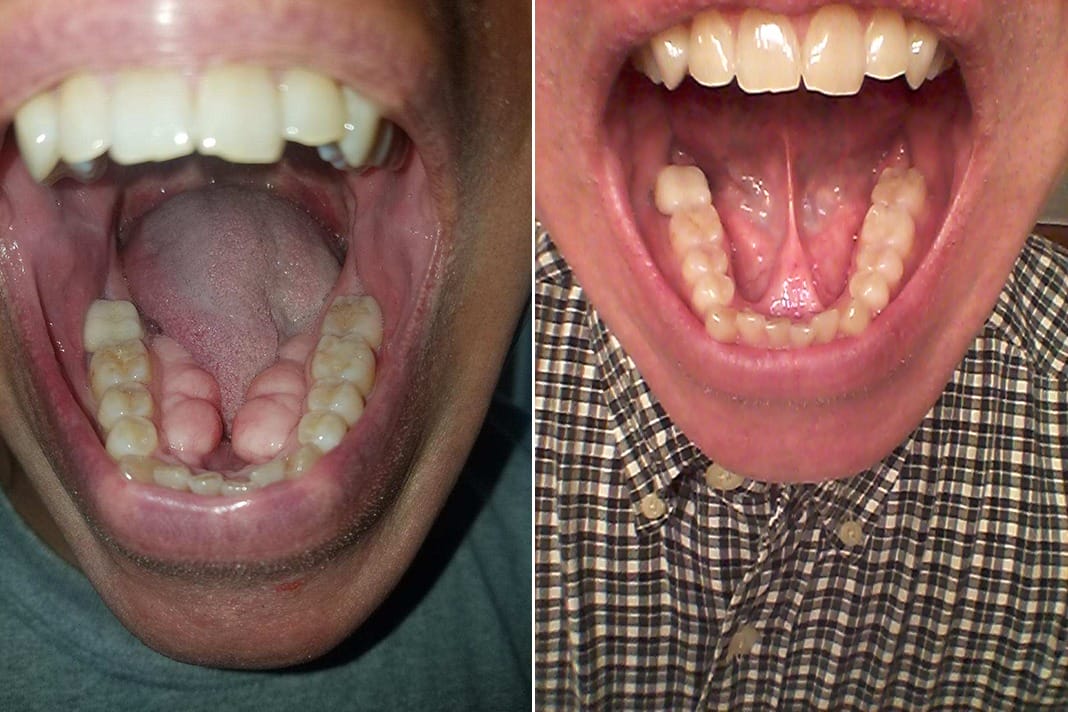
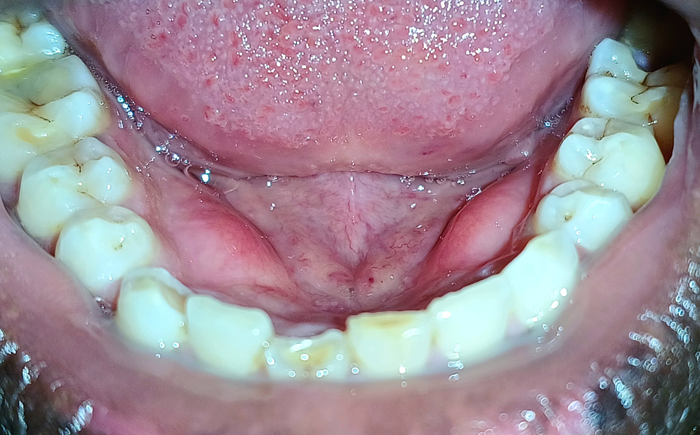
Mandibular tori represent a common, benign anatomical variation found in the oral cavity. Often discovered incidentally during routine dental examinations, these bony protuberances manifest as harmless growths on the inner surface of the lower jaw, near the tongue. While their precise etiology remains a subject of ongoing research, factors such as genetics, masticatory stress (chewing forces), and possibly bruxism (teeth grinding) are commonly implicated. Although generally asymptomatic, mandibular tori can range significantly in size, shape, and number, sometimes presenting as a single nodule or as multiple lobulated masses. For the most part, these formations require no intervention, but their presence takes on new significance when considering the advancements in modern dental technology, which not only aids in their accurate identification and monitoring but also informs strategies for managing any associated complications.

The Digital Frontier of Diagnosis and Monitoring
The advent of digital technologies has revolutionized the way dental professionals diagnose and monitor oral conditions, including mandibular tori. Precision, efficiency, and patient comfort have been significantly enhanced, moving beyond traditional methods to offer more comprehensive insights.
Advanced Imaging Technologies
Contemporary dental practice relies heavily on sophisticated imaging techniques that provide detailed visualizations of the oral anatomy, far surpassing the capabilities of conventional radiography.
- Digital Radiography: This technology has largely replaced film-based X-rays, offering numerous benefits. Digital X-rays expose patients to significantly less radiation, provide immediate image capture for instant review, and allow for easy storage, retrieval, and electronic transmission of images. For mandibular tori, digital radiographs can confirm their bony nature, assess their general size, and help differentiate them from other intraoral lesions. The ability to enhance images digitally also aids in clearer visualization of their boundaries and internal structure.
- Cone Beam Computed Tomography (CBCT): A cornerstone of modern dental imaging, CBCT provides three-dimensional (3D) volumetric data of the maxillofacial region. Unlike 2D radiographs, CBCT offers unparalleled insight into the exact size, morphology, and spatial relationship of mandibular tori to adjacent vital structures, such as nerves, blood vessels, and tooth roots. This 3D perspective is indispensable for thorough pre-surgical planning, especially if the tori are large or complex. It allows clinicians to accurately measure the volume of the tori, map their contours, and anticipate potential challenges during removal, thereby minimizing risks and improving surgical predictability.
- Intraoral Scanners: These handheld devices capture highly accurate 3D digital impressions of the oral cavity without the need for traditional, messy impression materials. For mandibular tori, intraoral scanners can create precise digital models of the patient’s dentition and soft tissues, including the tori. These models serve as an excellent baseline for monitoring changes in tori size or shape over time. Furthermore, the digital files can be used for designing custom prosthetic appliances that accommodate the tori, or for planning their removal by providing a precise digital representation of the surgical field.
AI in Early Detection and Analysis
Artificial intelligence (AI) is increasingly being integrated into dental diagnostics, promising a new era of enhanced accuracy and efficiency, particularly in the analysis of imaging data related to conditions like mandibular tori.
- AI-Assisted Radiographic Analysis: Machine learning algorithms are being trained on vast datasets of dental radiographs to identify anomalies, including the subtle presence of mandibular tori. AI can help flag potential tori for closer inspection by the clinician, improving early detection, especially in cases where they are small or easily overlooked. This serves as a valuable second opinion, reducing the chance of misdiagnosis or delayed recognition.
- Automated Measurement and Tracking: AI systems can accurately measure the dimensions of mandibular tori from digital images and track their growth or changes over successive examinations. This capability provides objective, quantifiable data that aids clinicians in making informed decisions about monitoring frequency or intervention. Automated tracking minimizes human error and ensures consistency in assessment.
- Predictive Analytics for Complications: By analyzing patterns in patient data—including imaging, medical history, and genetic predispositions—AI may one day contribute to predictive models that assess the likelihood of mandibular tori causing complications (e.g., interference with denture fabrication, mucosal ulceration). Such insights could help in proactive management and personalized patient care plans.
Tech-Enabled Management and Intervention
While most mandibular tori are managed conservatively, situations arise where surgical removal becomes necessary. Modern technology plays a pivotal role in making these interventions safer, more predictable, and less invasive.
Digital Surgical Planning and Guidance
Surgical removal of mandibular tori, though often straightforward, benefits immensely from digital planning, especially when the growths are large, multi-lobulated, or in close proximity to sensitive anatomical structures.
- CAD/CAM for Surgical Guides: Computer-Aided Design/Computer-Aided Manufacturing (CAD/CAM) technologies enable the fabrication of custom surgical guides. Based on CBCT data and intraoral scans, these guides are 3D-printed to precisely fit over the patient’s existing teeth or soft tissue, guiding the surgeon’s instruments to the exact location and depth required for bone removal. This minimizes collateral damage to surrounding tissues, enhances precision, and reduces surgical time and post-operative complications.
- Virtual Surgery Simulations: Before even touching a scalpel, surgeons can perform “virtual surgeries” using 3D models derived from CBCT scans. This allows them to plan the osteotomy (bone cut), visualize the bone reduction, and anticipate potential challenges in a simulated environment. Such simulations provide invaluable practice, refine surgical techniques, and allow for the optimization of the surgical approach, ensuring greater confidence and a smoother procedure in reality.
- Minimally Invasive Techniques: Technology facilitates less invasive approaches to tori removal. Piezoelectric surgical devices, for instance, use ultrasonic vibrations to selectively cut bone while preserving soft tissues like nerves and blood vessels, offering a safer alternative to traditional rotary instruments. Endoscopic assistance, though less common for tori removal, further exemplifies how technology enables direct visualization and precise manipulation in constricted spaces.
Materials Science and Prosthodontic Innovations
The interface between mandibular tori and dental prostheses is a critical area where advancements in materials science and digital prosthodontics have made significant impacts.
- Advanced Biocompatible Materials: For patients requiring removable prosthetics like dentures, the presence of large mandibular tori can pose a challenge to fit and stability. Modern materials for denture bases, such as enhanced acrylics or flexible polymers, are designed for better adaptation and comfort. When tori are surgically removed, the underlying bone may be contoured to allow for optimal prosthesis fit, and these new materials can adapt better to the altered anatomy.
- 3D Printing of Custom Appliances: Digital dentistry, through 3D printing, allows for the creation of highly customized dental appliances. If tori interfere with the fit of a denture or a nightguard, a precise digital impression captured by an intraoral scanner can be used to design and 3D print an appliance that perfectly accommodates the tori, either by creating relief areas or by contouring the appliance around them. This approach offers superior comfort, retention, and function compared to traditional fabrication methods.
- Digital Impression Systems for Precise Fit: The accuracy of digital impressions captured by intraoral scanners ensures that any prosthetic designed to interact with or avoid mandibular tori fits with extreme precision. This eliminates the inaccuracies inherent in traditional impression materials, leading to better-fitting dentures, retainers, or other oral devices, and significantly reducing the need for multiple adjustments.
Telehealth and Remote Consultation for Oral Health
The integration of telehealth into dentistry has expanded access to care and transformed how patients interact with their dental providers, particularly relevant for conditions like mandibular tori that often require ongoing monitoring rather than immediate intervention.
- Virtual Consultations: For patients with asymptomatic mandibular tori, telehealth platforms allow for initial assessments or follow-up consultations to be conducted remotely. Patients can submit high-resolution intraoral photographs or videos, and in some cases, share their previous digital imaging data securely with their dentist. This enables the clinician to evaluate the tori without the patient needing to travel, saving time and reducing logistical burdens. It is particularly beneficial for monitoring stable, non-problematic tori, identifying any changes that might warrant an in-person visit.
- Secure Data Sharing and Collaboration: Modern telehealth solutions are built on secure, encrypted platforms that comply with privacy regulations, facilitating the safe exchange of patient images, diagnostic reports, and treatment plans between patients and providers, or among specialists. This seamless data flow supports collaborative care and second opinions without geographical constraints.
- Remote Monitoring of Oral Changes: For patients with known mandibular tori, remote monitoring systems, potentially leveraging patient-uploaded images or even AI-powered analysis of oral scans performed at home (in future iterations of consumer-grade devices), could provide continuous tracking of tori growth or associated mucosal changes, enabling timely intervention if complications arise.
The Future of Tori Management: Emerging Technologies
The trajectory of technological innovation suggests even more transformative approaches to understanding and managing conditions like mandibular tori.
- Augmented Reality (AR) and Virtual Reality (VR) in Dentistry: These immersive technologies are already finding applications in surgical training, allowing dental students and seasoned practitioners to practice complex procedures in a risk-free virtual environment. For tori removal, AR could overlay 3D anatomical data directly onto the patient during surgery, providing real-time guidance and enhancing precision. VR could also be used for patient education, allowing individuals to visualize their tori and understand proposed treatments in an interactive manner.
- Nanotechnology for Targeted Interventions: While speculative for mandibular tori, nanotechnology holds promise for future medical interventions. In the context of benign bony growths, nano-scale drug delivery systems could potentially target specific cellular pathways to modulate bone growth, offering a non-surgical approach to manage excessively large tori, or even prevent their progression.
- Further Integration of IoT Devices: The Internet of Things (IoT) could lead to smart oral health devices that continuously monitor intraoral conditions, including subtle changes in bone structure or soft tissue health, and transmit data to dental professionals. While sophisticated for tori, simpler applications might involve smart toothbrushes detecting areas of chronic irritation due to tori and alerting the user and their dentist.
In conclusion, while mandibular tori are a straightforward anatomical feature, their management is increasingly sophisticated due to rapid technological advancements. From highly detailed 3D imaging and AI-assisted diagnostics to digitally guided surgical interventions and personalized prosthetic solutions, technology is transforming how these common oral growths are understood, monitored, and addressed, ultimately enhancing patient care and outcomes in the realm of oral health.
aViewFromTheCave is a participant in the Amazon Services LLC Associates Program, an affiliate advertising program designed to provide a means for sites to earn advertising fees by advertising and linking to Amazon.com. Amazon, the Amazon logo, AmazonSupply, and the AmazonSupply logo are trademarks of Amazon.com, Inc. or its affiliates. As an Amazon Associate we earn affiliate commissions from qualifying purchases.