
The mitotic spindle, a complex and dynamic cellular machine, is fundamental to life itself, orchestrating the precise segregation of chromosomes during cell division. While its core function is biological, the relentless pursuit of understanding and manipulating this intricate process has propelled significant advancements across various technological domains. From cutting-edge imaging techniques to AI-driven analytical platforms and sophisticated genetic engineering tools, technology is continually reshaping our grasp of the mitotic spindle, offering unprecedented insights into cellular health and disease, and paving the way for innovative therapeutic strategies.
The Biological Imperative: Mitotic Spindle as a Critical Cellular Machine
At its essence, the mitotic spindle is a transient, self-assembling macromolecular structure composed primarily of microtubules, motor proteins, and associated regulatory factors. Its formation is a hallmark of mitosis, the process by which a eukaryotic cell divides into two identical daughter cells. The spindle’s primary role is to ensure that each daughter cell receives a complete and accurate set of chromosomes, a feat crucial for genetic stability and the proper development and maintenance of multicellular organisms.

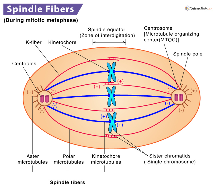
The assembly of the mitotic spindle begins with the duplication of the centrosome, the primary microtubule-organizing center in animal cells. These two centrosomes then migrate to opposite poles of the cell, forming the poles of the spindle. Microtubules, dynamic protein polymers, polymerize from these poles, forming three main types: astral microtubules (interacting with the cell cortex), interpolar microtubules (interdigitating in the spindle midzone), and kinetochore microtubules (attaching to specialized protein structures on each sister chromatid called kinetochores). These kinetochore microtubules apply forces that align chromosomes at the cell’s equator and then pull them apart, ensuring their faithful segregation. Any errors in this process can lead to aneuploidy (abnormal chromosome number), a common feature of cancer cells and developmental disorders. Given its critical role, the mitotic spindle has become a prime target for technological innovation aimed at both fundamental discovery and therapeutic intervention.
Technological Advancement in Visualizing and Analyzing the Mitotic Spindle
Understanding the mitotic spindle’s dynamic nature and intricate structure requires highly specialized technological approaches. Modern microscopy, combined with artificial intelligence and computational modeling, has revolutionized our ability to observe, quantify, and simulate its behavior with unprecedented detail.
High-Resolution Microscopy: Peering into the Nanoscale World
Traditional light microscopy provided initial glimpses of the mitotic spindle, but its diffraction limit restricted resolution, obscuring many critical details. The advent of super-resolution microscopy techniques has shattered these barriers, allowing scientists to visualize cellular structures at the nanoscale. Stimulated Emission Depletion (STED) microscopy, Photoactivated Localization Microscopy (PALM), and Stochastic Optical Reconstruction Microscopy (STORM) employ sophisticated optical principles to bypass the diffraction limit, achieving resolutions down to tens of nanometers. These technologies enable researchers to precisely localize individual spindle components, track microtubule polymerization and depolymerization in real-time, and observe the interactions between kinetochores and microtubules with exquisite clarity.
Beyond these point-scanning or single-molecule localization methods, light-sheet microscopy and lattice light-sheet microscopy have transformed the study of live cell dynamics. By illuminating only a thin plane of the sample, these techniques drastically reduce phototoxicity and photobleaching, allowing for long-term, high-speed 3D imaging of the entire mitotic spindle within a living cell. This capability provides crucial insights into the spatio-temporal dynamics of spindle assembly and function, often over multiple cell cycles. Furthermore, advancements in cryo-electron tomography (cryo-ET) are now pushing the boundaries even further, providing near-atomic resolution 3D reconstructions of spindle components and their interactions in their native cellular context, revealing previously unseen structural details.
AI and Image Analysis: Decoding Complex Dynamics
The massive datasets generated by high-resolution microscopy demand sophisticated analytical tools. Artificial intelligence, particularly machine learning and deep learning algorithms, has emerged as an indispensable technology for processing, segmenting, and quantifying complex mitotic spindle images. AI-powered image analysis software can automatically identify and track individual microtubules, centrosomes, and chromosomes across thousands of images or frames of a video, far surpassing the speed and accuracy of manual analysis.
Deep learning models, trained on vast datasets of annotated spindle images, can detect subtle abnormalities in spindle structure or dynamics that might be missed by human observers. For instance, these algorithms can quantify microtubule density, measure spindle length and width, track chromosome movement velocities, and even predict the outcome of a mitotic event based on early-stage spindle morphology. This technological capability allows researchers to conduct high-throughput screens for genetic perturbations or chemical compounds affecting spindle function, accelerating drug discovery and mechanistic studies. By reducing subjective bias and significantly increasing the scale of analysis, AI tools transform raw imaging data into quantifiable biological insights, driving a deeper understanding of spindle regulation and dysfunction.
Computational Modeling and Simulation: Predicting Spindle Behavior
Complementing experimental observations, advanced computational modeling and simulation technologies provide a powerful platform for understanding the underlying physical principles governing mitotic spindle assembly and function. By creating in silico models, researchers can test hypotheses, predict behaviors, and explore scenarios that are difficult or impossible to reproduce experimentally.
These models range from coarse-grained simulations that represent microtubules and motor proteins as simplified units to highly detailed, spatially explicit models that incorporate the mechanics of individual protein interactions. Software frameworks enable the simulation of microtubule growth and shrinkage dynamics, motor protein force generation, and the overall self-organization of the spindle. For example, some models predict how variations in motor protein activity or microtubule stability influence spindle length, chromosome alignment, or segregation accuracy. These computational tools also aid in interpreting complex experimental data, by revealing emergent properties of the spindle system that arise from the collective interactions of its components. They are instrumental in validating existing theories and generating new, testable predictions, forming a vital feedback loop with experimental science.
Engineering and Manipulating the Mitotic Spindle: A Biotech Frontier
Beyond observation, the ability to precisely engineer and manipulate the mitotic spindle, or the factors that control it, represents a significant technological leap with profound implications for biotechnology and medicine. This involves leveraging genetic tools and advanced screening platforms for both fundamental research and therapeutic development.
Genetic Engineering Tools: Unraveling Functional Roles
Technologies such as CRISPR-Cas9 genome editing, RNA interference (RNAi), and gene overexpression systems have revolutionized our ability to precisely manipulate the genes encoding mitotic spindle components and their regulators. CRISPR-Cas9 allows for targeted knockout, knockdown, or modification of specific genes, enabling researchers to study the precise function of individual proteins within the spindle. By generating cell lines or organisms with altered spindle proteins, scientists can systematically dissect the contributions of various factors to spindle assembly, dynamics, and force generation.
RNAi technology, which selectively silences gene expression, offers another powerful approach to perturb spindle components. By reducing the levels of specific proteins, researchers can observe the resulting phenotypes, such as spindle collapse, chromosome segregation errors, or altered mitotic timing. These genetic tools are not only crucial for basic research but also serve as platforms for identifying potential drug targets by systematically perturbing genes involved in spindle formation and observing the cellular consequences.
Drug Discovery and Therapeutic Targeting: Fighting Disease at the Cellular Level
The mitotic spindle has long been a critical target for anti-cancer chemotherapy. Drugs like taxanes (e.g., paclitaxel) and vinca alkaloids (e.g., vincristine) are potent spindle poisons, acting by stabilizing or destabilizing microtubules, respectively. This disrupts normal spindle function, preventing cell division and leading to apoptosis in rapidly dividing cancer cells. However, these traditional drugs often come with significant side effects due to their lack of specificity for cancer cells.
Modern technological platforms are addressing these challenges. High-throughput screening (HTS) allows for the rapid testing of thousands of compounds against cell lines to identify new molecules that selectively interfere with spindle function. Rational drug design, guided by structural biology and computational chemistry, enables the development of more specific inhibitors that target particular spindle proteins or pathways unique to cancer cells. In silico drug discovery leverages computational models to predict the binding affinity of potential drug candidates to spindle proteins, accelerating lead compound identification and optimization. Furthermore, advances in personalized medicine, utilizing technologies like genomic sequencing and transcriptomics, aim to identify which patients are most likely to respond to spindle-targeting therapies based on their tumor’s specific genetic profile, moving towards more effective and less toxic treatments.
Synthetic Biology and Bioengineering: Building and Rebuilding Cellular Machinery
The burgeoning field of synthetic biology is beginning to explore the possibility of building and modifying biological systems from the ground up. In the context of the mitotic spindle, this translates to efforts to reconstitute functional spindle components in vitro or even design simplified, synthetic versions of the spindle. Cell-free systems, where purified proteins are combined in a test tube, allow researchers to study spindle assembly and mechanics in a highly controlled environment, free from the complexities of a living cell. This bioengineering approach helps to delineate the minimal requirements for spindle function and allows for the precise manipulation of individual factors. While still in its nascent stages, the ability to engineer and control spindle-like structures opens avenues for understanding the fundamental principles of self-organization in biological systems and could eventually lead to novel biotechnological applications, such as engineered cellular therapeutics or diagnostic tools.
The Future of Mitotic Spindle Research: Integration and Innovation
The trajectory of mitotic spindle research is increasingly characterized by the integration of diverse technological disciplines. The future promises an even more holistic understanding, driven by synergistic advancements across multiple fronts.
Multi-omics Integration: A Holistic View of Spindle Regulation
One of the most exciting future directions involves integrating multi-omics data (genomics, transcriptomics, proteomics, metabolomics) with advanced imaging and computational models. Technologies that enable comprehensive profiling of gene expression, protein interactions, and metabolic states alongside real-time observation of spindle dynamics will provide an unprecedented, holistic view of how the mitotic spindle is regulated at multiple biological layers. AI and sophisticated bioinformatics tools will be critical for correlating these vast datasets, identifying novel regulatory networks, and uncovering subtle interdependencies that influence spindle fidelity and overall cell division outcomes. This integrated approach will undoubtedly reveal new biomarkers for disease and novel targets for therapeutic intervention.
Miniaturization and Automation: High-Throughput Discovery
The ongoing trend towards miniaturization and automation will profoundly impact mitotic spindle research. Microfluidic devices, which enable the precise manipulation of fluids at the microscale, allow for high-throughput screening of cellular responses to various perturbations while minimizing sample and reagent consumption. Automated microscopy platforms, combined with robotic handling and AI-driven image acquisition and analysis, will allow researchers to screen tens of thousands of conditions or compounds in a single experiment. This significantly accelerates the pace of discovery, making it feasible to systematically explore vast parameter spaces for understanding spindle function in health and disease. Such automation is crucial for advancing personalized medicine, where individual patient cells might be tested against a panel of drugs to predict efficacy.

AI-driven Drug Optimization and Predictive Models: Smarter Therapies
The increasing sophistication of AI will lead to the development of powerful predictive models for spindle-targeting therapies. Future AI systems, trained on comprehensive datasets encompassing molecular structures, cell biology, clinical trial outcomes, and patient genetic profiles, will be able to predict the efficacy and toxicity of novel spindle-targeting agents with remarkable accuracy. These AI-driven tools will not only accelerate the drug discovery pipeline but also enable the design of highly personalized therapeutic strategies, minimizing side effects and maximizing treatment success. Envision AI that can simulate drug interactions with specific spindle protein variants in a patient’s tumor, guiding the selection of the most effective compound for their unique biological context. The continuous feedback loop between experimental data generation and AI model refinement will ensure that our understanding and manipulation of the mitotic spindle continue to evolve, ushering in an era of smarter, more targeted biotechnological solutions.
aViewFromTheCave is a participant in the Amazon Services LLC Associates Program, an affiliate advertising program designed to provide a means for sites to earn advertising fees by advertising and linking to Amazon.com. Amazon, the Amazon logo, AmazonSupply, and the AmazonSupply logo are trademarks of Amazon.com, Inc. or its affiliates. As an Amazon Associate we earn affiliate commissions from qualifying purchases.