
The Digital Anatomy of Parasitic Infestation: Identifying Ear Mites Through Clinical Observation
When addressing the biological reality of Otodectes cynotis—the microscopic arachnid commonly known as the ear mite—we must look beyond mere folklore and into the systematic indicators of infestation. For pet owners and feline health enthusiasts, identifying these parasites is not just a matter of observation; it is a diagnostic process that bridges the gap between superficial symptoms and clinical reality. Ear mites are highly contagious, microscopic, eight-legged organisms that thrive in the dark, waxy environment of the feline ear canal. They feed on epidermal debris and ear oils, causing significant localized irritation and a distinct physical appearance that is often described by veterinary professionals as resembling “coffee grounds.”
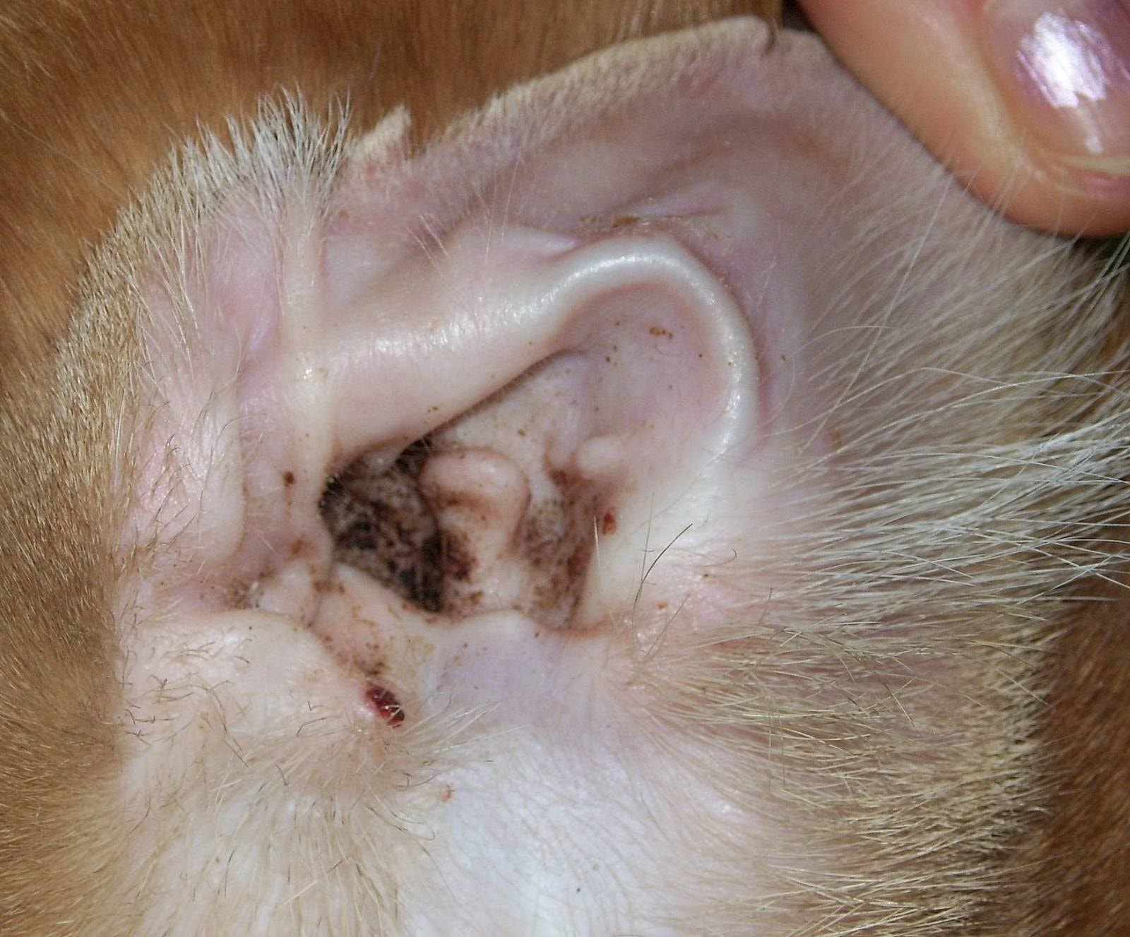
Understanding what ear mites look like requires a twofold approach: identifying the physical manifestation of the waste material within the ear and recognizing the behavioral markers that precede a visual inspection. While the mites themselves are virtually invisible to the naked eye, their biological byproduct is unmistakable.
Visual Indicators and the “Coffee Ground” Phenomenon
The most reliable visual cue for an ear mite infestation is the accumulation of dark, granular debris within the pinna (the outer ear) and the vertical ear canal. If you have ever wondered what the evidence of these parasites looks like, picture used coffee grounds or dark, dried tea leaves. This specific appearance is the result of a mixture of mite excrement, dead skin cells, and dried blood caused by the irritation of the mite’s feeding activity.
The Role of Ear Canal Inflammation
When mites invade, the body responds with an inflammatory process. This causes the internal tissue of the ear to become engorged and hyper-sensitive. The resulting discharge is not merely “dirty”; it is an exudate. In a healthy cat, ear wax is typically light-colored, thin, and minimal. When mites are present, this substance thickens and turns dark brown or black. If this substance is left unaddressed, it creates a hospitable environment for secondary bacterial and fungal infections, which further complicates the diagnostic visual.
Distinguishing Mites from General Debris
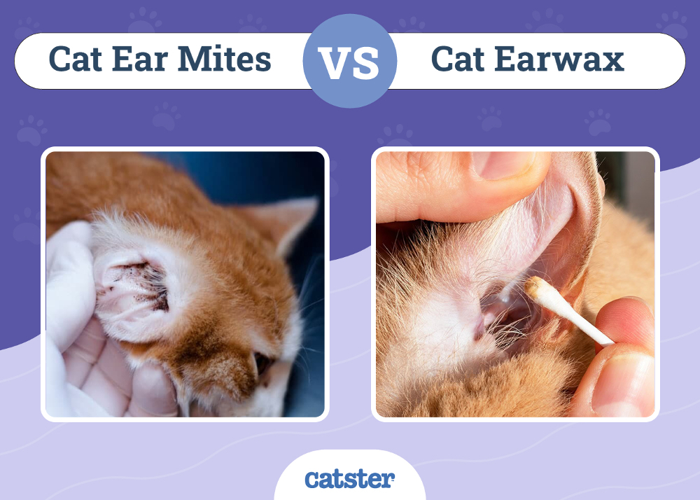
It is critical to distinguish between simple ear wax and a parasitic infestation. General ear debris is usually oily or waxy and lacks the crusty, granular texture associated with mites. A hallmark visual sign of an infestation is the persistence of this granular matter despite regular gentle cleaning. If the “coffee grounds” return rapidly after cleaning, it is a high-probability indicator that the life cycle of the Otodectes cynotis is currently active within the canal.
Behavioral and Physical Manifestations of Infestation
While the visual debris is the primary “look” of the infestation, the way the cat holds itself provides secondary visual cues that confirm the suspicion. Ear mites are profoundly irritating, and the physical response from the cat often manifests as a distorted ear shape or persistent postural irregularities.
Postural Indicators
Observe the cat’s head carriage. A cat with a severe infestation will frequently tilt its head toward the affected side. This is a visual symptom of deep-seated discomfort or the disruption of balance due to inner ear inflammation. Furthermore, the base of the ear may appear raw, red, or inflamed due to the cat’s own attempts to scratch the area. You may notice “crusty” skin patches around the back of the ears, which are often the result of self-inflicted trauma from the cat’s hind claws.
The “Ear Scratch” Visual
When observing a cat, look for the repetitive, aggressive scratching of the ears or the frantic shaking of the head. This is the physiological expression of the “crawling” sensation caused by the mites moving along the delicate skin of the ear canal. When the cat shakes its head, it is often trying to dislodge the mites or the accumulating debris. Seeing a cat constantly engaging in these behaviors is a visual prompt that the internal ear environment is compromised.
Diagnostic Procedures and Microscopic Verification
Because the ear mites themselves are so small—measuring roughly 0.3 to 0.5 millimeters—they cannot be definitively identified through simple visual inspection of the outer ear by a layman. To truly “see” the mites, one must employ clinical diagnostic tools. This is where the gap between observation and verification is bridged.
Clinical Microscopy
A veterinarian uses an otoscope to view the depths of the canal, but for a positive identification, they will perform a swab of the debris. When this sample is placed under a microscope on a slide, the mites become clearly visible. They appear as small, white, spider-like creatures moving sluggishly against the dark background of the ear wax. This is the only way to be 100% certain of an infestation, as other conditions, such as yeast infections or localized dermatitis, can sometimes mimic the granular, dark appearance of mite debris.
Professional Intervention vs. Home Observation
While it is helpful for pet owners to know what to look for, attempting to treat an infestation based purely on a visual guess can be counterproductive. Many over-the-counter treatments are oil-based or contain ingredients that can be toxic if the eardrum has been perforated by the mites. Identifying the look is the first step, but seeking professional confirmation ensures that the correct medication is administered, preventing the condition from escalating into a middle or inner ear infection (otitis media or interna), which can lead to neurological symptoms.
Maintaining Feline Ear Health Through Observation
Prevention and early detection rely on the consistency of your observations. Understanding that the “look” of ear mites is a combination of dark, crusty debris and inflammatory redness allows you to intervene before the cat suffers from chronic discomfort.
The Importance of Routine Checks
Make ear inspection a part of your regular grooming routine. Gently fold back the ear flap (the pinna) and shine a light into the opening. A healthy ear should be light pink, devoid of strong odor, and free of any dark, granular discharge. If you see any deviation from this, you are looking at the potential early stages of an infestation.

Environmental Considerations
Ear mites do not just live on the cat; they can exist in the environment for short periods and are easily transmitted between pets in the same household. Therefore, the “look” of an infestation in one cat should lead to the immediate observation of all other household pets. If one animal has mites, it is almost certain that the others have been exposed, even if they are not yet showing the classic “coffee ground” visual symptoms.
By maintaining a vigilant eye on the condition of your cat’s ears and understanding that the appearance of dark, crusty debris is the primary visual signature of these parasites, you can ensure a rapid response. The objective is to identify the signs early, avoid the potential for secondary complications, and restore the cat to a state of comfort and cleanliness. Through consistent observation, you serve as the first line of defense in protecting your pet from the persistent irritation of the Otodectes cynotis mite.
aViewFromTheCave is a participant in the Amazon Services LLC Associates Program, an affiliate advertising program designed to provide a means for sites to earn advertising fees by advertising and linking to Amazon.com. Amazon, the Amazon logo, AmazonSupply, and the AmazonSupply logo are trademarks of Amazon.com, Inc. or its affiliates. As an Amazon Associate we earn affiliate commissions from qualifying purchases.