The human body, a marvel of biological engineering, relies on a sophisticated interplay of systems to perform its myriad functions. Among these, muscle tissue stands out as a cornerstone of movement, posture, and internal bodily processes. While often discussed in biological or physiological contexts, understanding the underlying technological intricacies that govern muscle tissue’s formation, function, and even its augmentation offers a fascinating lens through which to view this fundamental biological component. From the molecular machinery to the advanced diagnostic and therapeutic tools employed by modern medicine and sports science, technology plays an indispensable role in deciphering and harnessing the power of muscle. This exploration delves into the technological underpinnings of muscle tissue, examining its composition, the digital tools used to study it, and the cutting-edge innovations shaping its future.
The Molecular Motors: Nanotechnology’s Role in Muscle Structure
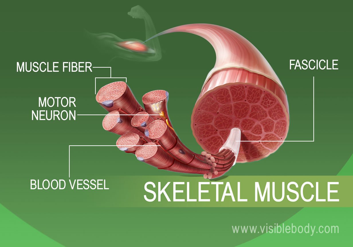
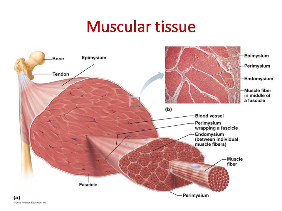
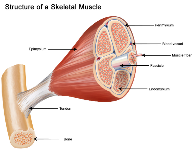
At its most fundamental level, muscle tissue is a testament to elegant biological engineering, powered by intricate molecular machines. Understanding these components requires sophisticated technological tools capable of operating at the nanoscale. The primary structural and functional units of muscle are myofibrils, which are themselves composed of repeating units called sarcomeres. These sarcomeres are the powerhouses of contraction, and their precise arrangement and the interactions within them are orchestrated by a suite of specialized protein molecules.
Actin and Myosin: The Filamentous Engines
The workhorses of muscle contraction are the thick filaments of myosin and the thin filaments of actin. These proteins are not static structures; they are dynamic and interact in a highly controlled, cyclical manner. Myosin heads, protruding from the myosin filament, possess the ability to bind to actin filaments. Fueled by adenosine triphosphate (ATP), a molecular energy currency, these myosin heads undergo a conformational change, pulling the actin filaments closer together – a process known as the sliding filament theory. This coordinated sliding results in the shortening of the sarcomere, and consequently, the contraction of the entire muscle.
Technological advancements have been crucial in visualizing and understanding these molecular interactions. Cryo-electron microscopy (Cryo-EM), for instance, allows researchers to capture snapshots of these protein complexes in near-native states, providing unprecedented detail of their three-dimensional structures. By analyzing these structures, scientists can discern how drugs might interact with these proteins, how genetic mutations affect their function, and how to design artificial muscle-like materials. Furthermore, atomic force microscopy (AFM) can be used to probe the mechanical properties of individual protein filaments and even their interactions, offering insights into the forces involved in muscle contraction at the single-molecule level.
Titin and Nebulin: The Structural Scaffolding
Beyond the primary contractile proteins, other molecules like titin and nebulin play critical roles in maintaining the structural integrity and precise assembly of the sarcomere. Titin, the largest known protein, acts as a molecular spring, anchoring the myosin filaments to the Z-lines of the sarcomere and contributing to muscle elasticity. Nebulin, a giant protein associated with actin filaments, is thought to act as a ruler, ensuring the correct length of actin filaments during muscle development.
The study of these large, complex proteins has also been facilitated by advanced imaging and sequencing technologies. High-throughput sequencing allows for rapid and accurate determination of the genes encoding these proteins, providing clues about their evolutionary history and functional variations. Advanced computational modeling and simulation are increasingly employed to predict how these large proteins fold, interact, and contribute to the overall mechanics of the sarcomere, bridging the gap between sequence data and physical function. These technologies are not just for basic research; they are the foundation for understanding diseases characterized by muscle weakness or structural defects.
The Digital Blueprint: Imaging and Diagnostic Technologies in Muscle Research
Understanding the macroscopic and microscopic architecture of muscle tissue, as well as detecting abnormalities, relies heavily on sophisticated imaging and diagnostic technologies. These tools allow for non-invasive visualization, quantitative analysis, and early detection of conditions affecting muscle health. The integration of artificial intelligence (AI) and machine learning (ML) is revolutionizing how this data is interpreted, leading to more accurate diagnoses and personalized treatment plans.
Magnetic Resonance Imaging (MRI) and Ultrasound: Visualizing Muscle in Action
Magnetic Resonance Imaging (MRI) is a cornerstone of modern medical imaging, offering detailed cross-sectional views of muscle tissue without the use of ionizing radiation. Advanced MRI sequences can differentiate between various types of muscle tissue, identify inflammation, edema, tumors, and tears. Techniques like diffusion tensor imaging (DTI) are being adapted to study the microstructure of muscle, providing insights into fiber orientation and integrity, which is crucial for evaluating sports injuries and neurological conditions affecting muscle control.
Ultrasound imaging, known for its real-time capabilities and portability, is also invaluable in assessing muscle tissue. It allows for dynamic evaluation of muscle contractions, detection of tears, hematomas, and the presence of scar tissue. Increasingly, AI algorithms are being integrated into ultrasound systems to automate measurements, enhance image quality, and assist in lesion detection, making it a more powerful diagnostic tool. The ability to visualize muscle in motion provides crucial functional information that static imaging techniques cannot capture.
Electromyography (EMG) and Nerve Conduction Studies: Probing Neuromuscular Function
Beyond structural imaging, understanding how muscles are activated and controlled requires investigating the electrical activity of nerves and muscles. Electromyography (EMG) measures the electrical potential generated by skeletal muscles. Needle EMG involves inserting a fine electrode into the muscle to record its electrical activity at rest and during contraction. Surface EMG, using electrodes placed on the skin, can assess broader muscle activation patterns.
Nerve conduction studies (NCS), often performed alongside EMG, measure the speed and strength of electrical signals traveling along nerves. Together, EMG and NCS are essential for diagnosing a wide range of neuromuscular disorders, including peripheral neuropathies, myopathies, and nerve entrapments. The technological sophistication of EMG equipment has advanced significantly, allowing for more precise recordings and detailed analysis of nerve and muscle function, often aided by specialized software that can automatically detect abnormalities and quantify signal characteristics.
AI-Powered Analysis: Unlocking Deeper Insights
The sheer volume of data generated by these imaging and diagnostic modalities presents a significant challenge. This is where AI and ML are making transformative contributions. Algorithms trained on vast datasets of muscle images and EMG recordings can now identify subtle patterns that might be missed by the human eye. For instance, AI can automatically segment muscle regions, quantify fat infiltration or muscle atrophy, and predict disease progression with remarkable accuracy. In sports science, AI can analyze biomechanical data from EMG and motion capture to optimize training regimens and prevent injuries. These technologies are not merely automating tasks; they are augmenting human expertise, leading to more efficient and accurate diagnoses and personalized approaches to muscle health.
The Future of Muscle: Bioengineering and Performance Enhancement Technologies
The continuous advancements in technology are not only helping us understand muscle tissue better but are also paving the way for novel approaches to repair, regenerate, and even enhance muscle function. From regenerative medicine to advanced prosthetics and performance-enhancing devices, technology is at the forefront of shaping the future of muscle.
Tissue Engineering and Regenerative Medicine: Rebuilding Muscle
For individuals suffering from significant muscle loss due to injury, disease, or aging, tissue engineering offers a promising solution. This field leverages scaffolds, often made from biocompatible polymers or decellularized extracellular matrix, onto which muscle stem cells (myoblasts) are seeded. These cells are then cultured under specific conditions, often involving electrical or mechanical stimulation, to promote their differentiation and fusion into functional muscle fibers.
3D bioprinting is an exciting frontier in tissue engineering, allowing for the precise placement of cells and biomaterials to create complex muscle constructs with organized fiber orientation. This technology holds the potential to create patient-specific muscle grafts for transplantation, minimizing rejection and maximizing integration. Furthermore, research into gene therapy and CRISPR-Cas9 gene editing is exploring ways to correct genetic defects that cause muscle diseases or to enhance muscle growth and repair at the molecular level. These technologies represent a paradigm shift from merely treating symptoms to actively regenerating and rebuilding damaged or lost muscle tissue.
Biomaterials and Wearable Technology: Augmenting Muscle Function
Beyond regeneration, technology is enabling the augmentation of existing muscle function. Advanced biomaterials, such as responsive polymers and hydrogels, are being developed for use in muscle implants or as drug delivery systems to enhance muscle repair and growth. These materials can be designed to release growth factors or other therapeutic agents in a controlled manner, optimizing the healing process.
Wearable technologies are also playing an increasingly significant role. Exoskeletons, robotic suits that provide external support and power, are revolutionizing rehabilitation for individuals with severe mobility impairments, effectively augmenting their muscle strength and endurance. Smaller, more sophisticated wearable sensors can continuously monitor muscle activity, fatigue levels, and biomechanics, providing real-time feedback to athletes for performance optimization and injury prevention. Myoelectric prosthetics, controlled by the electrical signals from remaining muscles, are becoming increasingly sophisticated, offering a more intuitive and natural extension of the wearer’s own body, blurring the lines between biological and artificial muscle.
The relentless march of technological innovation, from the nanoscale manipulation of proteins to the macro-scale engineering of robotic limbs, continues to deepen our understanding and expand our capabilities concerning muscle tissue. As we continue to unravel the complexities of this vital biological component through a technological lens, the future promises even greater advancements in muscle health, repair, and augmentation.
aViewFromTheCave is a participant in the Amazon Services LLC Associates Program, an affiliate advertising program designed to provide a means for sites to earn advertising fees by advertising and linking to Amazon.com. Amazon, the Amazon logo, AmazonSupply, and the AmazonSupply logo are trademarks of Amazon.com, Inc. or its affiliates. As an Amazon Associate we earn affiliate commissions from qualifying purchases.