
Computed Tomography (CT) scans have revolutionized medical diagnostics, offering unparalleled insights into the intricate structures of the human body. When it comes to the paranasal sinuses – those air-filled cavities within the bones of the skull surrounding the nasal passages – a CT scan acts as a sophisticated digital explorer, revealing a wealth of information that is often invisible to the naked eye or less advanced imaging techniques. This article delves into the technological capabilities of sinus CT scans and explores the detailed anatomical and pathological information they unveil, emphasizing the advanced imaging processing and diagnostic power that define this crucial medical technology.
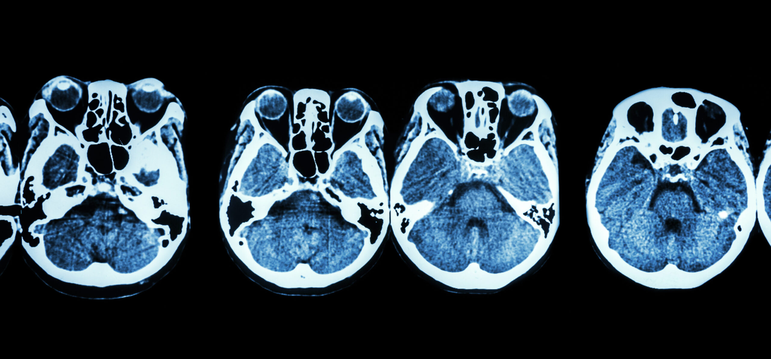
The Technological Foundation: How CT Scans Visualize Sinuses
At its core, a CT scan is a triumph of applied physics and computer science. It utilizes X-rays, but in a far more sophisticated manner than traditional radiography. Instead of capturing a single, flat image, a CT scanner employs a rotating X-ray source and a detector array that encircles the patient. As the X-ray beam passes through the body, it is attenuated by different tissues to varying degrees, with denser materials like bone absorbing more radiation than softer tissues or air. The detectors measure this differential absorption from multiple angles.
Data Acquisition and Reconstruction: From Raw Signals to Detailed Images
The process begins with data acquisition. The CT gantry, a doughnut-shaped structure, rotates around the patient, acquiring hundreds or thousands of thin X-ray “slices.” These slices represent the attenuation values of the tissues at specific points in the anatomy. The raw data, a complex stream of numbers, is then transmitted to a powerful computer workstation.
Here, advanced algorithms, often leveraging sophisticated mathematical techniques like filtered back-projection or iterative reconstruction, come into play. These algorithms process the raw attenuation data from numerous angles and reconstruct it into cross-sectional images of the sinuses. This reconstruction process essentially “stitches together” the information from each slice, creating detailed, two-dimensional images that can be viewed in axial, coronal, and sagittal planes. This ability to view the sinuses from different perspectives is a key technological advantage of CT over other imaging modalities.
Image Refinement and Visualization: Enhancing Diagnostic Clarity
Modern CT scanners produce incredibly high-resolution images. However, the raw reconstructed images may not always be optimal for diagnostic interpretation. Sophisticated image processing software plays a crucial role in enhancing these images for clarity and detail. This includes techniques such as:
- Windowing: CT images are displayed using a grayscale that represents different tissue densities. Windowing allows radiologists to adjust the range of Hounsfield Units (HU) displayed, effectively “tuning” the image to highlight specific tissues. For sinus CTs, a “bone window” is typically used to clearly delineate the bony structures and any abnormalities within them, while a “soft tissue window” can be employed to visualize the mucosal lining, mucus, and fluid.
- Multiplanar Reformation (MPR): Beyond the initial axial slices, MPR allows for the creation of images in any plane – coronal (front to back), sagittal (side to side), or even oblique. This is incredibly valuable for understanding the spatial relationships of sinus anatomy and pathology. For instance, a coronal view is essential for assessing the ostia (openings) of the sinuses, while sagittal views can reveal anterior-posterior extent of disease.
- 3D Reconstructions: Advanced software can create three-dimensional (3D) models of the sinuses from the CT data. These renderings provide an intuitive, almost surgical-like view of the anatomy, helping to visualize complex relationships and communicate findings effectively to other medical professionals or even patients.
The technological sophistication in data acquisition, reconstruction, and visualization is what allows a CT scan to reveal such precise details about the sinuses, forming the bedrock of accurate diagnosis.
Revealing Anatomical Nuances and Variations
The primary strength of a CT scan in the context of sinus imaging lies in its ability to visualize the complex bony architecture and the soft tissues within and around the nasal passages with remarkable detail. This allows for the identification of both normal anatomical variations and subtle abnormalities.
Bony Anatomy and Ostial Patency
CT scans excel at depicting the intricate bony walls of the paranasal sinuses, including the frontal, ethmoid, maxillary, and sphenoid sinuses. They can precisely identify the boundaries of each sinus cavity, the thickness of the bony walls, and the presence of any congenital anomalies such as hypoplasia (underdevelopment) or aplasia (absence) of a sinus.
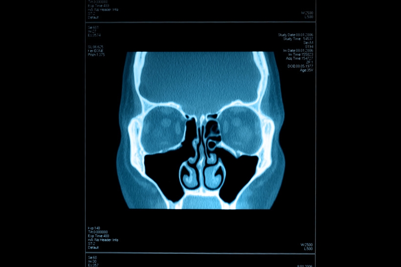
A critical aspect of sinus health is the patency of the ostia – the small openings that connect each sinus to the nasal cavity, allowing for drainage and ventilation. CT is invaluable in assessing these ostia. It can reveal if they are narrowed or completely blocked by thickened mucosa, polyps, or bony septations. This information is crucial for understanding the cause of recurrent sinusitis or chronic sinus issues. The thin slices and multiplanar capabilities allow for precise localization and measurement of any ostial obstruction.
Soft Tissue Details: Mucosa, Polyps, and Fluid
While CT is primarily known for its bone visualization capabilities, modern scanners with advanced multi-detector technology also provide excellent detail of the soft tissues within the sinuses. Radiologists can clearly visualize the mucosal lining of the sinuses.
- Mucosal Thickening: A hallmark of sinusitis, mucosal thickening appears as a smooth, often symmetrical increase in the soft tissue density lining the sinus walls. CT can quantify the degree of thickening and its distribution, helping to differentiate between acute and chronic inflammatory changes.
- Polyps: Nasal polyps, benign overgrowths of inflamed nasal or sinus mucosa, are readily identifiable on CT scans. They appear as soft tissue masses within the sinus cavities or nasal passages, often with a characteristic polypoid or lobulated appearance. CT helps determine the size, number, and location of polyps, which is vital for surgical planning.
- Fluid and Pus: Accumulation of fluid or pus within the sinuses, a common finding in acute sinusitis, appears as areas of increased density within the air-filled cavities. CT can differentiate between fluid and thickened mucosa, which can sometimes appear similar on other imaging modalities.
By meticulously examining these anatomical details, a CT scan provides a comprehensive blueprint of the sinus landscape, laying the groundwork for accurate diagnosis.
Unveiling Pathological Conditions: Diagnosing Sinus Diseases
The detailed visualization provided by CT scans makes them indispensable for diagnosing a wide spectrum of sinus-related pathologies, ranging from common infections to more complex tumors.
Inflammatory Conditions: Sinusitis and its Complications
The most frequent reason for ordering a sinus CT is to investigate sinusitis. CT scans can differentiate between various types of sinusitis:
- Acute Bacterial Sinusitis: CT findings often include mucosal thickening, air-fluid levels (indicating pus or fluid accumulation), and ostial obstruction. The distribution of these findings can help pinpoint the affected sinuses.
- Chronic Sinusitis: This is characterized by more generalized and often severe mucosal thickening, sometimes with bony remodeling or thickening of the sinus walls. The presence of polyps is also a common finding in chronic sinusitis, particularly in the ethmoid sinuses.
- Allergic Fungal Sinusitis: While not definitively diagnosed by CT alone, CT findings can be suggestive. This condition may present with significant mucosal thickening and inspissated (thickened) mucus, often with sinus opacification.
Beyond simple inflammation, CT is crucial for identifying complications of sinusitis, such as:
- Orbital Cellulitis/Abscess: Extension of infection into the tissues surrounding the eye. CT can visualize inflammatory changes in the orbit and identify fluid collections.
- Intracranial Complications: Though rare, severe sinusitis can lead to intracranial spread of infection. CT is essential for detecting complications like epidural or subdural abscesses, or even meningitis, by visualizing associated inflammatory changes in the brain and meninges.
Structural Abnormalities and Tumors
CT scans are also highly effective in detecting and characterizing structural abnormalities and neoplasms (tumors) within the sinuses.
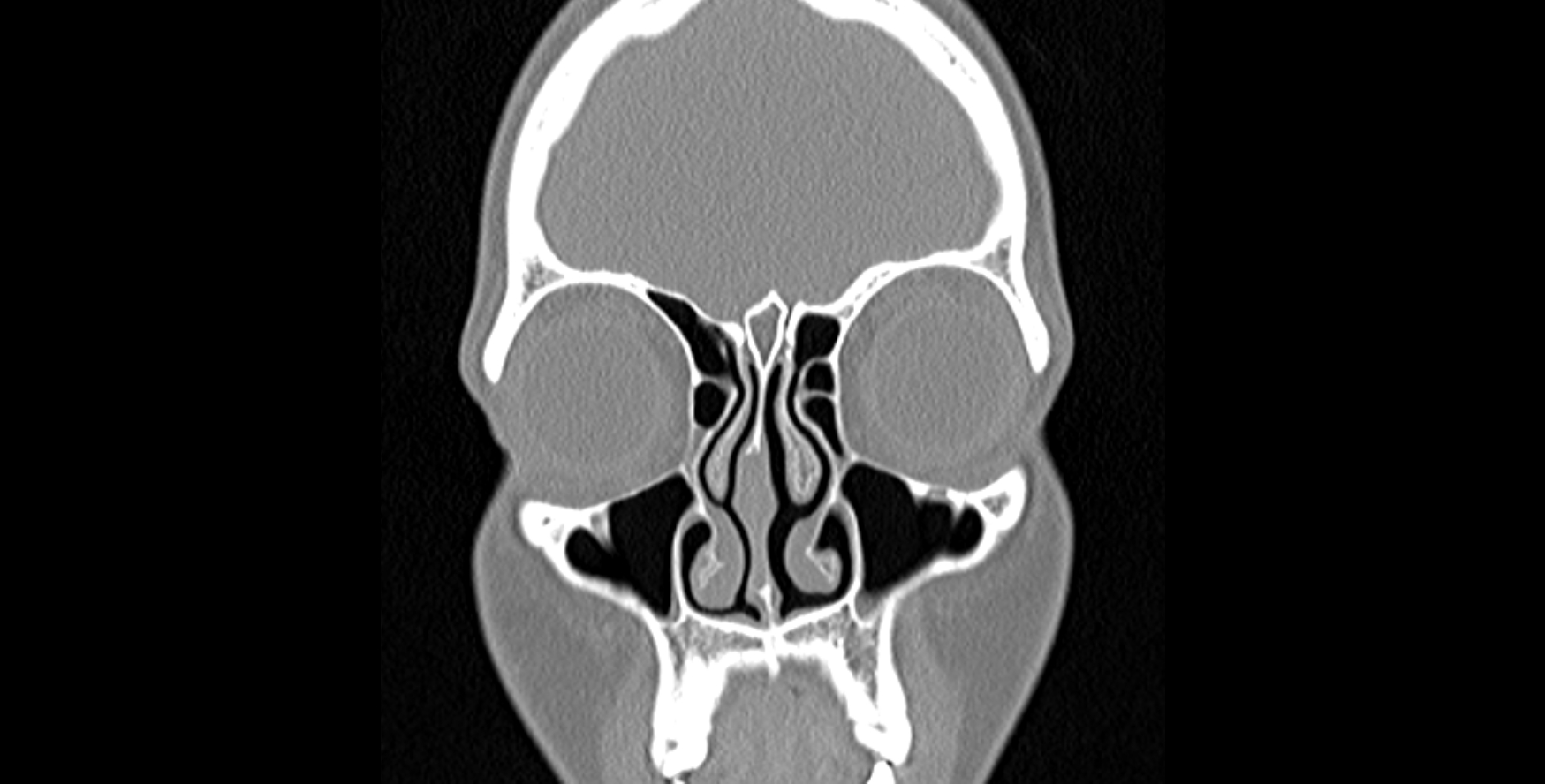
- Septal Deviations and Concha Bullosa: A deviated nasal septum can impede airflow and drainage, potentially contributing to sinusitis. A concha bullosa is an anatomical variation where a turbinate (a bony structure in the nasal cavity) pneumatizes (fills with air), which can sometimes obstruct sinus ostia. CT clearly visualizes these structural variations.
- Benign Tumors: Conditions like inverted papillomas, although benign, can be locally aggressive and destructive. CT can identify their presence, size, and extent, and importantly, can demonstrate their effect on the surrounding bony structures.
- Malignant Tumors: CT is a primary tool for the initial evaluation and staging of sinus cancers. It can delineate the extent of the tumor, identify involvement of adjacent structures (orbit, cranial base, infratemporal fossa), and assess for lymph node involvement. The ability to distinguish between soft tissue masses and bony erosion is critical in cancer diagnosis.
The technological prowess of CT scanning allows for the early and accurate detection of these diverse pathological processes, guiding appropriate treatment strategies and significantly improving patient outcomes. The continuous advancement in CT technology, including faster scan times, lower radiation doses, and enhanced image processing, further solidifies its role as a cornerstone in the diagnostic armamentarium for sinus diseases.
aViewFromTheCave is a participant in the Amazon Services LLC Associates Program, an affiliate advertising program designed to provide a means for sites to earn advertising fees by advertising and linking to Amazon.com. Amazon, the Amazon logo, AmazonSupply, and the AmazonSupply logo are trademarks of Amazon.com, Inc. or its affiliates. As an Amazon Associate we earn affiliate commissions from qualifying purchases.