The intersection of medical science and consumer technology has fundamentally altered the way we monitor our bodies. While the question “what does ear fluid look like” was once reserved for a professional consultation in a sterile clinic, the advent of high-definition smart otoscopes, artificial intelligence (AI), and telemedicine has brought this diagnostic capability into the palm of the consumer’s hand. Today, identifying the visual characteristics of ear fluid is less about a cursory glance and more about high-resolution data capture and algorithmic interpretation.
This digital evolution is not merely a convenience; it is a shift in the diagnostic paradigm. By leveraging sophisticated sensors and machine learning models, we can now distinguish between various types of middle ear effusions with a level of precision that was previously impossible for non-professionals. Understanding what ear fluid looks like in the digital age requires an exploration of the hardware that captures it, the software that analyzes it, and the data security frameworks that protect this sensitive information.
The Evolution of Otoscopy: From Manual Inspections to High-Definition Imagery
For over a century, the traditional otoscope remained largely unchanged—a simple magnifying glass coupled with a light source. However, the tech sector has recently revolutionized this tool, transforming it into a sophisticated digital imaging device. These smart otoscopes are the primary interface through which we now observe the internal environment of the ear.
Bridging the Gap: The Rise of Consumer Smart Otoscopes
The consumerization of medical hardware has led to the development of “smart” otoscopes designed for home use. These devices, often featuring ultra-thin probes and LED arrays, connect via Wi-Fi or Bluetooth to smartphone applications. When a user asks what ear fluid looks like, these devices provide a literal answer in 1080p or 4K resolution. Companies like TytoCare and various Silicon Valley startups have refined the optics of these tools, ensuring that the visual data captured is of clinical grade. This democratization of hardware allows parents and individuals to monitor conditions like Otitis Media (middle ear infection) in real-time, capturing images of fluid behind the eardrum to share with specialists via the cloud.
Resolution and Clarity: Why Visual Data Accuracy Matters
In the digital realm, the “look” of ear fluid is defined by pixels and color accuracy. High-definition sensors allow for the visualization of the light reflex on the tympanic membrane and the subtle nuances of fluid levels behind it. If the resolution is too low, the distinction between a healthy, pearly-gray eardrum and one with amber-colored serous fluid can be lost. Modern tech-driven otoscopes utilize multi-element lens systems and advanced image processing chips to reduce noise and enhance contrast, ensuring that the “visual signature” of the fluid is accurately represented for both human eyes and AI algorithms.
Leveraging AI and Machine Learning in Ear Fluid Identification
Capturing a high-quality image is only the first step. The true technological breakthrough lies in how software interprets those images. Artificial Intelligence is now being trained to recognize the visual markers of ear fluid, providing a layer of diagnostic support that minimizes human error.
Computer Vision: Training Algorithms to Detect Middle Ear Effusion
The question of what ear fluid looks like is being answered by neural networks trained on millions of clinical images. Computer vision algorithms are designed to identify specific patterns: the bulging of the eardrum, the presence of air-fluid levels, and the opacity of the fluid itself. By categorizing fluid as serous (clear/watery), mucoid (thick/cloudy), or purulent (pus-like), AI models can provide a probability score for different types of infections. This tech does not just show the user what the fluid looks like; it provides a data-driven interpretation of the visual evidence, identifying subtle discolorations that a human might overlook.
Real-Time Diagnostics: The Future of Remote ENT Consultations
Integration with telemedicine platforms is the logical extension of this technology. When a smart otoscope captures an image of ear fluid, the software can automatically flag “areas of interest” for a remote physician. This synchronous data sharing means that the visual characteristics of the fluid—its color, viscosity as inferred by movement (pneumatic otoscopy simulation), and volume—are transmitted instantly. This reduces the need for physical appointments and allows for rapid intervention. The tech stack involved here includes low-latency video streaming protocols and edge computing, where the initial analysis is performed on the device itself to provide immediate feedback to the user.
Data Interpretation: Visualizing Variations in Ear Fluid for the Digital Age
To understand what ear fluid looks like through a digital interface, one must understand the spectrum of colors and textures that sensors are calibrated to detect. In the tech-heavy world of digital health, these are often referred to as “visual biomarkers.”
Serous vs. Purulent: Digitizing the Visual Spectrum
Digital sensors categorize ear fluid based on light absorption and reflection.
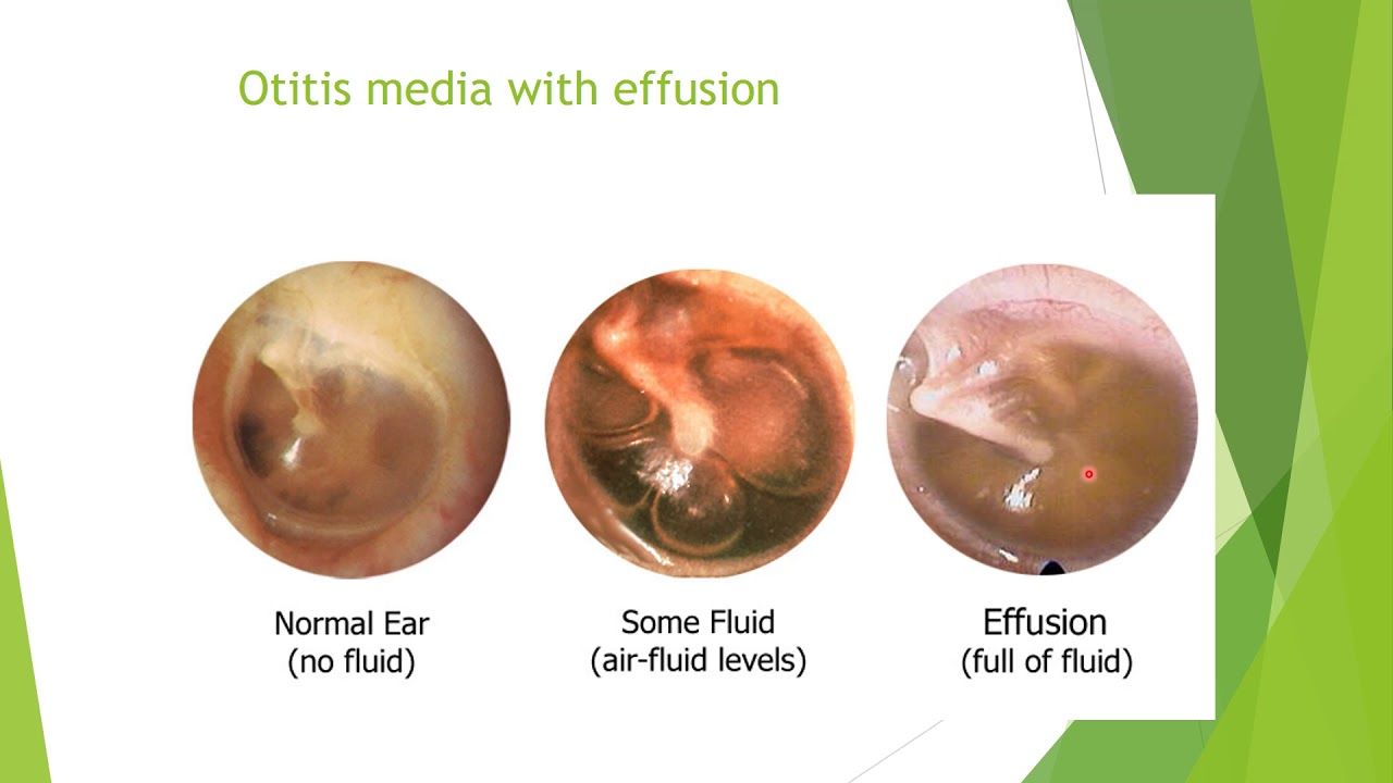
- Serous Fluid: On a digital screen, this typically appears as a thin, yellowish, or straw-colored liquid. It often presents with “bubbles” or a distinct “water line” (air-fluid level) visible through a semi-transparent eardrum.
- Purulent Fluid: This is identified by the software as a dense, opaque, and often white or creamy substance. The software looks for a lack of transparency in the tympanic membrane and a shift in the “cone of light” reflection.
- Mucoid Fluid: Often referred to as “glue ear,” this appears as a dull, gray, or dark amber substance that lacks the mobility of serous fluid.
Modern diagnostic apps use color-correction algorithms to ensure that the yellow of a serous effusion isn’t confused with the amber of earwax (cerumen), a common hurdle in automated ear health tech.
Integrating Visual Data with Electronic Health Records (EHR)
The visualization of ear fluid is increasingly being integrated into a broader digital health ecosystem. When an image is captured, it is not just stored in a gallery; it is tagged with metadata—time, temperature, and symptom logs—and pushed to an Electronic Health Record (EHR). This allows for longitudinal tracking. For example, a “Money” or “Tech” executive monitoring their child’s health can see a visual timeline of how the fluid has changed over 72 hours. This trend analysis is a core feature of modern health-tech platforms, turning a single image of ear fluid into a comprehensive data point in a patient’s medical history.
The Cybersecurity of Personal Health Data in Digital Otology
As we move toward a world where we “see” ear fluid via internet-connected devices, the security of that visual data becomes a paramount technological concern. An image of the inner ear is considered Protected Health Information (PHI), and its transmission must be guarded by robust security protocols.
End-to-End Encryption in Telemedicine Apps
When a smart otoscope transmits an image of ear fluid to a smartphone or a cloud server, it must utilize end-to-end encryption (E2EE). This ensures that the visual data cannot be intercepted by unauthorized parties. Tech developers in the medical space prioritize AES-256 encryption and Secure Socket Layer (SSL) certificates to maintain the integrity of the diagnostic session. For the user, knowing what their ear fluid looks like is only valuable if that information remains private and secure within the healthcare provider’s ecosystem.
Ethical Considerations in AI-Driven Medical Analysis
There is a significant tech-ethical debate regarding the use of AI in identifying ear fluid. Developers must ensure that the datasets used to train these algorithms are diverse, preventing “algorithmic bias” where the software might misidentify fluid in patients of different ethnicities due to variations in ear canal structure or skin tone. Furthermore, as these tools become more autonomous, the industry must navigate the balance between “tech-assisted” and “tech-driven” diagnosis, ensuring that a human professional is always in the loop for final clinical decisions.

Future Trends: The Intersection of Wearables and Aural Monitoring
The future of answering “what does ear fluid look like” may move away from hand-held devices toward continuous monitoring via wearables. We are already seeing the emergence of “Hearables”—smart earbuds equipped with sensors that can monitor more than just heart rate.
Future iterations of these devices could use infrared sensors or miniature ultrasound transducers to detect fluid buildup behind the eardrum without the need for a camera. This would move the diagnostic process from a reactive “look” to a proactive “alert.” Instead of a user checking their ear because of pain, their smart earbuds might send a notification to their phone: “Fluid detected in the middle ear; 85% probability of serous effusion. Schedule a virtual consultation?”
This shift represents the pinnacle of health technology: a transition from visual inspection to continuous, invisible data surveillance. As we refine these sensors and the AI that powers them, our understanding of ear fluid will become less about its visual appearance and more about the digital signature it leaves on our personal health networks. The “look” of the fluid is merely the interface for a much deeper, data-driven insight into human wellness.
aViewFromTheCave is a participant in the Amazon Services LLC Associates Program, an affiliate advertising program designed to provide a means for sites to earn advertising fees by advertising and linking to Amazon.com. Amazon, the Amazon logo, AmazonSupply, and the AmazonSupply logo are trademarks of Amazon.com, Inc. or its affiliates. As an Amazon Associate we earn affiliate commissions from qualifying purchases.