
In the rapidly evolving landscape of medical technology, few procedures exemplify the intersection of biological precision and engineering innovation as clearly as the DIEP (Deep Inferior Epigastric Perforator) flap surgery. While often categorized simply as a medical procedure, DIEP flap reconstruction is, in reality, a high-tech feat of microsurgery that relies on advanced imaging, precision instrumentation, and sophisticated physiological monitoring. As we move deeper into an era of personalized medicine, understanding the “how” behind this surgery reveals a fascinating world of technological advancement that has transformed patient outcomes and surgical standards.
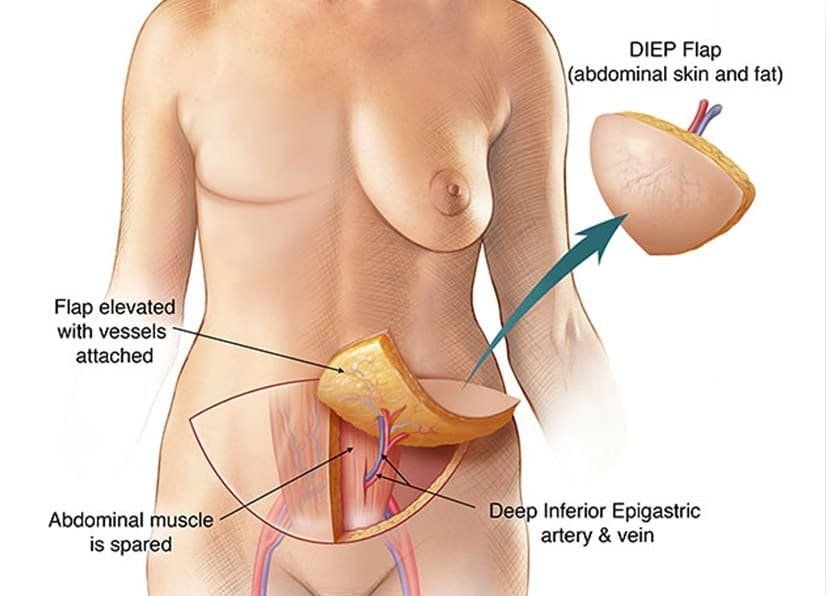
This article explores DIEP flap surgery through the lens of technology, examining the hardware, software, and digital breakthroughs that make this complex tissue transfer possible.
The Micro-Surgical Engine: Hardware and Precision Engineering
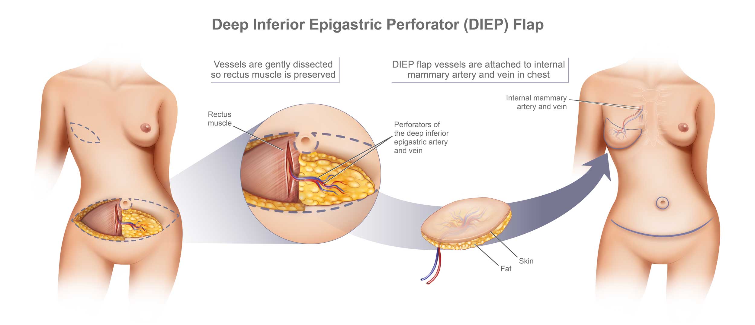
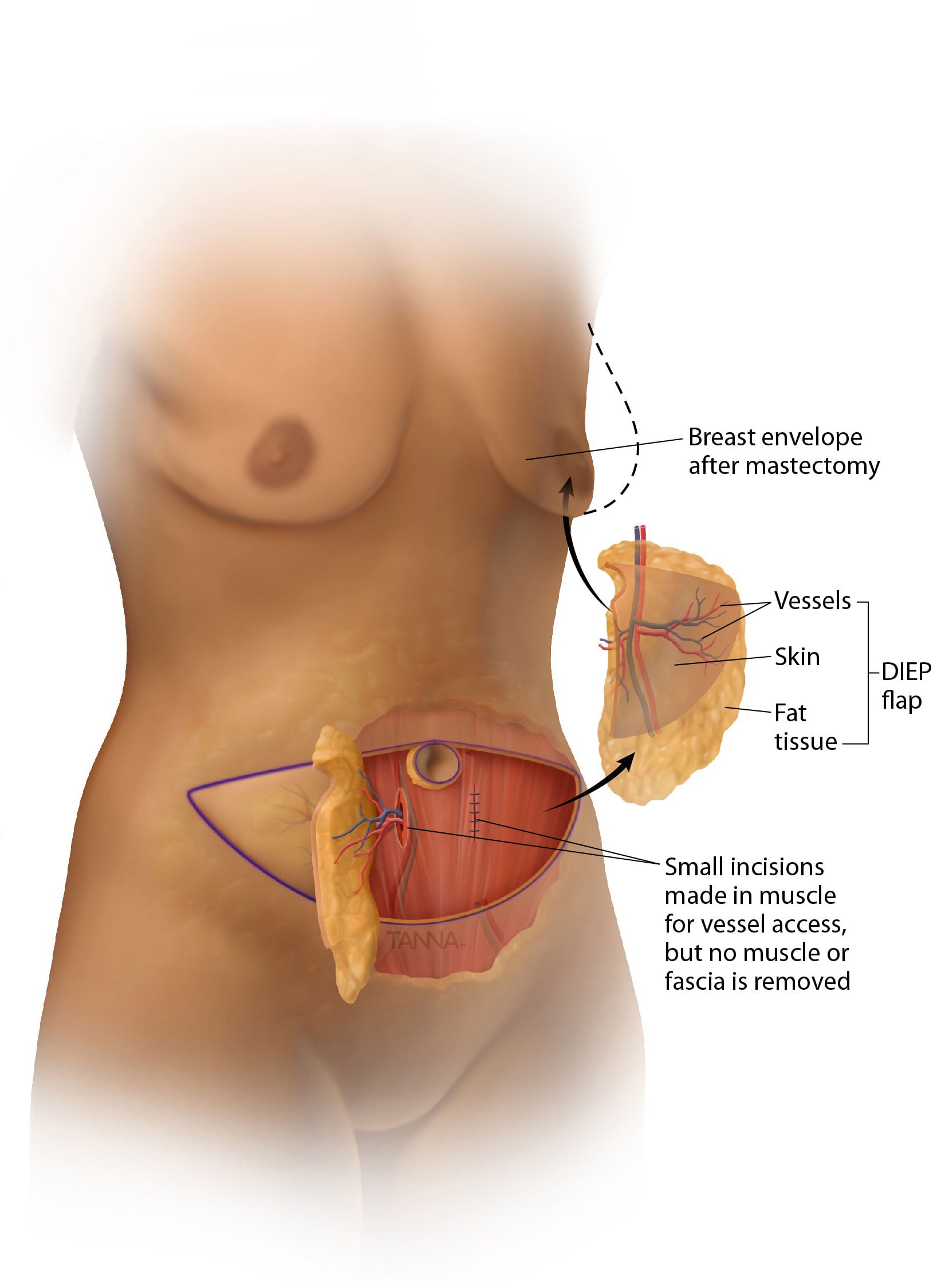
At its core, DIEP flap surgery is a masterclass in microsurgical engineering. Unlike older techniques that required the removal of entire muscle groups, the DIEP flap utilizes technology to isolate a single, microscopic blood vessel—a perforator—to sustain transferred tissue. This “muscle-sparing” approach is only possible because of the evolution of microsurgical hardware.
High-Definition Visualization: The Operating Microscope
The cornerstone of DIEP flap technology is the high-powered surgical microscope. Modern units from industry leaders like Zeiss or Leica provide not just magnification, but high-definition optical clarity and integrated digital overlays. Surgeons work under 20x to 40x magnification to perform anastomosis—the surgical connection of blood vessels that are often less than 2 millimeters in diameter.
These microscopes now feature integrated robotic assistance, allowing for stabilized movement and the elimination of human tremors. Some newer models even incorporate “Heads-Up Display” (HUD) technology, where the surgeon can see real-time data or pre-operative scans superimposed onto their field of view.
Precision Instrumentation and Suture Technology
The tools used in DIEP flap surgery are triumphs of metallurgy and design. Microsurgical forceps, needle holders, and scissors are engineered to work with sutures finer than a human hair (typically 9-0 or 10-0 nylon). The development of these non-reactive, ultra-fine synthetic polymers has drastically reduced the risk of vascular thrombosis (clotting) at the site of the connection.
Furthermore, the introduction of mechanical coupling devices—essentially “micro-staplers” for veins—has utilized high-precision engineering to speed up the vascular connection process, reducing the time the tissue is “cold” (without blood flow) and improving overall flap survival rates.
Advanced Imaging and AI-Driven Pre-Surgical Planning
One of the greatest challenges in DIEP flap surgery is the “mapping” of the patient’s unique vascular anatomy. No two humans have the same blood vessel layout in their abdomen. Technology has moved this from a “guess-and-check” process in the operating room to a precise digital simulation before the first incision is ever made.
Computed Tomographic Angiography (CTA) and Perforator Mapping
The use of high-resolution CTA scans is perhaps the most significant technological leap in reconstructive planning. By injecting a contrast dye and using a specialized CT scanner, software can generate a three-dimensional map of the abdominal wall’s blood vessels.
This data is then processed by specialized software to identify the “dominant” perforator—the specific vessel that provides the strongest blood supply to the skin and fat. By utilizing this digital map, surgeons can reduce “donor site morbidity,” ensuring they only harvest what is necessary and preserving as much of the patient’s physical integrity as possible.
Virtual Surgical Planning and 3D Modeling
Beyond simple mapping, surgeons are now utilizing 3D modeling software to perform “virtual” surgeries. Using the patient’s CTA data, AI-driven tools can simulate how a specific flap of tissue will fit into the reconstructive site.
These 3D models allow the surgical team to calculate the exact volume of tissue needed to match the patient’s natural anatomy. In some cutting-edge institutions, these models are even 3D printed to create physical templates that the surgeon can use in the operating room, ensuring an “engineered” fit that was previously impossible to achieve by eye alone.
Intraoperative and Post-Operative Monitoring Technologies
The success of a DIEP flap depends entirely on “perfusion”—the continuous flow of oxygenated blood through the new connections. If a vessel kinks or clots, the tissue will fail. Technology has provided a suite of monitoring tools that act as an early-warning system for the surgical team.
Indocyanine Green (ICG) Angiography
One of the most impressive tech integrations in the modern OR is ICG fluorescence imaging. By injecting a fluorescent dye (Indocyanine Green) into the patient’s bloodstream and using a specialized near-infrared camera (such as the SPY Elite system), surgeons can see blood flow in real-time on a digital monitor.
The tissue glows green where the blood flow is healthy and appears dark where it is lacking. This allows the surgeon to prune away “at-risk” tissue before the surgery is finished, significantly lowering the rate of fat necrosis and post-operative complications. It is a perfect example of how imaging technology removes the guesswork from biological reconstruction.
Digital Perfusion Monitoring: The “Smart Flap”
The 48 to 72 hours following surgery are the most critical. Traditionally, nurses monitored the flap by looking at skin color and temperature. Today, digital technology has automated this process.
Devices like the “Cook-Swartz” Doppler probe use a tiny ultrasonic crystal attached directly to the blood vessel connection. This probe sends a wireless signal to a bedside monitor that produces an audible “whoosh-whoosh” sound of the blood flow. If the flow stops or slows, an alarm sounds instantly. Newer wearable sensors are also being developed that measure tissue oxygen saturation (StO2) through the skin, providing a constant stream of digital data to a smartphone app, allowing surgeons to monitor their patients remotely and with high precision.
The Future of Biotechnical Reconstruction
As we look toward the future, the technology of DIEP flap surgery is expected to merge with the fields of regenerative medicine and robotics, moving beyond simply “moving” tissue to “engineering” it.
Robotics and Microsurgical Automation
While most DIEP flaps are currently performed by human hands, the integration of robotic platforms like the Da Vinci system is gaining traction. Robotics offer a level of stability and range of motion that exceeds the human wrist. Future iterations of these platforms are expected to include AI-assisted suturing, where the robot can autonomously perform the repetitive, high-precision task of sewing a vessel, reducing surgeon fatigue and potentially increasing the speed of the procedure.
Towards 3D Bioprinting and Synthetic Scaffolds
The ultimate technological goal is to move away from harvesting tissue from the patient’s own body (autologous transfer). Researchers are currently experimenting with 3D bioprinting, where a patient’s own cells are used to “print” a tissue flap on a synthetic, biodegradable scaffold.
While we are not yet at the stage where a DIEP flap can be fully 3D printed, the technology is moving toward “hybrid” reconstructions. In the future, we may see the use of 3D-printed vascular grafts that simplify the connection process, or bio-engineered “growth factors” that are applied to the flap to accelerate the integration of the new tissue into the patient’s body.
Data Analytics and Outcome Optimization
Finally, the “Big Data” revolution is hitting the reconstructive field. By aggregating the surgical data, imaging maps, and recovery stats of thousands of DIEP flap patients, machine learning algorithms are beginning to predict which patients are at higher risk for specific complications. This allows for “preventative tech” interventions—customizing the surgical approach based on a digital risk profile generated by AI.
Conclusion
DIEP flap surgery is a testament to how far technology has advanced the field of human medicine. It is no longer just a “surgery” in the traditional sense; it is a complex intersection of micro-engineering, digital imaging, and real-time physiological data analysis.
From the high-definition optics of the surgical microscope to the AI-driven vascular mapping of CTA scans, every step of the DIEP flap process is supported by a sophisticated tech stack. As robotics and bioprinting continue to mature, the procedure will only become safer, more precise, and more accessible, cementing its place as a pinnacle of technological achievement in the 21st century. For the patient, this means that the restoration of their body is no longer a matter of chance, but a result of high-precision engineering.
aViewFromTheCave is a participant in the Amazon Services LLC Associates Program, an affiliate advertising program designed to provide a means for sites to earn advertising fees by advertising and linking to Amazon.com. Amazon, the Amazon logo, AmazonSupply, and the AmazonSupply logo are trademarks of Amazon.com, Inc. or its affiliates. As an Amazon Associate we earn affiliate commissions from qualifying purchases.