
The intersection of clinical diagnostics and advanced computing has transformed how we interpret the smallest components of human biology. In the modern laboratory environment, the question of “what are epithelial cells in urinalysis” is no longer just a manual observation made by a technician with a microscope. Instead, it has become a complex data-processing challenge addressed by high-resolution imaging, artificial intelligence (AI), and automated flow cytometry. As we move further into the era of Health-Tech, the identification and quantification of these cells serve as a primary case study in how machine learning can enhance diagnostic accuracy and clinical workflow efficiency.
The Evolution of Automated Urinalysis Technology
For decades, identifying epithelial cells—the “bricks and mortar” of the urinary tract—was a manual, labor-intensive process. Technicians would centrifuge urine samples, place the sediment on a slide, and manually count cells across various fields of view. Today, the tech landscape has shifted toward fully automated systems that integrate hardware and software to provide rapid, standardized results.
Automated Flow Cytometry and Fluorescence
One of the cornerstone technologies in modern urinalysis is fluorescent flow cytometry. This technology works by passing a thin stream of the urine sample through a laser beam. As cells pass through, they are labeled with fluorescent dyes that bind to specific cellular components, such as DNA or cell membranes.
The system measures the “scatter” of light—forward scatter (indicating size) and side scatter (indicating internal complexity or granularity). For epithelial cells, this tech is crucial because it allows the software to differentiate between large, flat squamous cells and smaller, more complex renal tubular cells. By digitizing the physical properties of each cell, the system creates a scattergram, providing a high-level data visualization that allows clinicians to see the “health” of the urinary tract at a glance.
Digital Morphological Analysis
Parallel to flow cytometry is the development of Automated Digital Microscopy (ADM). Systems like the Iris iR2000 or the Sysmex UN-series utilize high-speed cameras to capture tens of thousands of images per sample. The software then uses proprietary algorithms to isolate individual objects.
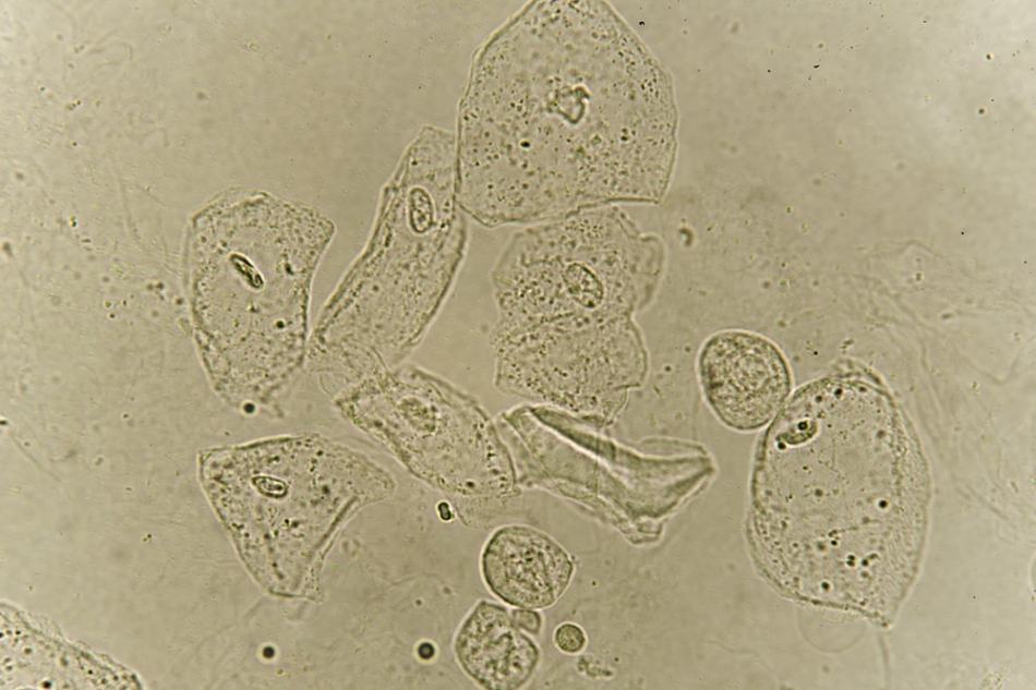
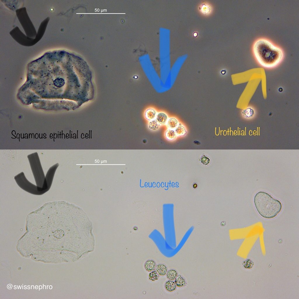
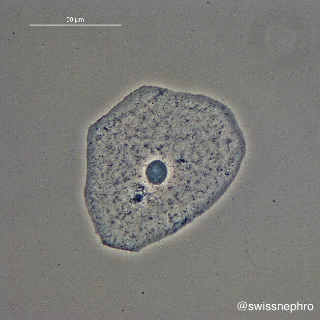
In this technological framework, an epithelial cell isn’t just a biological entity; it is a set of pixels with specific edge detection parameters. The tech must distinguish between squamous epithelial cells (often indicating a contaminated sample) and transitional or renal tubular cells (which may indicate pathology). The shift from human eyesight to digital pixel analysis has significantly reduced the coefficient of variation in lab results, ensuring that “cell counts” are consistent across different diagnostic centers.
AI and Machine Learning in Cellular Classification
The true “tech” revolution in urinalysis lies in the software layers that interpret captured images. Machine learning, specifically Deep Learning (DL) and Convolutional Neural Networks (CNNs), has become the gold standard for classifying epithelial cells.
Training Neural Networks for Morphological Precision
To accurately identify what an epithelial cell is within a messy urine matrix (which might also contain crystals, bacteria, and debris), AI models are trained on massive datasets. These datasets consist of millions of labeled images.
A CNN functions by breaking down an image of a cell into specific features: the ratio of the nucleus to the cytoplasm, the irregularity of the cell border, and the texture of the internal contents. For instance, a Renal Tubular Epithelial (RTE) cell is technologically distinct because of its smaller size and larger nucleus compared to a squamous cell. AI models can now identify these nuances with a level of precision that exceeds the average human technician, particularly when fatigue or high sample volume becomes a factor in a busy lab.
Reducing “Noise” and False Positives
One of the greatest hurdles in diagnostic software is “noise”—objects that look like cells but are actually artifacts, such as starch granules from gloves or air bubbles. Advanced software filters use “Feature Extraction” to ignore these non-biological artifacts. By applying logical thresholds, the technology ensures that the reported epithelial cell count reflects the actual biological state of the patient rather than technical interference. This level of computational filtering is what allows modern labs to process hundreds of samples per hour with minimal human intervention.
Data Integration and the Cloud-Based Pathology Ecosystem
As urinalysis becomes more digitized, the data generated by these machines is being integrated into broader health information systems (HIS) and Electronic Health Records (EHR). This connectivity is transforming epithelial cell counts from isolated numbers into actionable longitudinal data.
The Role of APIs in Diagnostic Telemetry
Modern urinalysis hardware is rarely a standalone silo. Through Application Programming Interfaces (APIs), the data from a urinalysis machine is instantly pushed to a cloud-based server where it can be analyzed against the patient’s historical data.
In a tech-forward clinical setting, if a machine detects a sudden spike in Renal Tubular Epithelial cells—a potential marker for acute kidney injury—the system doesn’t just print a report. It triggers a high-priority alert in the physician’s dashboard. This real-time telemetry is a hallmark of the “Smart Hospital,” where software acts as a secondary layer of diagnostic safety, ensuring that critical findings are not buried in a sea of routine results.
Cybersecurity and Data Integrity in Labs
With the rise of cloud-integrated diagnostics comes the critical need for digital security. Epithelial cell data, while seemingly benign, is part of a patient’s Protected Health Information (PHI).
Tech companies specializing in lab equipment are now prioritizing end-to-end encryption and blockchain-based audit trails to ensure that the data generated by the microscope hasn’t been tampered with or intercepted. As we move toward AI-driven diagnostics, the “provenance” of the data—knowing exactly which machine produced a result and which version of the AI algorithm was used for classification—is becoming a standard requirement for regulatory compliance.
The Future: Smartphone Integration and Point-of-Care (POC) Tech
The final frontier for urinalysis technology is the transition from the centralized lab to the palm of the hand. We are currently seeing a surge in Point-of-Care (POC) devices that bring epithelial cell analysis into the home or the small clinic.
Miniaturized Optical Sensors and Computer Vision
Startups in the med-tech space are developing smartphone-based urinalysis kits. These kits often involve a dipstick that is photographed using the phone’s high-resolution camera. While traditional dipsticks cannot measure epithelial cells (as they only measure chemical markers), new “lab-on-a-chip” technologies are emerging.
These devices use microfluidics to trap cells in a viewing chamber, which the smartphone camera then analyzes using a mobile version of the same CNNs used in large-scale lab equipment. The tech challenge here is significant: the software must account for varying lighting conditions, different camera qualities, and the “shaky hands” of the user. However, advancements in mobile processing power are making it possible to run complex computer vision tasks locally on the device, providing instant feedback on whether a sample is clean or requires professional follow-up.
Personalized Health Tech and Preventative Monitoring
The long-term vision for this technology is a shift from reactive to proactive medicine. By integrating urinalysis tech into daily or weekly routines—much like a smart scale or a wearable heart monitor—users can generate a “baseline” for their urinary health.
If the AI detects an unusual trend in cell shedding (an increase in transitional epithelial cells, for example), it can suggest a professional consultation before symptoms even manifest. This democratization of high-end diagnostic technology represents the pinnacle of Health-Tech: turning complex biological indicators into simple, understandable data points that empower the individual.
Conclusion: The Silicon Lens of Modern Medicine
Understanding what epithelial cells are in a urinalysis today requires an appreciation for the silicon and software that identify them. We have moved beyond the era of simple observation into an era of high-dimensional data analysis. From the flow cytometers that digitize cell structures to the AI models that classify them with superhuman accuracy, technology has become the primary lens through which we view human health.
As these tools become more connected, more mobile, and more intelligent, the “urinalysis” of the future will likely be a seamless, automated part of our digital lives. The epithelial cell, once a mere speck on a glass slide, is now a vital data point in the global movement toward precision medicine and tech-enabled wellness. Through the continued evolution of hardware and software, the clarity of our diagnostic vision will only continue to sharpen, offering a more profound understanding of the human body through the power of technology.
aViewFromTheCave is a participant in the Amazon Services LLC Associates Program, an affiliate advertising program designed to provide a means for sites to earn advertising fees by advertising and linking to Amazon.com. Amazon, the Amazon logo, AmazonSupply, and the AmazonSupply logo are trademarks of Amazon.com, Inc. or its affiliates. As an Amazon Associate we earn affiliate commissions from qualifying purchases.