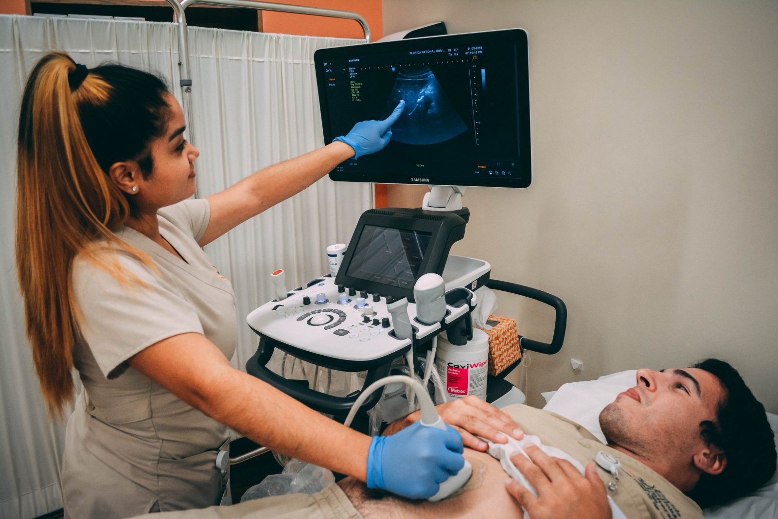
In the rapidly evolving landscape of modern medicine, the role of an ultrasonographer has shifted from a traditional clinical position to that of a high-tech data specialist. While the foundational principles of ultrasound—using high-frequency sound waves to visualize internal structures—remain the same, the tools and software used to execute this task have undergone a digital revolution. An ultrasonographer is no longer just a medical professional; they are a technical operator managing some of the most sophisticated hardware and artificial intelligence (AI) software in the healthcare sector.
To understand what an ultrasonographer is today, one must look through the lens of technology. They are the human interface between complex wave physics and diagnostic clarity, utilizing advanced signal processing, cloud computing, and automated algorithms to provide a window into the human body.
The Evolution of Diagnostic Imaging: From Sound Waves to Data Streams
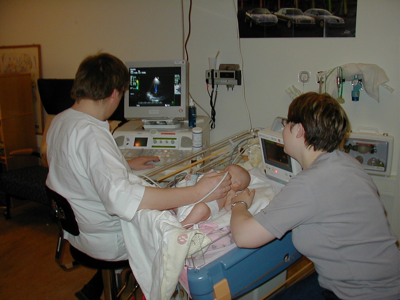
At its core, the work of an ultrasonographer is a feat of engineering. The profession relies on the “transducer,” a device that utilizes the piezoelectric effect to convert electrical energy into mechanical sound waves and back again. However, the modern version of this tech is a far cry from the analog systems of the late 20th century.
The Physics of Transducers and Piezoelectric Advances
The modern ultrasonographer works with transducers that utilize high-density single-crystal technology. Unlike traditional ceramics, these single crystals allow for a wider bandwidth and higher sensitivity. This technological leap enables the professional to achieve deeper penetration without sacrificing resolution, a common hurdle in older hardware. By manipulating these frequencies, the ultrasonographer manages “axial” and “lateral” resolution, essentially acting as a real-time editor of a biological data stream.
Digital Beamforming and High-Resolution Visualization
Modern ultrasound machines utilize digital beamformers to control the timing of electrical pulses sent to the transducer elements. This allows the ultrasonographer to “steer” the sound beam electronically rather than mechanically. The tech behind this involves massive parallel processing power, capable of handling billions of calculations per second to produce a coherent image. For the professional, this means the ability to toggle between various modes—like B-mode (brightness), M-mode (motion), and Color Doppler—with instantaneous software responsiveness.
The AI Revolution in Sonography
Perhaps the most significant shift in the niche of medical technology is the integration of Artificial Intelligence and Machine Learning into ultrasound platforms. For the ultrasonographer, AI is not a replacement but a powerful co-pilot that enhances diagnostic accuracy and workflow efficiency.
Automated Measurements and Predictive Analytics
One of the most time-consuming aspects of an ultrasonographer’s job is taking precise measurements, such as the circumference of a fetal head or the volume of a cardiac chamber. Modern software suites now include “Auto-OB” or “Auto-EF” (Ejection Fraction) tools. These AI-driven applications use pattern recognition to identify anatomical landmarks and perform measurements automatically. This reduces human error and ensures that the data is reproducible, regardless of which technician is operating the machine.
Image Enhancement and Noise Reduction Software
The “noise” or “speckle” inherent in ultrasound images has historically been a barrier to clear diagnosis. Modern tech addresses this through sophisticated spatial compounding and speckle reduction algorithms. These software tools analyze multiple frames from different angles in real-time, combining them to eliminate artifacts and sharpen edges. The ultrasonographer must be adept at calibrating these settings, balancing the “smoothness” of an image against the need for raw, unfiltered data to detect subtle pathologies.
Point-of-Care Ultrasound (POCUS) and the Rise of Portable Tech
The image of an ultrasonographer pushed behind a massive, refrigerator-sized machine is becoming obsolete. The “Tech” niche of sonography is currently defined by the miniaturization of hardware, often referred to as Point-of-Care Ultrasound (POCUS).
Smartphone-Integrated Ultrasound Gadgets
The emergence of “Ultrasound-on-a-chip” technology has changed the game. Companies have replaced traditional piezoelectric crystals with Silicon Photonics or Capacitive Micromachined Ultrasonic Transducers (CMUTs). This allows an entire ultrasound system to be shrunk down to a handheld probe that plugs into an iPhone or tablet. The ultrasonographer utilizing this tech relies heavily on mobile app interfaces and cloud-based image processing to conduct exams in emergency rooms, ambulances, or even remote field locations.
Breaking Barriers in Remote and Rural Medicine
With the rise of 5G connectivity and portable hardware, ultrasonographers are now engaging in “tele-sonography.” This involves using high-speed data transmission to stream live ultrasound feeds to specialists thousands of miles away. The technician on the ground uses specialized software that allows a remote doctor to guide their hand movements or adjust the machine’s gain and depth settings via the cloud. This technological synergy is bridging the gap in global healthcare, making diagnostic imaging accessible in tech-deprived regions.
The Intersection of 3D/4D Imaging and Virtual Reality
As processing power has increased, the ultrasonographer’s output has moved from flat, 2D slices to complex 3D volumes. This shift represents one of the most exciting frontiers in medical tech, blending traditional sonography with the world of computer graphics and volumetric rendering.
Beyond 2D: The Complexity of Volumetric Rendering
To create a 3D ultrasound, the ultrasonographer uses a specialized probe that captures a “volume” of data rather than a single plane. The software then performs “surface rendering” or “multiplanar reconstruction” (MPR). This requires the professional to understand the geometry of data acquisition, ensuring that the “voxels” (volume pixels) are captured with enough density to create a realistic reconstruction. In a 4D scan, time is added as the fourth dimension, requiring the hardware to process high-frame-rate volumetric data in real-time.
VR and AR Applications in Surgical Planning
The data captured by an ultrasonographer is increasingly being exported into Virtual Reality (VR) and Augmented Reality (AR) environments. For example, a complex cardiac scan can be converted into a 3D digital model that a surgeon explores using a VR headset before an operation. The ultrasonographer acts as the primary data gatherer for these simulations, ensuring the fidelity of the digital twin matches the patient’s actual anatomy. This intersection of sonography and immersive tech is redefining the “tech stack” required for modern diagnostic professionals.
Cybersecurity and Data Management in Medical Imaging
As ultrasound machines have become connected devices, the role of the ultrasonographer has expanded to include a fundamental understanding of digital security and data architecture. Every image captured is a piece of sensitive biometric data that must be managed within a complex digital ecosystem.
Cloud-Based PACS and Collaborative Diagnostics
Modern medical facilities utilize Picture Archiving and Communication Systems (PACS). An ultrasonographer must be proficient in navigating these databases, ensuring that images are correctly tagged with metadata, DICOM (Digital Imaging and Communications in Medicine) standards, and patient identifiers. The shift toward cloud-based PACS allows for seamless collaboration, where the ultrasonographer uploads a study to a secure server, and an AI algorithm or a remote radiologist analyzes it instantly.
Protecting Sensitive Biometric Data in a Digital Age
With the rise of cyberattacks on healthcare infrastructure, the “Tech” side of sonography involves maintaining the integrity of the data pipeline. This includes understanding encrypted transmissions and the importance of secure logins on ultrasound consoles. As machines become more integrated with hospital Wi-Fi networks and IoT (Internet of Things) ecosystems, the ultrasonographer must be vigilant about the digital footprint of their equipment, ensuring that the proprietary software and patient data remain shielded from external threats.

Conclusion
What is an ultrasonographer? In the modern era, they are the navigators of the medical technological frontier. They are professionals who must master the physics of sound, the logic of AI algorithms, the portability of mobile hardware, and the security of cloud-based data systems.
As we look toward the future, the role will only become more tech-centric. With the development of wearable ultrasound patches that monitor the heart continuously and the integration of haptic feedback “robotic” sonography, the ultrasonographer stands at the center of a digital health revolution. They are no longer just “taking a picture”; they are managing a sophisticated technological workflow that translates raw physical energy into life-saving digital insights. For anyone interested in the intersection of cutting-edge tech and human biology, the role of the modern ultrasonographer represents the pinnacle of diagnostic innovation.
aViewFromTheCave is a participant in the Amazon Services LLC Associates Program, an affiliate advertising program designed to provide a means for sites to earn advertising fees by advertising and linking to Amazon.com. Amazon, the Amazon logo, AmazonSupply, and the AmazonSupply logo are trademarks of Amazon.com, Inc. or its affiliates. As an Amazon Associate we earn affiliate commissions from qualifying purchases.