
In the intricate world of medical diagnostics, two powerful imaging technologies, Positron Emission Tomography (PET) and Magnetic Resonance Imaging (MRI), stand out for their ability to peer inside the human body without invasive surgery. While both are indispensable tools for clinicians, they operate on fundamentally different principles and provide distinct types of information. Understanding these technological distinctions is crucial for appreciating their respective roles in diagnosing diseases, monitoring treatment, and advancing medical science. This article delves into the core mechanisms, applications, and operational nuances of PET scans and MRIs, framing them within the broader context of cutting-edge medical technology.
Understanding the Core Technologies
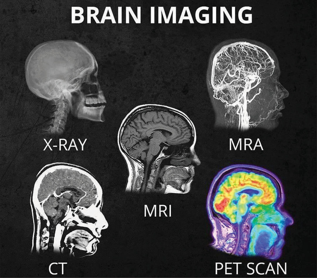
At their heart, PET and MRI represent two different philosophies of medical imaging: one focused on function at a molecular level, and the other on structure with unparalleled anatomical detail.
PET Scan: Unveiling Metabolic Activity
A PET scan is a nuclear medicine imaging technique that provides insights into the physiological and metabolic functions of tissues and organs. Its technological prowess lies in its ability to detect biochemical changes that can indicate disease processes often before structural changes become evident.
The operational principle of a PET scan revolves around radioactive tracers. A small amount of a short-lived radioactive material, often bound to a substance like glucose (forming fluorodeoxyglucose, or FDG), is injected into the patient’s bloodstream. This tracer emits positrons, which are antimatter particles. When a positron encounters an electron in the body’s tissues, they annihilate each other, producing two gamma rays that travel in opposite directions.
These gamma rays are detected by a ring of sensors around the patient. Sophisticated computer algorithms then reconstruct a 3D image based on the timing and location of these annihilation events. Areas of higher metabolic activity, such as rapidly growing tumors or inflamed tissues, will accumulate more of the tracer, thus showing up as “hot spots” on the PET image. The technology essentially maps where the body is actively using the tracer, providing a dynamic view of cellular activity. Key components include cyclotrons for producing the necessary short-lived radioisotopes and advanced detector rings with complex signal processing capabilities.
MRI: Detailed Anatomical Insights
Magnetic Resonance Imaging, on the other hand, is a non-invasive medical imaging technique that utilizes powerful magnetic fields and radio waves to generate detailed images of organs, soft tissues, bone, and virtually all other internal body structures. Unlike PET, MRI does not involve ionizing radiation, making it a safer option for repeated scans, particularly for sensitive populations.
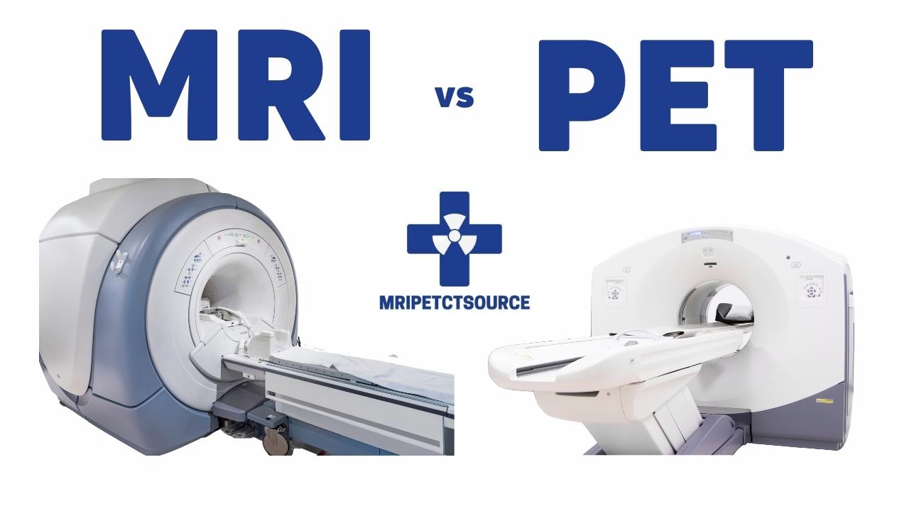
The technological foundation of MRI rests upon the manipulation of hydrogen atoms within the body’s water molecules. The patient is placed inside a large, powerful superconducting magnet that creates a strong, uniform magnetic field. This field aligns the protons (which are essentially tiny magnets) within the hydrogen atoms in the body. Next, a radiofrequency (RF) current is pulsed through the patient, knocking these aligned protons out of alignment. When the RF pulse is turned off, the protons relax back into alignment with the main magnetic field, releasing energy in the form of radio signals.
Different tissues (e.g., bone, fat, muscle, fluid) contain different amounts of water and, consequently, different densities of hydrogen atoms. They also relax at different rates. The MRI scanner’s computer detects these varying signals and uses them to construct highly detailed cross-sectional images. The contrast between different soft tissues provided by MRI is exceptional, making it an invaluable tool for visualizing structures like the brain, spinal cord, muscles, ligaments, and internal organs. Key components are the immensely powerful superconducting magnets (measured in Teslas) and precisely tuned RF coils that emit and detect radio signals.
Divergent Applications and Diagnostic Strengths
The fundamental differences in their underlying technology dictate their primary applications and diagnostic strengths. One reveals processes; the other unveils structures.
When PET Takes the Lead: Functional Assessment
PET’s unique ability to visualize metabolic and biochemical activity makes it indispensable in specific diagnostic scenarios where understanding cellular function is paramount.
- Oncology: PET scans are widely used in cancer management. They can detect cancerous cells, which typically have higher metabolic rates (glucose uptake) than healthy cells, often before a tumor becomes large enough to be seen on other imaging modalities like CT or MRI. PET helps in initial cancer detection, staging (determining the extent of cancer spread), monitoring the effectiveness of treatment (showing changes in metabolic activity post-therapy), and detecting recurrence. The technological advantage here is its capacity to pinpoint abnormal cellular function rather than just structural anomalies.
- Neurology: In neurological disorders, PET can assess brain activity. It’s crucial for diagnosing conditions like Alzheimer’s disease (by detecting amyloid plaques or reduced glucose metabolism), identifying seizure foci in epilepsy, and evaluating Parkinson’s disease. Specific tracers can target neurotransmitter systems or protein aggregates linked to these conditions.
- Cardiology: PET can evaluate myocardial viability (assessing blood flow to heart muscle), helping determine if damaged heart tissue is still alive and could benefit from revascularization procedures.
When MRI Excels: Structural Precision
MRI’s unparalleled ability to provide exquisite detail of soft tissues without ionizing radiation makes it the gold standard for structural imaging in numerous medical fields.
- Neurological Disorders: MRI offers superior visualization of the brain and spinal cord. It’s the preferred method for diagnosing conditions such as brain tumors, strokes, multiple sclerosis (MS) lesions, aneurysms, and spinal cord injuries. The high-resolution images allow clinicians to pinpoint the exact location and extent of structural abnormalities.
- Musculoskeletal Injuries: For injuries involving ligaments, tendons, cartilage, and bone marrow, MRI is unparalleled. It can accurately diagnose tears in knee ligaments (ACL, meniscus), shoulder rotator cuffs, and spinal disc herniations, providing critical information for surgical planning.
- Soft Tissue Imaging: MRI is excellent for imaging internal organs like the liver, kidneys, pancreas, and reproductive organs, as well as blood vessels (MR angiography) without requiring contrast agents in many cases. Its ability to differentiate between various types of soft tissues is a key technological advantage.
Operational Aspects and Patient Experience
Beyond the science, the practical aspects of undergoing a PET or MRI scan also differ significantly, impacting patient experience and logistical considerations for healthcare providers.
Procedural Differences: From Preparation to Scan Duration
The journey through a PET scan typically involves a few key steps. After an initial consultation and preparation (often fasting), the patient receives an intravenous injection of the radioactive tracer. There’s then a waiting period (usually 30-90 minutes) during which the tracer circulates and is absorbed by tissues. The actual scan time can range from 15 to 60 minutes, depending on the area being imaged. A critical consideration for PET is the radiation exposure from the injected tracer, although the dose is generally low and considered safe for diagnostic purposes. Patients are often advised to avoid close contact with pregnant women or young children for a few hours post-scan.
MRI procedures are distinct. Patients must remove all metallic objects, and those with certain metallic implants (pacemakers, some clips, cochlear implants) may be unable to undergo an MRI due to the powerful magnetic field. Patients may receive an intravenous contrast agent (e.g., gadolinium-based) to enhance specific tissue visibility, although this is not always necessary. The patient lies on a table that slides into a large, tube-like scanner. The scan itself is often lengthy, ranging from 30 to 90 minutes, and can be quite noisy due to the rapid switching of magnetic gradients. This noise, combined with the enclosed space, can induce claustrophobia in some patients, necessitating open MRI machines or sedation in certain cases. The absence of ionizing radiation is a significant advantage in terms of long-term safety.
Technological Constraints and Considerations
Both technologies come with their own set of operational and cost implications driven by their sophisticated hardware and software.
- PET Constraints: The need for short-lived radioactive tracers means that PET centers often need access to a nearby cyclotron or a frequent delivery system, adding to the logistical complexity and cost. The limited half-life of tracers like FDG (around 110 minutes) dictates careful scheduling and rapid processing. The higher capital investment in both the cyclotron and the PET scanner, combined with the ongoing cost of tracer production, makes PET scans generally more expensive than MRI.
- MRI Constraints: The primary constraint of MRI is its strong magnetic field. This necessitates strict screening for metal objects and implants, as ferrous materials can be violently attracted to the magnet or malfunction within the field, posing serious safety risks. The immense power required to sustain the superconducting magnets and the sophisticated cooling systems (often liquid helium) contribute to the high operational costs. The size of the scanner bore can be an issue for larger or claustrophobic patients, though “open MRI” designs exist, often with some compromise in field strength or image quality.
Complementary Roles in Modern Diagnostics
Despite their differences, PET and MRI are not always competing technologies. Increasingly, they are seen as complementary, providing a more complete picture when used in conjunction or, in some cases, integrated into a single device.
Hybrid Systems: PET-MRI and PET-CT
The most significant technological advancement in this domain is the advent of hybrid imaging systems.
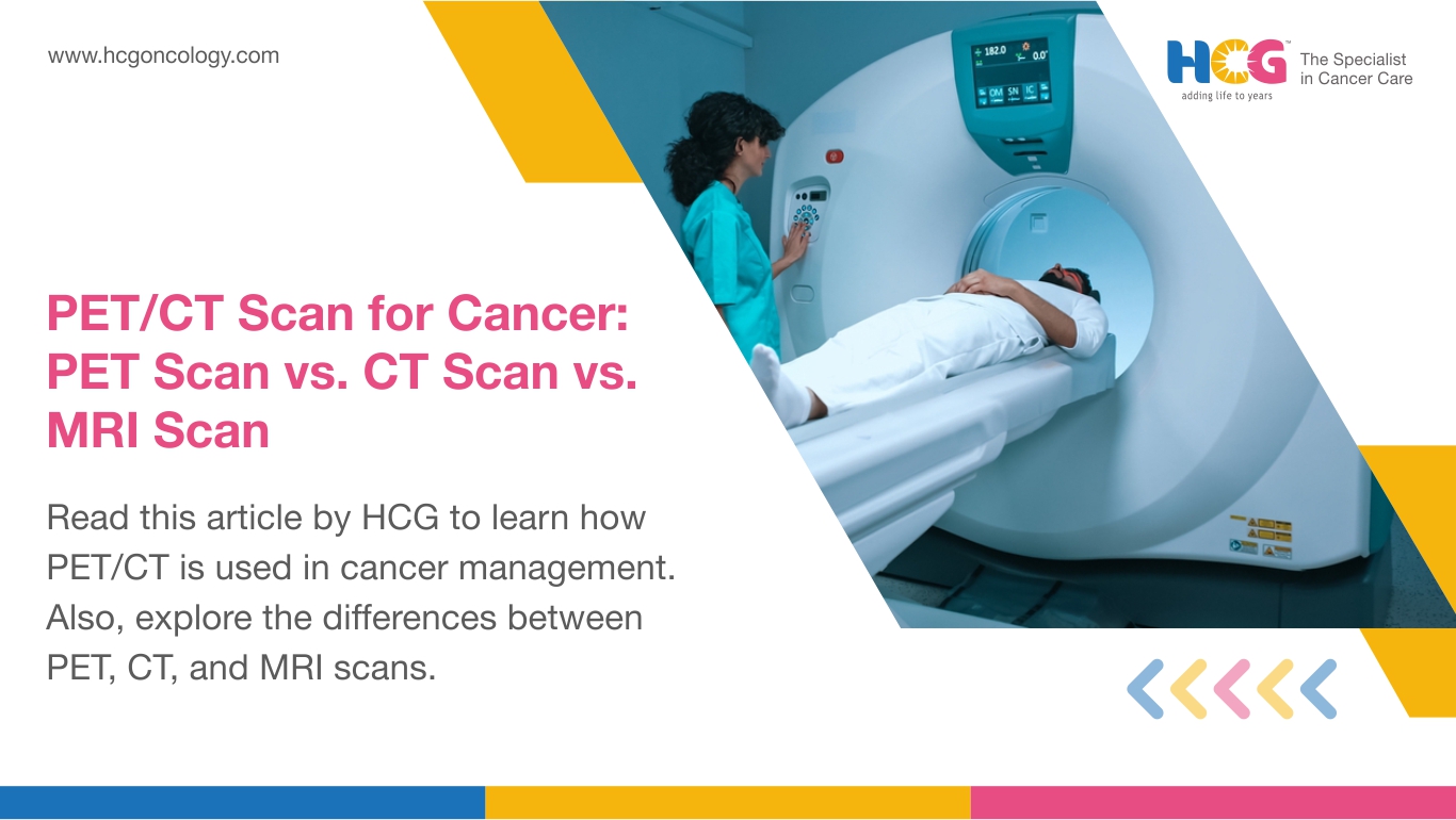
- PET-CT: This is the most common hybrid system, combining the functional insights of PET with the anatomical detail of Computed Tomography (CT). A patient undergoes both scans sequentially in the same machine, and the images are fused, providing both metabolic activity and precise anatomical location. This is particularly valuable in oncology for accurately staging tumors and guiding biopsies. The integration of two distinct scanning technologies into one workflow presents significant engineering challenges but offers immense diagnostic power.
- PET-MRI: The more recent and technologically complex integration is the PET-MRI scanner. This system aims to combine the superior soft-tissue contrast of MRI with the metabolic information of PET in a single, simultaneous acquisition. This integration is challenging because the strong magnetic fields of MRI can interfere with the PET detectors. Overcoming these challenges has led to innovative detector designs compatible with high magnetic fields. PET-MRI is particularly promising for neurological and pediatric imaging, where minimizing radiation exposure and maximizing soft-tissue detail are crucial.
The Future of Medical Imaging Technology
The trajectory of medical imaging technology points towards greater integration, precision, and intelligence.
- AI Integration for Image Analysis: Artificial intelligence and machine learning are rapidly transforming how PET and MRI images are processed and interpreted. AI algorithms can help with noise reduction, image reconstruction, automated lesion detection, and quantitative analysis, leading to faster, more accurate diagnoses and reduced variability among readers.
- Advancements in Scanner Speed and Resolution: Ongoing research focuses on developing faster scanning sequences for MRI, reducing patient discomfort and motion artifacts. For PET, advancements in detector technology are leading to improved sensitivity and resolution, allowing for the detection of smaller lesions and reduced tracer doses.
- Minimally Invasive Techniques: The goal is always to gather the most diagnostic information with the least burden on the patient. Future developments may include even more precise targeting of tracers, advanced image processing for virtual biopsies, and integrating these modalities with real-time interventional procedures.
In conclusion, PET and MRI, while both powerful windows into the human body, offer fundamentally different perspectives. PET illuminates the metabolic functions that underpin life and disease, while MRI provides unparalleled structural clarity. Together, and especially in their hybrid forms, they represent the pinnacle of modern diagnostic imaging technology, continuously evolving to provide clinicians with ever more precise tools to understand, diagnose, and treat complex medical conditions.
aViewFromTheCave is a participant in the Amazon Services LLC Associates Program, an affiliate advertising program designed to provide a means for sites to earn advertising fees by advertising and linking to Amazon.com. Amazon, the Amazon logo, AmazonSupply, and the AmazonSupply logo are trademarks of Amazon.com, Inc. or its affiliates. As an Amazon Associate we earn affiliate commissions from qualifying purchases.