
The Magnetic Resonance Imaging (MRI) machine is a cornerstone of modern diagnostic medicine, offering unparalleled insights into the human body’s soft tissues, organs, bone marrow, and virtually every internal structure. While its diagnostic capabilities are revolutionary, for many, the initial encounter with an MRI machine is a visual one, often shrouded in a mix of curiosity and apprehension. Far from a simple X-ray machine, an MRI scanner is a complex piece of engineering, distinct in its appearance and function. Understanding its visual anatomy not only demystifies the experience but also offers a glimpse into the sophisticated technology at its core.
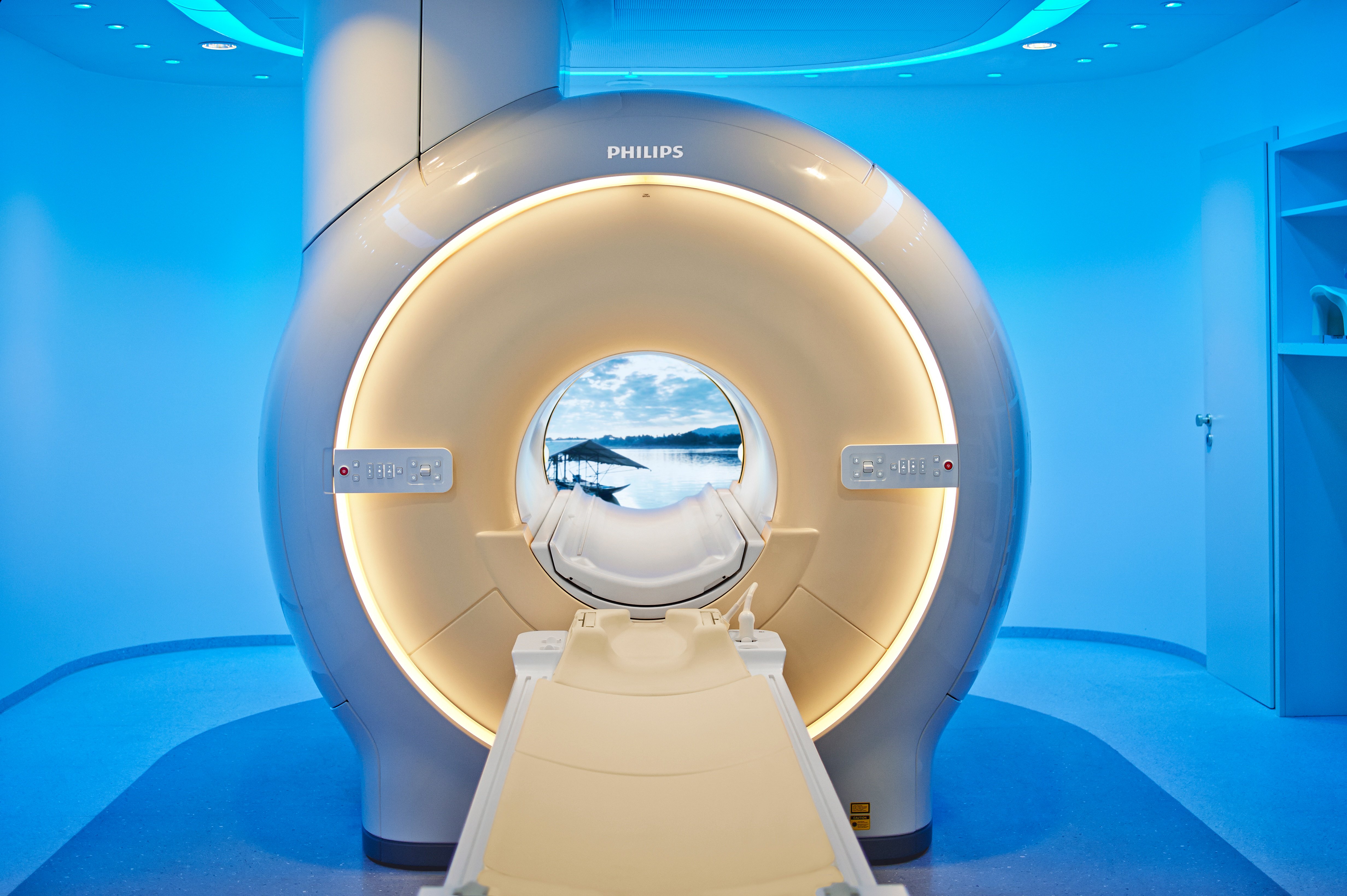
At first glance, the most common image of an MRI machine is that of a large, cylindrical tube, often referred to as a “bore” or “tunnel,” into which a patient slides. However, this is just one manifestation of MRI technology. Modern advancements have led to a variety of designs, each tailored to specific diagnostic needs or patient comfort considerations. Regardless of its form, every MRI machine houses a powerful magnet, a radiofrequency coil system, gradient coils, and a sophisticated computer system, all working in concert to generate incredibly detailed images without using ionizing radiation. This article will delve into the visual characteristics of MRI machines, exploring their core components, design variations, and the technological rationale behind their imposing yet intricate appearance.
The Core Components: A Visual Breakdown
To truly understand what an MRI machine looks like, it’s essential to break down its primary visual and functional components. Each part plays a critical role, and its design reflects its specific technical purpose.
The Gantry: Tunnel or Open?
The gantry is perhaps the most recognizable part of an MRI machine. In its traditional form, it’s a large, cylindrical tunnel, often several feet long and wide enough to accommodate a human body. This “closed-bore” design is characteristic of high-field MRI scanners, where a strong, uniform magnetic field is crucial for image quality. The interior of this tunnel can appear somewhat stark, though modern designs often incorporate lighting, mirrors, or even small screens to reduce feelings of claustrophobia.
Alternatively, some MRI machines feature an “open” gantry. Instead of a closed tunnel, these machines have a flat top and bottom magnet, creating a more expansive space around the patient. This design, often resembling a large, C-shaped or U-shaped structure, significantly reduces feelings of confinement and is particularly beneficial for claustrophobic patients, children, and larger individuals. While some open MRIs have lower field strengths compared to closed bores, newer wide-bore and high-field open designs are bridging this gap, offering both comfort and diagnostic accuracy.
The Patient Table: Movement and Comfort
Extending from the gantry, or positioned within its open space, is the patient table. This motorized table is designed to slide smoothly into and out of the MRI bore, positioning the patient precisely within the magnetic field. Visually, it’s typically a padded, relatively narrow bed, sturdy enough to support patients of various weights. Control panels near the table allow technicians to adjust its height and movement. Patients often lie on their back, but depending on the exam, they may be positioned on their side or stomach. The table’s design prioritizes both stability during scanning and the patient’s comfort during what can be a lengthy procedure.
The Control Room: The Operator’s Hub
Separated from the scanner room by a thick, shielded window is the control room. This is where the MRI technologist operates the machine. Visually, it resembles a sophisticated computer workstation, featuring multiple monitors displaying real-time images, patient vitals, and complex software interfaces. Keyboards, mice, and specialized control panels are all part of this setup, allowing the technologist to program scan sequences, monitor the patient, and process the acquired data. The shielding is crucial here, as it protects the electronic equipment and personnel from the powerful magnetic field and radiofrequency pulses emitted by the scanner.
Ancillary Equipment: Coils and Cooling Systems
Beyond the main structure, several vital ancillary components contribute to the MRI’s overall appearance and function. Various “coils” – specialized receivers and transmitters – are placed around the body part being imaged (e.g., head coil, knee coil, spinal coil). These coils are often intricate, custom-molded plastic structures that fit snugly around the anatomy, resembling helmets, sleeves, or blankets. They are crucial for sending and receiving radiofrequency signals, enhancing image detail.
Less visible but equally critical are the cooling systems. High-field MRI machines use superconducting magnets, which must be cooled to extremely low temperatures using cryogens like liquid helium. This necessitates external chilling units, pipes, and vents, which are typically hidden behind the machine’s exterior casing or in an adjacent technical room. These systems ensure the magnet’s superconductivity and prevent overheating.
Varieties of MRI Scanners: Beyond the Standard Tunnel
The appearance of an MRI machine is not monolithic; different designs cater to specific diagnostic needs and patient preferences. Understanding these variations helps to paint a complete picture of what an MRI machine can look like.
Closed-Bore (Tunnel) MRI: The Traditional Design
This is the archetypal MRI machine. It features a long, cylindrical tunnel, typically about 60-70 cm (24-28 inches) in diameter. The patient is slid into this enclosed space. These machines are known for their high magnetic field strengths (1.5 Tesla, 3 Tesla, and even higher), which translate to superior image resolution and faster scan times. The closed design is essential for maintaining the homogeneity of the strong magnetic field, which is critical for image quality, especially for detailed neurological or musculoskeletal studies. While effective, the enclosed nature can be challenging for claustrophobic or anxious patients.
Open MRI: Addressing Claustrophobia and Size
Open MRI scanners present a stark visual contrast to their closed-bore counterparts. Instead of a tunnel, they feature two large, flat magnetic plates positioned above and below the patient table, leaving the sides completely open. This design significantly reduces the feeling of confinement, making it a preferred option for claustrophobic patients, obese individuals who may not fit into a closed bore, and children who might benefit from having a parent nearby. Earlier open MRIs often had lower field strengths (e.g., 0.3T to 0.7T), which could result in slightly longer scan times or less detailed images for certain examinations. However, advancements have led to “wide-bore” MRIs, which are still cylindrical but have a much larger diameter (typically 70 cm or more) and shorter length, offering a blend of higher field strength and improved patient comfort, often seen as a hybrid between true open and traditional closed systems.
Upright MRI: Specialized Positioning
A less common but visually distinct type is the upright MRI. As its name suggests, this machine allows patients to be scanned in a weight-bearing position—standing, sitting, or even bending. Visually, it might resemble a large, open box or a specialized chair with magnetic components surrounding the patient’s torso or head. This design is particularly useful for imaging conditions that are exacerbated by gravity, such as spinal instability, back pain that worsens when standing, or certain joint issues. The ability to scan in a functional position provides unique diagnostic insights that supine imaging might miss.
Extremity MRI: Focused Imaging
For examinations confined to specific limbs, such as a hand, wrist, foot, or ankle, an extremity MRI offers a smaller, more focused solution. Visually, these machines are much more compact than full-body scanners. They often feature a small opening or a dedicated compartment where the limb is placed, while the rest of the patient remains outside the magnetic field. This design is less intimidating, more comfortable, and typically less expensive to operate, making it an efficient choice for localized musculoskeletal imaging.

The Science Behind the Appearance: How It Works Visually
The visual form of an MRI machine is not arbitrary; it’s a direct consequence of the physics and engineering required to generate its powerful magnetic field and detect subtle changes within it. Understanding the underlying technology helps explain why these machines look the way they do.
Powerful Magnets: The Invisible Force Shaping the Design
The most dominant visual feature of an MRI—its large, often imposing structure—is dictated by the massive magnet it contains. In closed-bore systems, this magnet creates a uniform magnetic field that runs along the length of the bore. The cylindrical shape is ideal for generating a consistent and powerful field around the patient. For superconducting magnets, this immense magnet coil requires substantial insulation and a cryostat system to maintain extremely low temperatures (near absolute zero). This cooling apparatus adds to the bulk and cylindrical appearance of the machine, as it must house the cryogens and their associated plumbing. In open MRI systems, the magnets are typically positioned above and below the patient, dictating the characteristic “C” or “U” shape. The size and configuration of these magnet components directly influence the overall footprint and visual presence of the scanner.
Radiofrequency Coils: Signal Reception and Transmission
While not always visible from the outside, the design of various radiofrequency (RF) coils plays a crucial role in the scanner’s overall aesthetic when preparing a patient. These coils, which look like specialized plastic casings, often molded to fit specific body parts (e.g., a “cage” for the head, a “sleeve” for a knee), are placed directly around the area to be imaged. They are responsible for transmitting RF pulses into the patient’s body and then detecting the faint signals emitted by hydrogen atoms within the tissues. Their form factor is a balance between optimal signal reception (requiring close proximity to the anatomy) and patient comfort.
Gradient Coils: Spatial Encoding and Noise
Inside the main magnet bore, beneath the visible interior lining, are gradient coils. These coils generate rapidly changing magnetic fields that allow the MRI system to pinpoint the exact location of signals within the body. While not directly visible, their operation has a significant auditory impact—the loud banging, tapping, and whirring noises heard during an MRI scan are produced by the rapid switching of electrical currents through these gradient coils. The engineering design of these coils and their housing contributes to the internal structure of the bore and the need for robust vibration dampening materials.
Cooling Systems: Maintaining Superconductivity
For high-field MRI scanners, the powerful main magnet is superconducting, meaning it operates with zero electrical resistance only at extremely low temperatures. This necessitates a cryogenic cooling system, typically involving liquid helium, to keep the magnet coils supercooled. The extensive network of pipes, insulation layers, and a cryostat—often housed within the scanner’s casing—contributes significantly to the machine’s large, enclosed appearance. External chiller units might also be located in a separate equipment room, feeding cooling fluids to maintain the cryostat’s efficiency. These systems are critical, not just for function but also for the machine’s physical integrity and safety.
The Patient Experience: Interaction with the Machine
For the patient, the visual and sensory interaction with the MRI machine is a significant part of the diagnostic process. Understanding this interaction from a design perspective highlights the engineering considerations for both functionality and user experience.
Entering the Bore: Visualizing the Space
As a patient is positioned on the table and slides into the gantry, their perception of the machine shifts. In a closed-bore MRI, the immediate visual is the circular opening and the enclosed tunnel. Modern machines often incorporate mirrors to allow patients to see outside the bore, or project calming images and videos onto the interior ceiling to mitigate claustrophobia. Lighting within the bore can range from standard clinical white light to softer, adjustable ambient illumination. The visual narrowness of the tunnel, though intimidating for some, is a direct result of optimizing the magnetic field’s homogeneity for precise imaging. In an open MRI, the experience is visually different, with a much wider field of view, and often the ability to see the ceiling or surroundings, offering a less enclosed feeling.
Noise and Vibration: Auditory and Tactile Aspects
While not a visual characteristic, the intense noise and vibrations during an MRI scan are an undeniable sensory component of interacting with the machine. These sounds—ranging from rhythmic thumps to high-pitched whines—are caused by the rapid switching of currents in the gradient coils. Visually, this means robust housing and structural design are necessary to contain and manage these mechanical forces. Patients are always provided with earplugs or noise-canceling headphones, and the visual cues of the technician monitoring from the control room through a window serve as a reassurance during this noisy process.
Safety Features: Alarms and Communication
Safety is paramount in MRI design, and some features are visually apparent. Emergency stop buttons are typically placed within easy reach of the patient (e.g., a “squeeze ball”) and clearly marked. Intercom systems, often with a visible microphone and speaker within the bore, allow direct communication between the patient and the technologist, providing visual confirmation of connection. These elements, though small, are critical design considerations that visually communicate patient safety and control within the powerful environment of the scanner.
Evolution of MRI Design: Towards Patient-Centric Innovation
The appearance of MRI machines is continuously evolving, driven by technological advancements, clinical demands, and a growing emphasis on patient comfort and accessibility.
Compact Designs: Space Optimization
Historically, MRI machines required vast, shielded rooms. However, advancements in magnet technology, particularly in self-shielding magnets, have led to more compact designs. While still large, newer machines can fit into smaller spaces, reducing the infrastructure requirements for hospitals and imaging centers. This “shrinking footprint” is not always visually obvious from the patient’s perspective, but it signifies a significant engineering achievement, making MRI technology more accessible.
Enhanced Comfort Features: Lighting, Music, and Visuals
Recognizing the anxiety many patients experience, modern MRI designs increasingly integrate features aimed at improving the patient experience. The interior of the bore might include customizable LED lighting, allowing patients to choose calming colors. Built-in sound systems can play music or natural soundscapes through noise-canceling headphones. Some advanced systems project soothing visuals or even “virtual windows” onto the interior of the bore, transforming the potentially intimidating tunnel into a more pleasant environment. These design elements actively work to counteract the inherent visual challenges of the closed-bore system.

AI Integration: Guiding Future Aesthetics and Functionality
While not directly influencing the physical appearance of the gantry itself, the integration of Artificial Intelligence (AI) and machine learning is profoundly impacting the ‘look’ of the interface and the diagnostic output. AI-powered software can optimize scan parameters, reduce scan times (meaning less time inside the machine for the patient), and improve image reconstruction quality. This leads to a more streamlined and visually intuitive user interface for the technologists and ultimately better, clearer diagnostic images for physicians. As AI continues to advance, it may even influence the design of next-generation coils and patient positioning systems, further refining the overall visual and interactive experience with the MRI machine.
In conclusion, an MRI machine is a marvel of engineering, and its visual characteristics are a direct manifestation of its intricate technology. From the imposing gantry to the precision patient table, and the myriad of specialized coils, every aspect of its appearance serves a vital purpose. While the traditional closed-bore design remains prevalent for its imaging prowess, the evolution towards open, wide-bore, and even upright systems demonstrates a concerted effort to balance diagnostic excellence with patient comfort. As technology advances, the MRI machine continues to transform, promising even more sophisticated and patient-friendly designs in the future, all while maintaining its critical role in unlocking the mysteries within the human body.
aViewFromTheCave is a participant in the Amazon Services LLC Associates Program, an affiliate advertising program designed to provide a means for sites to earn advertising fees by advertising and linking to Amazon.com. Amazon, the Amazon logo, AmazonSupply, and the AmazonSupply logo are trademarks of Amazon.com, Inc. or its affiliates. As an Amazon Associate we earn affiliate commissions from qualifying purchases.