
The human heart relies on a sophisticated electrical conduction system to maintain a rhythmic, life-sustaining pulse. When this internal circuitry malfunctions, the result can be a catastrophic cessation of blood flow. In medical terminology, four specific rhythm disturbances are categorized as fatal—or potentially fatal—because they immediately compromise the heart’s ability to pump oxygenated blood to the brain and vital organs. Understanding these conditions is the cornerstone of emergency cardiac care and clinical intervention.
Ventricular Fibrillation: The Electrical Storm
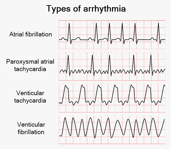
Ventricular Fibrillation (VFib) is arguably the most critical rhythm disturbance encountered in clinical practice. It represents the ultimate breakdown of organized electrical activity within the heart’s lower chambers, the ventricles.
The Mechanism of Chaos
In a healthy heart, the ventricles contract in a synchronized fashion to push blood into the systemic circulation. During VFib, however, the ventricles cease their coordinated contraction. Instead of a rhythmic beat, the muscle fibers undergo rapid, uncoordinated, and chaotic twitching. Electrically, the heart is essentially “shivering.” Because the ventricles are not contracting, cardiac output drops to zero instantly.
Clinical Implications and Treatment
The clinical presentation of VFib is sudden cardiac arrest. The patient loses consciousness immediately, stops breathing, and lacks a palpable pulse. Survival hinges on the “Chain of Survival” protocol. Because the heart is in a state of chaotic electrical noise, the only definitive treatment is immediate electrical defibrillation. By delivering a high-energy shock to the myocardium, the defibrillator depolarizes the entire heart muscle simultaneously, providing the natural pacemaker—the sinoatrial node—a chance to regain control and reset the rhythm.
Pulseless Ventricular Tachycardia: The High-Speed Failure
While Ventricular Tachycardia (VT) can sometimes be tolerated for short bursts in stable patients, it often devolves into a Pulseless Ventricular Tachycardia (pVT). This state is a recognized fatal arrhythmia that necessitates immediate life-saving intervention.
Understanding Rapid Depolarization
Ventricular Tachycardia occurs when an ectopic focus within the ventricles begins firing at an abnormally high rate, often exceeding 120 beats per minute. When the rate becomes so rapid that the heart chambers have no time to fill with blood during diastole, the heart essentially “spins its wheels.” Even if the electrical rhythm is regular, the mechanical efficiency is so severely compromised that the pulse disappears.
Distinguishing Stable from Unstable
Medical professionals distinguish between monomorphic and polymorphic VT. Regardless of the morphology, if the patient becomes pulseless, the condition is treated as an emergency equivalent to cardiac arrest. The physiological result is identical to VFib: systemic hypoperfusion leading to rapid cerebral hypoxia. Just as with VFib, the primary intervention is defibrillation, as the heart has effectively stopped performing its function as a pump despite its electrical activity.
Pulseless Electrical Activity: The Mechanical Disconnect

Pulseless Electrical Activity (PEA) is perhaps the most enigmatic of the fatal arrhythmias. It is characterized by a paradox: the heart’s electrical monitor displays a rhythm that looks relatively organized or even near-normal, yet the patient has no detectable pulse.
The Physiology of the “Silent” Heart
In PEA, the heart’s conduction system is functioning, but the cardiac muscle is failing to respond. This can occur for several reasons, often categorized by the “Hs and Ts” of advanced cardiac life support. Common culprits include severe hypovolemia (lack of blood volume), tension pneumothorax, cardiac tamponade, or massive pulmonary embolism. The electrical signal is present, but the physical heart muscle is either empty of blood, physically compressed, or experiencing severe biochemical distress that prevents contraction.
Managing the Underlying Cause
Unlike VFib or pVT, PEA is not treated with defibrillation, as the heart is already depolarizing. Instead, the focus is on identifying and reversing the underlying mechanical or chemical insult. Effective management requires high-quality cardiopulmonary resuscitation (CPR) to maintain minimal perfusion while medical teams search for the cause—such as administering fluids for hypovolemia or performing needle decompression for a tension pneumothorax. It is a race against time to restore the mechanical coupling between the electrical stimulus and the muscle’s mechanical action.
Asystole: The Electrical Silence
Asystole is the finality of cardiac rhythm disturbances. Often referred to colloquially as a “flatline,” it represents the total absence of electrical activity within the heart.
The Breakdown of Pacemaker Function
Asystole occurs when all internal pacemakers of the heart fail. The heart is no longer sending out the electrical signals required to stimulate contraction. There is no rhythm, no pulse, and no cardiac output. While it is the most common terminal rhythm in many cardiac arrests, it is also the most challenging to reverse.
The Challenges of Resuscitation
Because there is no electrical activity to “reset” or “synchronize,” defibrillation is entirely ineffective in cases of asystole. Attempts to shock a flatline are useless and potentially harmful. Treatment centers exclusively on high-quality chest compressions and the administration of pharmacological agents, primarily epinephrine. Epinephrine works to stimulate the adrenergic receptors in the heart, potentially increasing coronary perfusion pressure and restoring enough myocardial energy to jumpstart a rhythm. The prognosis for asystole is typically poor, as it usually represents the end stage of a prolonged period of hypoxia or metabolic decay.
Integrated Approaches to Cardiac Emergency
The successful management of these four fatal arrhythmias requires a systematic, evidence-based approach. The evolution of cardiac care has shifted from reactive measures to proactive, standardized protocols that emphasize speed and quality of intervention.
The Role of Rapid Response
In all four conditions, the common denominator is the cessation of cerebral perfusion. The brain begins to suffer irreversible damage within four to six minutes of oxygen deprivation. Therefore, the initiation of bystander CPR—or advanced life support in a clinical setting—is the most critical variable in determining patient outcome. By manually circulating oxygenated blood to the brain and heart, responders buy the necessary time to address the specific rhythm disorder.
Technological Advancements in Monitoring
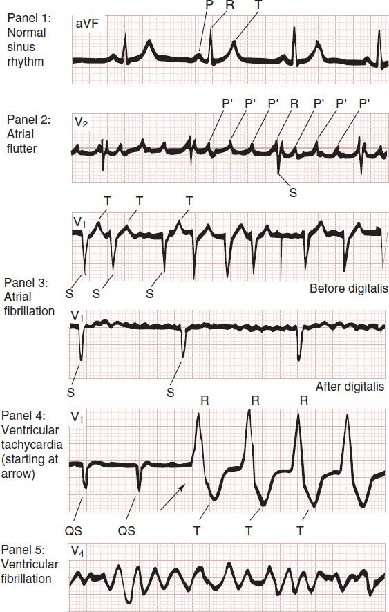
Modern medical technology has significantly improved the detection and management of these arrhythmias. Automated External Defibrillators (AEDs) placed in public spaces have democratized the ability to treat VFib and pVT. Furthermore, sophisticated cardiac monitoring systems in intensive care units provide real-time telemetry, allowing clinicians to identify “pre-arrest” rhythms—such as frequent premature ventricular contractions or periods of unstable tachycardia—before they devolve into one of the four fatal arrhythmias.
Clinical Vigilance
Ultimately, the four fatal arrhythmias are not just isolated events; they are the terminal points of various underlying pathologies. Whether it is an electrolyte imbalance, an acute myocardial infarction, or drug toxicity, the underlying cause must be identified concurrently with the resuscitation effort. By understanding the electrical and mechanical properties of the heart, healthcare providers and first responders are better equipped to navigate the chaos of cardiac arrest. The mastery of these four rhythms remains the definitive standard for life-saving expertise in the modern medical landscape.
aViewFromTheCave is a participant in the Amazon Services LLC Associates Program, an affiliate advertising program designed to provide a means for sites to earn advertising fees by advertising and linking to Amazon.com. Amazon, the Amazon logo, AmazonSupply, and the AmazonSupply logo are trademarks of Amazon.com, Inc. or its affiliates. As an Amazon Associate we earn affiliate commissions from qualifying purchases.