
For over a century, the chest X-ray has stood as a cornerstone of medical diagnosis. From revealing broken bones to detecting pneumonia or tuberculosis, its ability to peer into the human body non-invasively revolutionized healthcare. However, the seemingly simple question, “what will a chest X-ray show?” is evolving dramatically in the 21st century, propelled by relentless technological innovation. We are moving far beyond the grainy film images of yesteryear, entering an era where artificial intelligence, advanced digital sensors, and integrated data systems are transforming a traditional diagnostic tool into a powerhouse of precision and predictive insight.

Today, understanding what a chest X-ray will show us is less about the basic anatomical structures and more about the intricate, often invisible, details that sophisticated technology can extract. This isn’t just an incremental improvement; it’s a fundamental shift in how we approach diagnosis, driven by technology trends that promise faster, more accurate, and more accessible healthcare outcomes. This article delves into the technological marvels that are redefining the capabilities of chest X-rays, exploring the software, AI tools, and digital advancements that are unveiling a future where diagnostic imaging offers unprecedented clarity.
The Evolution of X-Ray Technology: From Film to Digital Zenith
The journey of the X-ray machine from a rudimentary device producing shadow images on photographic plates to a high-fidelity digital scanner is a testament to persistent technological advancement. This evolution has been critical in enhancing what an X-ray can show both diagnostically and operationally.
Digital Radiography’s Foundational Shift
The most significant leap from traditional film-based radiography was the advent of digital radiography (DR). This technology replaces film with digital image receptors, such as charge-coupled devices (CCDs) or flat-panel detectors, which directly convert X-ray photons into an electrical signal. This signal is then processed by a computer to produce an image that can be viewed instantly on a monitor.
The advantages of digital radiography are manifold and fundamentally change what a chest X-ray reveals. Firstly, image quality is significantly improved, with greater dynamic range and contrast resolution, allowing radiologists to discern subtle abnormalities that might be missed on film. Secondly, reduced radiation dose is a critical patient safety benefit; digital systems are more sensitive, requiring less radiation to produce diagnostic-quality images. Thirdly, the immediacy of digital images means faster diagnoses and treatment initiation, crucial in emergency settings.
Furthermore, digital radiography ushered in the era of Picture Archiving and Communication Systems (PACS). PACS software allows for the storage, retrieval, and distribution of digital images across healthcare networks. This means a radiologist can access a patient’s entire imaging history, compare current scans with previous ones side-by-side, and even consult with specialists remotely, irrespective of geographical barriers. This interconnectivity not only streamlines workflows but also enriches the diagnostic process by providing comprehensive historical context, ultimately allowing the chest X-ray to show more by comparison and collaboration.
Beyond 2D: The Rise of Advanced Imaging Techniques
While standard chest X-rays provide a 2D projection, technological advancements are pushing the boundaries towards creating more comprehensive, quasi-3D insights. Computed Tomography (CT) scans, while distinct from traditional X-rays, exemplify the power of technology to generate cross-sectional images, offering far greater anatomical detail than a single X-ray shot. CT technology utilizes a rotating X-ray beam and multiple detectors to create a series of 2D images that a computer then reconstructs into detailed cross-sectional views, effectively presenting a 3D understanding of internal structures.
Newer X-ray modalities are also emerging, such as dual-energy X-ray and phase-contrast X-ray imaging. Dual-energy X-ray technology uses two different X-ray energies to differentiate materials based on their atomic number, allowing for the isolation of bone from soft tissue, or the detection of specific elements like iodine. This can help to show calcifications, lesions, or foreign bodies with greater clarity. Phase-contrast X-ray imaging, still largely experimental, exploits the phase shift of X-rays as they pass through tissues, offering superior soft tissue contrast compared to conventional absorption-based X-rays, potentially showing tumors or inflammation with unprecedented detail without the need for contrast agents. These innovations indicate a trajectory towards X-ray technologies that can convey a richer, multi-dimensional understanding of complex pathologies.
Artificial Intelligence: The New Diagnostic Lens
Perhaps no other technology has impacted the diagnostic capabilities of imaging more profoundly than Artificial Intelligence (AI). AI tools, particularly those leveraging machine learning and deep learning, are not just assisting radiologists; they are fundamentally changing what a chest X-ray can show by enhancing detection, interpretation, and predictive capabilities.
AI in Image Analysis: Enhancing Accuracy and Speed
AI algorithms, trained on vast datasets of X-ray images, can identify patterns, anomalies, and subtle indicators of disease that might elude the human eye or require significant time and effort for manual detection. For instance, AI software can automatically detect nodules, infiltrates, or effusions indicative of conditions like lung cancer, pneumonia, or congestive heart failure.
The impact here is twofold: enhanced accuracy and increased speed. AI can act as a “second reader,” flagging suspicious areas for a radiologist’s review, thereby reducing false negatives and improving diagnostic confidence. In high-volume settings, AI tools can prioritize critical cases, ensuring that patients with urgent findings receive immediate attention. This means a chest X-ray will show critical findings faster, allowing for earlier intervention and better patient outcomes. Deep learning models, a subset of AI, excel in tasks like segmentation (identifying and outlining specific structures or lesions) and classification (categorizing images based on disease presence), making the diagnostic process more objective and standardized.
Predictive Analytics and Early Disease Detection
Beyond mere detection, AI is enabling chest X-rays to show predictive insights. By analyzing an X-ray image in conjunction with a patient’s electronic health records (EHRs), demographic data, and historical imaging, AI algorithms can identify subtle biomarkers or risk factors that predict the future onset or progression of diseases. For example, AI might detect early signs of emphysema years before symptoms become apparent, or predict the likelihood of a heart attack based on subtle calcifications in the coronary arteries visible on a chest X-ray.
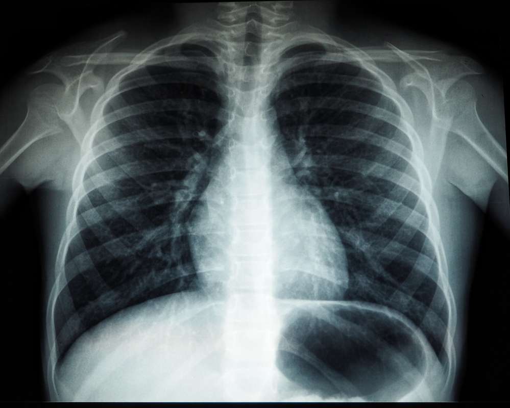
This capability transforms the chest X-ray from a reactive diagnostic tool into a proactive one. What it will show is not just the current state of a disease, but potentially its future trajectory. This paradigm shift holds immense promise for preventative medicine, allowing clinicians to intervene much earlier, personalize treatment plans, and potentially avert more severe health crises.
Integrating Data and Interoperability: A Holistic View
The true power of advanced X-ray technology is unleashed when it doesn’t operate in a vacuum. The integration of imaging data with other patient information through robust software platforms creates a holistic view that enhances diagnostic precision and care coordination.
The Power of PACS and EHR Integration
As mentioned, PACS revolutionized imaging management. However, the true synergy comes when PACS integrates seamlessly with Electronic Health Record (EHR) systems. An EHR aggregates all of a patient’s medical information—diagnoses, medications, lab results, clinical notes, and imaging reports—into a single digital record. When a chest X-ray is performed, its digital image and accompanying report are instantly accessible within the patient’s comprehensive EHR.
This integration means that a chest X-ray doesn’t just show an image; it shows an image in the context of a patient’s entire health story. A radiologist interpreting an X-ray can instantly review previous medical conditions, current medications that might affect findings, or relevant lab values. Clinicians can view the X-ray alongside patient symptoms and physical exam findings to make more informed decisions. This interoperability ensures that the information gleaned from the X-ray is maximized, leading to more accurate diagnoses and better-coordinated care.
Tele-radiology and Remote Diagnostics
Technology has also dissolved geographical barriers in diagnostics through tele-radiology. This application allows X-ray images taken anywhere in the world to be transmitted securely over networks to radiologists located thousands of miles away for interpretation. This capability is particularly vital for rural hospitals, underserved communities, or during off-hours, ensuring that expert diagnostic opinions are always available.
The impact on what a chest X-ray will show is profound: it ensures consistent, high-quality interpretation, regardless of the local availability of specialists. This democratizes access to advanced diagnostic capabilities, improving equity in healthcare. Tele-radiology platforms are often enhanced with collaboration tools, allowing multiple experts to review cases simultaneously and consult, further enriching the diagnostic process.
Emerging Technologies on the Horizon
The innovation in X-ray technology is far from over. The horizon is dotted with exciting advancements that promise to further transform what a chest X-ray can show, pushing the boundaries of non-invasive diagnosis.
Quantum Sensing and Novel Imaging Modalities
Future X-ray technologies are exploring principles beyond traditional absorption. Photon-counting CT (PCCT), for instance, uses detectors that count individual X-ray photons and measure their energy, offering superior image resolution, reduced noise, and the ability to differentiate between different materials within the body with unprecedented accuracy. This could allow a chest X-ray to show subtle tissue changes or early-stage disease with much greater clarity.
Research into terahertz imaging and functional X-ray imaging (e.g., dynamic imaging of lung mechanics) also points towards a future where X-rays might not just capture static anatomy but also physiological function in real-time. What a chest X-ray will show could expand from structural abnormalities to dynamic functional insights, offering a more complete picture of health and disease.
Augmented Reality/Virtual Reality in Training and Interpretation
The application of Augmented Reality (AR) and Virtual Reality (VR) is beginning to extend into medical imaging. For training purposes, AR/VR can provide immersive environments for medical students and residents to practice interpreting complex X-ray images and understand anatomical relationships in 3D.
In clinical practice, AR could overlay X-ray data onto a patient during a procedure, providing real-time guidance, or allowing radiologists to visualize complex 3D reconstructions of organs and pathologies in an immersive VR environment. This could help radiologists better conceptualize spatial relationships of lesions or tumors identified on X-rays, making interpretations more precise and potentially aiding in surgical planning. The X-ray image would no longer be confined to a flat screen; it would be an interactive, spatial data point.

Conclusion
The venerable chest X-ray, once a simple silhouette, is being reimagined by a confluence of advanced technologies. From the fundamental shift to digital radiography and the increasing sophistication of multi-dimensional imaging techniques to the revolutionary power of AI for analysis and prediction, technology is ceaselessly expanding what a chest X-ray will show us. Integration with EHRs and the widespread adoption of tele-radiology are ensuring that these insights are holistic and accessible.
As we look to the future, quantum sensing and immersive AR/VR applications promise even deeper levels of detail and interactivity. The trajectory is clear: diagnostic imaging is becoming more precise, predictive, and patient-centric, driven by innovation in software, AI tools, and advanced gadgetry. The modern chest X-ray is no longer just a window into the body; it’s an intelligent, interconnected gateway to understanding health and disease with unparalleled clarity, ultimately leading to better outcomes for patients worldwide. The collaboration between human medical expertise and cutting-edge technological prowess is charting a new course for diagnostics, where the invisible is continually unveiled.
aViewFromTheCave is a participant in the Amazon Services LLC Associates Program, an affiliate advertising program designed to provide a means for sites to earn advertising fees by advertising and linking to Amazon.com. Amazon, the Amazon logo, AmazonSupply, and the AmazonSupply logo are trademarks of Amazon.com, Inc. or its affiliates. As an Amazon Associate we earn affiliate commissions from qualifying purchases.