
In the rapidly evolving landscape of medical technology, two giants stand at the forefront of non-invasive diagnostics: Computed Tomography (CT) and Magnetic Resonance Imaging (MRI). To the casual observer, both machines look remarkably similar—large, donut-shaped structures that peer into the human body. However, from a technological and engineering perspective, they are as different as a high-speed digital camera is from a sophisticated sonar system.
Understanding the difference between CT scans and MRI requires an exploration of physics, hardware engineering, data processing, and the burgeoning role of artificial intelligence. These tools are not merely “cameras” for the body; they are complex computational platforms that transform physical signals into high-resolution digital data.
Hardware Architecture: Comparing the Mechanics of CT and MRI
At the core of these technologies lies a fundamental difference in how they interact with matter. The hardware requirements for each are dictated by the physical phenomena they exploit to generate an image.
The Physics of Ionizing Radiation in CT Scans
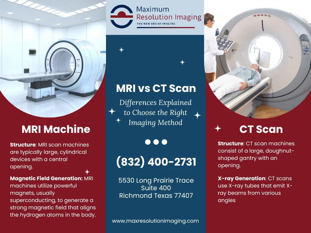
Computed Tomography is an advanced evolution of the traditional X-ray. A CT scanner consists of an X-ray tube that rotates rapidly around the patient. This tube emits a fan-shaped beam of ionizing radiation. On the opposite side of the gantry, high-speed digital detectors capture the photons that pass through the body.
The technological challenge in CT hardware design involves the “gantry speed.” Modern scanners rotate multiple times per second, subjecting the internal components to immense G-forces. The tech industry has pushed these limits to allow for “multi-slice” scanning, where hundreds of images are captured in a single rotation, enabling the 3D reconstruction of moving organs like the heart with millisecond precision.
Superconducting Magnets and Radiofrequency in MRI
In contrast, an MRI machine is a masterpiece of electromagnetic engineering. It does not use radiation. Instead, it relies on three main tech components: a primary static magnetic field (measured in Tesla), gradient coils, and radiofrequency (RF) coils.
The primary magnet is usually a superconductor cooled by liquid helium to near absolute zero. This creates a powerful magnetic field that aligns the protons in the patient’s body. The RF coils then emit pulses of radio waves (similar to those used in telecommunications) to flip these protons. When the pulse stops, the protons “relax” and emit energy. The hardware’s sensitivity to these minute energy changes determines the final image quality. This is a far more complex hardware environment than CT, requiring specialized shielding to prevent external electronic noise from interfering with the delicate signals.
Hardware Limitations and Environmental Constraints
From a gadgetry perspective, CT scanners are more “portable” (relative to MRI) and have fewer environmental restrictions. MRI machines, however, require “Faraday cages” to block outside radio interference and must be kept in rooms free of ferromagnetic materials. The engineering required to keep a 3-Tesla magnet stable while pulsing gradient coils—which creates the loud knocking sound familiar to MRI patients—is one of the most significant achievements in modern mechanical engineering.
The Computational Edge: Software, Algorithms, and Image Reconstruction
While the hardware captures the signal, the software is what creates the vision. Both CT and MRI rely heavily on advanced mathematical algorithms to turn raw sensor data into something a human can interpret.
Reconstructive Algorithms and Fourier Transforms
In CT scanning, the raw data consists of “projections” or shadows. To turn these into a 3-dimensional volume, the software utilizes a process called Filtered Back Projection (FBP) or, more recently, Iterative Reconstruction (IR). IR is a computationally intensive software process that compares the captured data against a simulated model to reduce “noise” and improve clarity. This allows for lower doses of radiation while maintaining high image quality—a major tech trend in “Green Imaging.”
MRI data processing is even more complex. The signals are collected in what engineers call “K-space,” a complex mathematical domain. The software uses a Fast Fourier Transform (FFT) to convert these frequency and phase signals into a spatial image. Because MRI can be programmed with different “sequences” (software protocols like T1, T2, or FLAIR), a single piece of hardware can be repurposed via software to look specifically at water, fat, or blood flow.
Resolution, Voxel Density, and Data Throughput
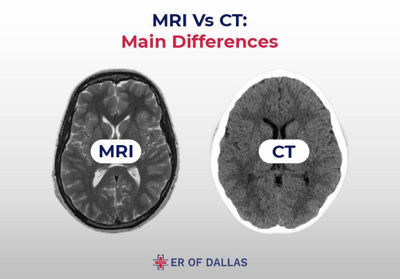
In the world of digital imaging, we talk about pixels. In medical tech, we talk about “voxels” (volume elements). A CT scan is the king of spatial resolution; it can identify tiny fractures in bone or calcifications in arteries with sub-millimeter precision.

MRI, however, dominates in “contrast resolution.” This refers to the software’s ability to distinguish between two similar types of soft tissue, such as a tumor versus healthy brain matter. The data throughput for a high-resolution MRI study can be massive, requiring high-speed bus architectures and specialized GPUs (Graphics Processing Units) to process the raw data into 3D renderings in real-time.
Multi-Planar Reformation (MPR)
Modern diagnostic software allows for Multi-Planar Reformation. Once the data is captured, the software can virtually “slice” the body in any direction—coronal, sagittal, or axial—without the patient moving. This is a feat of pure digital geometry, allowing clinicians to fly through virtual representations of a patient’s colon or blood vessels, a field known as virtual endoscopy.
The Integration of Artificial Intelligence and Machine Learning
The most significant trend in imaging technology today is the integration of Artificial Intelligence (AI). Both CT and MRI are being revolutionized by deep learning models that optimize every step of the process.
AI-Driven Image Enhancement and Denoising
One of the biggest hurdles in MRI technology is scan time; patients must remain still for 30 to 60 minutes. New AI tools, such as deep-learning-based reconstruction, can now predict what a high-resolution image should look like based on a shorter, lower-quality scan. This “upscaling”—similar to how DLSS works in high-end PC gaming—allows for faster scan times without sacrificing diagnostic integrity.
In CT technology, AI is used for “Denoising.” By training neural networks on millions of high-dose (perfect) scans and low-dose (noisy) scans, the software can now remove the visual “grain” from a low-radiation scan, making it look as clear as a traditional high-radiation scan. This is a monumental shift in digital security and patient safety.
Automated Pattern Recognition and CAD
Computer-Aided Detection (CAD) systems are now common in CT workflows. For instance, when a CT scan of the chest is performed, AI algorithms can automatically scan the data for lung nodules or pulmonary embolisms, flagging them for the human operator. In MRI, AI is being used to map the “fingerprinting” of tissues, identifying the specific molecular signatures of different types of multiple sclerosis lesions or prostate cancers faster than the human eye can.
Workflow Optimization and Predictive Maintenance
Beyond the images themselves, AI manages the “fleet” of scanners. Software now monitors the heat levels of CT tubes and the helium levels of MRI cryogens. Predictive analytics can alert engineers that a component is likely to fail before it actually does, ensuring that these multi-million dollar tech assets have maximum uptime.
Digital Security and Interoperability in Modern Imaging Tech
As medical imaging becomes increasingly digitized, it faces the same challenges as any other tech sector: data security, cloud integration, and interoperability.
DICOM Standards and Data Portability
The “language” of medical imaging is DICOM (Digital Imaging and Communications in Medicine). This protocol ensures that a CT scan taken on a GE machine can be read by software on a Siemens or Philips workstation. The tech industry has worked hard to ensure that these massive datasets (often hundreds of megabytes per study) can be compressed and transmitted without losing a single bit of diagnostic information.
Cybersecurity in the Medical Device Niche
As CT and MRI scanners are now networked devices, they are targets for cyberattacks. Manufacturers are now building “Digital Security” layers into the scanners themselves. This includes encrypted hard drives, secure boot protocols, and sandboxed operating systems to prevent the scanner from being a gateway for ransomware into a hospital’s broader network.
Cloud-Based Analysis and Telehealth
The latest evolution in CT and MRI tech is the move to the cloud. Instead of having a powerful computer located in the room next to the scanner, the raw data is increasingly sent to cloud-based GPU clusters. This allows for “Teleradiology,” where a specialist in London can analyze an MRI taken in a rural clinic in real-time, using advanced visualization tools hosted in the cloud. This democratization of high-tech diagnostics is perhaps the most impactful trend in the current decade.
Conclusion: The Future of Diagnostic Gadgetry
The difference between a CT scan and an MRI is not just a matter of “X-rays vs. Magnets.” It is a difference in engineering philosophy, data handling, and computational application. CT is the high-speed, high-resolution workhorse of the tech world, specializing in structure and speed. MRI is the sophisticated, software-driven analytical tool, specializing in tissue characterization and molecular detail.
As we move forward, the lines between these technologies may blur through “Hybrid Imaging” (like PET/CT or PET/MRI), but their core identities as triumphs of technological innovation remain. Whether through the refinement of superconducting materials or the deployment of next-generation AI, the evolution of CT and MRI continues to push the boundaries of what is possible at the intersection of technology and human health.
aViewFromTheCave is a participant in the Amazon Services LLC Associates Program, an affiliate advertising program designed to provide a means for sites to earn advertising fees by advertising and linking to Amazon.com. Amazon, the Amazon logo, AmazonSupply, and the AmazonSupply logo are trademarks of Amazon.com, Inc. or its affiliates. As an Amazon Associate we earn affiliate commissions from qualifying purchases.