
In the traditional medical landscape, the “color” of lung cancer was a matter of physical observation—the grayish-white mass of a tumor against the pinkish hue of healthy lung tissue, or the dark carbon deposits found in a smoker’s lungs. However, in the contemporary era of high-tech diagnostics, the question “what color is lung cancer” has shifted from the biological to the digital. Today, the color of malignancy is defined by the heatmaps of artificial intelligence, the spectral signatures of hyperspectral imaging, and the pixel-by-pixel analysis of digital pathology software.

As technology advances, our ability to visualize, identify, and categorize lung cancer is no longer limited by the biological constraints of the human eye. Instead, we rely on a sophisticated stack of software, hardware, and data science to render the invisible visible. This exploration dives into the technological ecosystem that defines the modern visual representation of lung cancer.
The Digital Visualization of Malignancy: Beyond the Human Eye
The evolution of medical imaging has transitioned from analog film to complex digital environments. In this context, “color” is often a functional tool used by software to assist radiologists in identifying anomalies that would otherwise be indistinguishable in a standard grayscale X-ray or CT scan.
From Grayscale to Heatmaps: How Software Interprets Density
Standard Computed Tomography (CT) scans generate data based on Hounsfield Units (HU), a scale that measures radiodensity. In its raw form, this data is displayed in shades of gray. However, modern diagnostic workstations utilize advanced “False Color” mapping. By assigning specific colors to different density ranges, software can highlight suspicious nodules. For instance, a software suite might render highly dense, calcified regions in blue while flagging lower-density, infiltrating soft tissues in bright orange or red.
This colorization isn’t just for aesthetics; it is a critical component of Computer-Aided Detection (CAD). Software packages like GE Healthcare’s Centricity or Siemens Healthineers’ Syngo.via use these chromatic overlays to draw a clinician’s eye to regions where the probability of malignancy is highest. In these digital environments, lung cancer is “colored” by the probability of its existence, often appearing as a vibrant “hot spot” on a multi-planar reconstruction.
PET/CT Fusion and Metabolic Coloration
The most iconic “color” of lung cancer in the tech world comes from Positron Emission Tomography (PET) scans fused with CT data. This technology measures the metabolic activity of cells. Cancer cells, which consume glucose at a higher rate than healthy cells, absorb a radiotracer called FDG.
The software then overlays this metabolic data onto the anatomical CT scan. The result is a vibrant, neon-colored map—typically ranging from bright yellow to deep red—that represents the Standardized Uptake Value (SUV). In this technological framework, the color of lung cancer is the color of energy consumption. The brighter the glow, the more aggressive the metabolic activity, providing tech-driven insights that guide biopsy and treatment planning.
Artificial Intelligence and the Chromatic Analysis of Cellular Structures
While macro-imaging provides a bird’s-eye view, the true frontier of “coloring” lung cancer lies in the microscopic realm of digital pathology. This is where Artificial Intelligence (AI) and Machine Learning (ML) are rewriting the rules of diagnosis.
Convolutional Neural Networks (CNNs) and Pattern Recognition
Artificial Intelligence tools, specifically Convolutional Neural Networks, are trained on millions of high-resolution images of lung tissue. These AI models don’t “see” color the way humans do; they analyze the numerical values of pixels. However, the output they provide to pathologists often involves complex color-coding.
When a biopsy slide is digitized using a high-throughput scanner, AI software can perform automated segmentation. It identifies the nuclei of cells, the cytoplasm, and the surrounding stroma. By applying digital stains, the software can differentiate between small-cell lung cancer (SCLC) and non-small cell lung cancer (NSCLC) with a degree of precision that exceeds manual observation. The “color” here is a digital label—a green highlight for a benign cluster, a red outline for a malignant one, and perhaps a yellow zone for tissue that requires human intervention.
Digital Pathology: The Algorithmic Transformation of Biopsy Slides
Digital pathology platforms, such as those developed by Paige.ai or Leica Biosystems, have revolutionized the traditional glass-slide workflow. In a traditional setting, a pathologist looks at tissue stained with Hematoxylin and Eosin (H&E), which turns nuclei purple and other structures pink.
Tech-driven pathology takes this a step further through “Multiplex Immunohistochemistry” (mIHC). This software-heavy approach allows for the simultaneous staining of multiple biomarkers on a single tissue section. Each biomarker is assigned a unique fluorescent color. Through spectral unmixing algorithms, the software can display a kaleidoscope of colors, each representing a different protein or genetic marker. In this high-tech view, lung cancer is a complex mosaic of colors that tells a story of genetic mutation and immune system evasion.

Hyperspectral Imaging: Finding the Invisible Spectrum
One of the most exciting trends in medical technology is the move toward hyperspectral imaging (HSI). While the human eye sees three bands of light (red, green, and blue), hyperspectral cameras can capture hundreds of narrow spectral bands across the electromagnetic spectrum.
Light Scattering and Molecular Fingerprinting
Every tissue type has a unique “spectral signature”—a way it reflects and absorbs light across different wavelengths. Lung cancer cells have a different chemical and structural makeup than healthy cells, meaning they reflect light differently.
Hyperspectral sensors, often integrated with robotic surgical systems like the Da Vinci or Ion platforms, can analyze these signatures in real-time. During a minimally invasive procedure, the surgeon can view a “spectral map” of the lung. Software processes the incoming light data and applies a color overlay to the live video feed. This allows the surgeon to see exactly where the tumor ends and healthy tissue begins. In this context, the “color” of lung cancer is an invisible molecular fingerprint that is translated by software into a visible, actionable guide.
The Role of GPU Acceleration in Real-Time Diagnostics
The computational demand of processing hyperspectral data or real-time AI video feeds is immense. This is where hardware companies like NVIDIA have entered the medical space. Through platforms like NVIDIA Clara, developers can use GPU-accelerated computing to process high-resolution diagnostic imagery with near-zero latency. The ability to render these “colors” of cancer in real-time is a triumph of hardware engineering, allowing for “smart” operating rooms where data-driven visuals guide every incision.
The Intersection of Data Security and Diagnostic Accuracy
As lung cancer diagnosis becomes a purely digital endeavor, the focus shifts to the infrastructure that supports these tools. The “color” of cancer is now stored in the cloud, analyzed by remote servers, and transmitted across global networks.
Protecting the Digital Biome: Cybersecurity in Oncology Tech
With the rise of cloud-based diagnostic tools, the security of medical imaging data (DICOM files) is paramount. Each high-resolution chest CT or digital pathology slide can be several gigabytes in size. When these files are uploaded to AI platforms for analysis, they must be protected by robust encryption and digital security protocols.
Blockchain technology is increasingly being explored as a method to ensure the integrity of diagnostic data. If an AI “colors” a region of a lung as malignant, that data becomes a permanent part of the patient’s digital record. Ensuring that this record cannot be tampered with or intercepted is a critical tech challenge. The future of oncology tech relies as much on cybersecurity experts as it does on radiologists.
The Rise of Federated Learning
To train AI to recognize the various “colors” and patterns of lung cancer, developers need massive datasets. However, patient privacy laws (like HIPAA or GDPR) make it difficult to aggregate this data in a single location. The tech solution is “Federated Learning.”
In this model, the AI algorithm travels to the data, rather than the data traveling to the algorithm. A central model is sent to various hospitals, where it “learns” from local imaging data and then returns only the learned insights (not the images themselves) to the central server. This allows AI tools to become incredibly proficient at identifying the chromatic nuances of lung cancer across diverse populations without ever compromising individual privacy.
The Future of Digital Diagnostics: Wearables and Real-Time Monitoring
The ultimate goal of medical technology is to move from reactive treatment to proactive monitoring. We are approaching a point where the “color” of lung cancer might be detected not by a massive CT scanner, but by a wearable sensor or a liquid biopsy analyzed by a handheld device.
Nanotechnology and “smart” biosensors are being developed to detect volatile organic compounds (VOCs) in a patient’s breath—often referred to as a “digital scent.” Software can then translate these chemical concentrations into a visual dashboard. Similarly, liquid biopsy tech uses high-throughput sequencing to find circulating tumor DNA (ctDNA) in the blood. The results are visualized through complex data-viz tools, where the presence of cancer is indicated by shifts in genetic frequency charts.
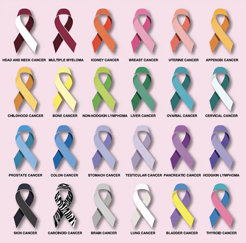
In conclusion, the question “what color is lung cancer” no longer has a simple answer. In the realm of technology, it is a fluid concept. It is the red glow of a PET scan, the purple-pink of a digitized biopsy, the neon overlay of a hyperspectral surgical feed, and the complex data points of a machine learning model. As software continues to evolve, our digital vision will only become sharper, allowing us to see the colors of malignancy earlier, more clearly, and with more life-saving precision than ever before.
aViewFromTheCave is a participant in the Amazon Services LLC Associates Program, an affiliate advertising program designed to provide a means for sites to earn advertising fees by advertising and linking to Amazon.com. Amazon, the Amazon logo, AmazonSupply, and the AmazonSupply logo are trademarks of Amazon.com, Inc. or its affiliates. As an Amazon Associate we earn affiliate commissions from qualifying purchases.