
In the rapidly evolving landscape of health technology, the term “angioma” has transitioned from a purely clinical descriptor to a focal point for high-tech diagnostic innovation. Traditionally defined as benign growths consisting of small blood vessels, angiomas—including cherry angiomas, spider angiomas, and cavernous angiomas—are now being reimagined through the lens of computer vision, machine learning, and advanced laser optics. As the tech industry continues to merge with the medical sector, understanding what angiomas are requires a deep dive into the software architectures and hardware breakthroughs that are redefining dermatological care and vascular analysis.

The intersection of software engineering and dermatology has turned the identification of these vascular clusters into a masterclass in pattern recognition. For developers and tech enthusiasts, the study of angiomas represents a unique challenge: how to differentiate a benign vascular growth from a malignant lesion using only digital inputs. This has led to the development of sophisticated AI tools that are setting new standards for diagnostic accuracy in modern clinics.
The Role of Computer Vision in Identifying Vascular Structures
At the heart of modern diagnostic tech lies computer vision, a branch of artificial intelligence that enables software to interpret and understand the visual world. When we ask “what are angiomas” in a digital context, we are essentially discussing the ability of Convolutional Neural Networks (CNNs) to map the specific pixel density and chromatic variance of a vascular lesion.
Training Neural Networks on Vascular Data Sets
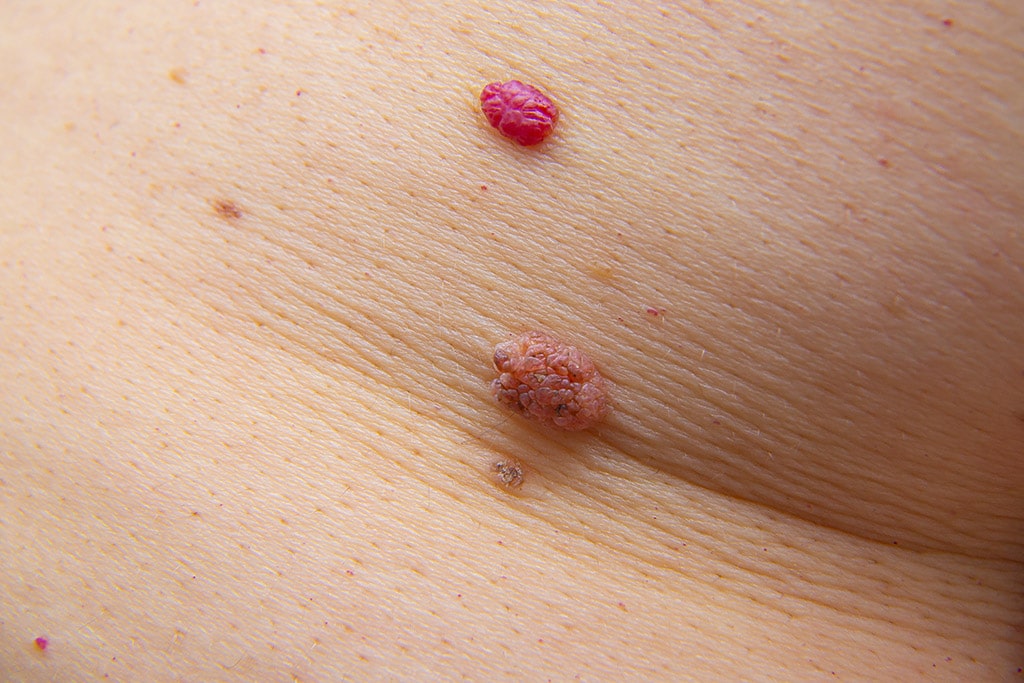
The identification of an angioma begins with the training phase. AI tools are fed millions of high-resolution images of various skin conditions. Developers use supervised learning to teach the algorithm that an angioma typically presents as a well-demarcated, ruby-red or purple papule. The software must be trained to distinguish these from hemangiomas or more dangerous precursors like nodular melanoma.
This training involves deep learning layers that analyze “feature hierarchies.” The first layer might detect edges and borders, the second layer identifies color saturation, and the third layer recognizes the specific “cherry-like” structure of a common angioma. By the time the software reaches a decision, it has processed the image through dozens of filters that no human eye could replicate with consistent precision.
Real-Time Diagnostic Apps and Edge Computing
The next frontier for this technology is the move toward “Edge Computing.” Rather than sending image data to a centralized cloud server, which can raise privacy concerns and introduce latency, modern diagnostic apps perform complex calculations directly on a smartphone’s GPU. This allows for real-time scanning. A user can hover their phone camera over a suspected angioma, and the local AI model provides an immediate classification based on its pre-trained weights. This shift to local processing is a major trend in digital security and user experience design, ensuring that sensitive biometric data never leaves the device.
Advanced Laser Hardware and the Engineering of Treatment
Understanding what angiomas are is only half the battle; the other half is the engineering required to treat them. In the tech world, the treatment of angiomas is a study in precision physics and gadgetry. The evolution from traditional surgical excision to non-invasive laser technology is a testament to the advancements in optical engineering.
Pulsed Dye Lasers (PDL) and Selective Photothermolysis
The gold standard for treating angiomas is the Pulsed Dye Laser (PDL). This device is a marvel of technology, utilizing a high-concentration organic dye as the gain medium. The tech works on the principle of selective photothermolysis. Essentially, the software controlling the laser is programmed to target a specific wavelength (usually 585 or 595 nm) that is absorbed by hemoglobin but not by the surrounding skin tissue.
This requires incredibly precise pulse-width modulation. If the pulse is too long, it causes thermal damage to the skin; if it is too short, it fails to coagulate the blood vessels within the angioma. Modern PDL gadgets feature integrated cooling systems—cryogen sprays that fire milliseconds before the laser—managed by high-speed microcontrollers to protect the epidermis while the laser obliterates the vascular growth.
The Rise of Multi-Platform Workstations

In contemporary medical-tech reviews, we are seeing a shift toward multi-platform workstations. These are the “all-in-one” gadgets of the dermatological world. A single workstation might combine Intense Pulsed Light (IPL), Nd:YAG lasers, and radiofrequency tools. For tech-focused practitioners, these platforms are managed via intuitive touchscreen interfaces that run specialized OS versions, allowing for customized treatment parameters based on the patient’s specific “angioma profile” stored in a digital database.
Digital Security and Data Privacy in Vascular Imaging
As we move toward a world where angiomas are tracked and diagnosed via apps and AI, digital security becomes a paramount concern. The “what” of angiomas now includes the “where” and “how” of the data they generate. Medical imaging data is a high-value target for cybercriminals, making the security architecture of these tech tools as important as their diagnostic capabilities.
Encryption and Compliance in Health-Tech Software
Any software designed to analyze angiomas must adhere to strict regulatory standards, such as HIPAA in the United States or GDPR in Europe. This means that the developers must implement end-to-end encryption for all image transmissions. In the world of SaaS (Software as a Service) for clinics, this often involves “zero-knowledge” architectures. This ensures that even the company providing the AI tool cannot see the patient’s images; the decryption keys remain solely in the hands of the medical provider.
Blockchain for Verifiable Medical Records
A growing trend in the “Money and Tech” crossover is the use of blockchain to store medical history. If an individual has multiple angiomas that need to be monitored for growth over several years, a blockchain-based ledger can provide an immutable record of these changes. This tech ensures that the diagnostic history of an angioma cannot be tampered with or lost during a transfer between clinics, providing a “single source of truth” for the patient’s vascular health.
The Future of AI-Driven Dermatology: Beyond Simple Detection
When we look at the trajectory of current technology, the question of “what are angiomas” will soon be answered by predictive analytics. We are moving beyond simple identification and toward a future where software can predict the appearance of these growths before they are visible to the naked eye.
Predictive Modeling and Genomic Tech
The next generation of health-tech will likely involve the integration of genomic data with visual AI. By analyzing a patient’s genetic markers alongside their digital skin maps, predictive software could alert users to a predisposition for certain types of angiomas. This represents a massive shift from reactive care to proactive, tech-enabled wellness.
Augmented Reality (AR) in Surgical Planning
For complex cases, such as deep-seated cavernous angiomas, Augmented Reality (AR) is becoming an essential tool. Surgeons can now use AR headsets like the HoloLens or specialized medical equivalents to overlay 3D MRI data onto the patient’s actual body. This “X-ray vision” allows the clinician to see the exact structure of the angioma beneath the skin in real-time, significantly reducing the risk of complications and improving the precision of the removal process.
Conclusion: The Digital Transformation of Vascular Health
Angiomas, once viewed as simple red spots on the skin, have become a catalyst for some of the most exciting innovations in the tech sector. From the deep learning models used to categorize them to the precision lasers used to erase them, the technology surrounding angiomas is a microcosm of the broader digital health revolution.
As software becomes more intelligent and hardware more precise, the line between technology and medicine will continue to blur. For the tech-savvy consumer or the developer looking for the next big challenge, the field of AI-driven vascular analysis offers a roadmap for how we will handle health and wellness in the 21st century. The study of angiomas is no longer just a medical pursuit; it is a digital endeavor that leverages the best of AI, cloud security, and optical engineering to improve human outcomes through superior technology.
aViewFromTheCave is a participant in the Amazon Services LLC Associates Program, an affiliate advertising program designed to provide a means for sites to earn advertising fees by advertising and linking to Amazon.com. Amazon, the Amazon logo, AmazonSupply, and the AmazonSupply logo are trademarks of Amazon.com, Inc. or its affiliates. As an Amazon Associate we earn affiliate commissions from qualifying purchases.