
In the contemporary landscape of digital health, the question of what a chest X-ray “looks like” has evolved from a simple visual observation into a complex interrogation of high-resolution data. For the modern technologist, radiologist, and software engineer, a chest X-ray showing pneumonia is no longer just a film held up to a light box; it is a sophisticated digital matrix. Through the lens of medical technology, pneumonia manifests as a series of pixel variances, density fluctuations, and algorithmic patterns that represent the battle between air-filled lung tissue and fluid-based infection.
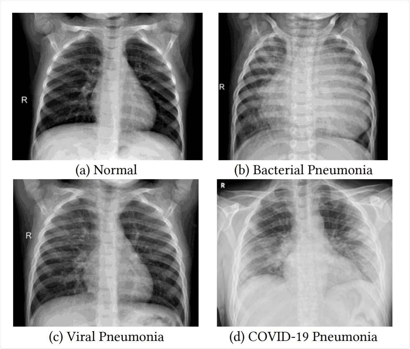
The transition from analog to digital radiography (DR) has fundamentally changed how we visualize thoracic pathology. Today, identifying pneumonia involves high-performance computing, sophisticated image-processing algorithms, and the integration of artificial intelligence to assist human interpretation.
The Digital Transformation of Thoracic Imaging
At its core, a modern chest X-ray is a product of advanced sensor technology. When an X-ray beam passes through a patient’s chest, it hits a digital detector—essentially a high-grade version of the sensor found in a digital camera, but tuned for high-energy radiation. In a healthy lung, the air allows most of the X-rays to pass through, hitting the detector with high intensity and resulting in dark or black pixels. However, when pneumonia is present, the air sacs (alveoli) fill with fluid, pus, or inflammatory cells. This denser material absorbs more radiation, preventing it from reaching the sensor.
From Film to Digital Radiography (DR)
The move from traditional silver-halide film to Direct Radiography (DR) and Computed Radiography (CR) has revolutionized the resolution available to clinicians. Digital detectors utilize thin-film transistor (TFT) arrays and scintillators to convert X-ray photons into electrical signals. This digital signal is then processed through a series of lookup tables (LUTs) to optimize contrast and brightness. In a case of pneumonia, these digital systems must be calibrated to distinguish between the subtle gray-scale differences of soft tissue, fluid, and bone. The result is a high-fidelity image where “consolidation”—the hallmark of pneumonia—appears as cloudy, opaque white patches against the dark background of the lungs.
The Bit Depth and Dynamic Range of Diagnostic Images
Unlike standard consumer JPEG images, which typically operate at 8-bit depth (256 shades of gray), medical X-rays are captured at 12-bit, 14-bit, or even 16-bit depths. This allows for thousands of shades of gray, providing the dynamic range necessary to see the “air bronchograms” characteristic of pneumonia. Technologically, this means that even if an image appears too dark or too light to the naked eye, the raw data contains enough information for software to “window” or “level” the image, bringing the hidden details of the infection into view without requiring a second exposure.
Leveraging AI and Deep Learning for Automated Diagnostics
One of the most significant trends in medical technology is the application of Artificial Intelligence (AI) to interpret what pneumonia looks like. With the rise of deep learning, specifically Convolutional Neural Networks (CNNs), software is now capable of identifying the visual markers of pneumonia with a precision that rivals experienced specialists.
Convolutional Neural Networks (CNNs) in Radiology
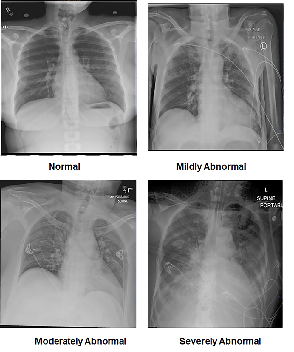
To an AI model, pneumonia is not a medical condition but a pattern of spatial frequencies and pixel intensities. Deep learning models are trained on massive datasets—such as the NIH ChestX-ray14 or the CheXpert dataset—containing hundreds of thousands of labeled images. These models learn to recognize “opacities,” “infiltrates,” and “consolidations.” By breaking the image down into layers, the software identifies edges, textures, and eventually complex shapes that correlate with the presence of infection. This automated “looking” allows for rapid triage in emergency departments, where the software can flag a suspicious X-ray for immediate review.
Computer-Aided Detection (CADe) and Diagnosis (CADx)
CAD systems act as a second set of eyes. When the software analyzes a chest X-ray for pneumonia, it often generates a “heatmap” or a “saliency map.” These visual overlays use color-coding (often red or yellow) to highlight the areas where the algorithm has detected high probabilities of fluid buildup. For the technologist managing these systems, the challenge lies in reducing “false positives”—where the software mistakes a normal anatomical structure or a technical artifact for pneumonia. Modern software suites now integrate these AI insights directly into the radiologist’s workflow, providing a seamless bridge between raw data and clinical decision-making.
Software Infrastructure: PACS, DICOM, and the Radiologist’s Digital Workspace
The visualization of pneumonia is also dependent on the software environment in which the image is viewed. The Picture Archiving and Communication System (PACS) and the Digital Imaging and Communications in Medicine (DICOM) standard form the backbone of this technological ecosystem.
The Role of DICOM Standards
DICOM is more than just a file format; it is a comprehensive protocol for handling, storing, printing, and transmitting medical imaging information. A chest X-ray file contains not only the pixel data but also a wealth of metadata, including the dose of radiation used, the orientation of the patient, and the technical specifications of the X-ray machine. This metadata is crucial when comparing a current X-ray to a previous “baseline” image to see if the pneumonia is clearing or worsening. Advanced viewing software allows for “syncing” these images, so a technologist can scroll through a patient’s history and see the exact same anatomical region side-by-side.
Advanced Visualization Tools in PACS
Within the PACS environment, several software tools help clarify what pneumonia looks like:
- Edge Enhancement: This algorithm sharpens the boundaries between different tissue densities, making it easier to see the fine lines of an infection.
- Inversion: By flipping the grayscale (making whites black and blacks white), certain patterns of interstitial pneumonia become more apparent to the human eye.
- Magnification and Pan: High-resolution monitors (often 3 to 5 megapixels) allow clinicians to zoom into the costophrenic angles or the retrocardiac space, where early-stage pneumonia often hides.
- 3D Reconstruction: In cases where a standard X-ray is inconclusive, software can sometimes assist in “stitching” multiple views or integrating with CT data to provide a more volumetric look at the lung’s condition.
Security, Scalability, and the Future of Cloud-Native Radiology
As the tech world moves toward cloud-based solutions, the way we store and share images of pneumonia is changing. The “look” of an X-ray is now something that can be accessed from a tablet in a remote clinic or a workstation in a major hospital, necessitating a robust digital infrastructure.
Cybersecurity in Medical Imaging
Because a chest X-ray is protected health information (PHI), the technology used to transmit it must be highly secure. End-to-end encryption, secure VPNs, and multi-factor authentication are standard components of the radiology tech stack. When a digital X-ray showing pneumonia is sent from an imaging center to a specialist, it travels through secure gateways that ensure the integrity of the data. If a single packet of data is corrupted during transmission, the “look” of the image could be altered, leading to a potential misdiagnosis—hence the importance of lossless compression algorithms like JPEG 2000 (standard in DICOM).

Edge Computing and Tele-radiology
The future of identifying pneumonia lies in edge computing. Instead of sending a massive 50MB DICOM file to a central server for AI analysis, the “intelligence” is moving closer to the source—the X-ray machine itself. Modern “smart” X-ray suites are equipped with onboard processing power that can run AI models locally. This means that within seconds of the image being captured, the machine can alert the technologist if it “sees” signs of pneumonia, potentially saving hours in critical care situations.
Furthermore, cloud-native PACS are allowing for massive scalability. During respiratory surges, such as during a pandemic, the ability to spin up virtualized instances of diagnostic software allows healthcare systems to handle a higher volume of images. This tech-heavy approach ensures that whether an X-ray is viewed in a rural clinic or a metropolitan hospital, the quality, clarity, and diagnostic accuracy remains consistent.
In conclusion, when we ask what a chest X-ray looks like with pneumonia, we are really asking about the state of modern medical technology. It is a visual representation of the intersection between physics, software engineering, and data science. From the silicon pixels of the detector to the neural networks of the AI, the “look” of pneumonia is a digital signature that the tech industry continues to refine, secure, and automate for the benefit of global health.
aViewFromTheCave is a participant in the Amazon Services LLC Associates Program, an affiliate advertising program designed to provide a means for sites to earn advertising fees by advertising and linking to Amazon.com. Amazon, the Amazon logo, AmazonSupply, and the AmazonSupply logo are trademarks of Amazon.com, Inc. or its affiliates. As an Amazon Associate we earn affiliate commissions from qualifying purchases.