In the realm of modern diagnostics, the chest X-ray remains one of the most frequently utilized and technologically significant tools in a clinician’s arsenal. While the basic concept—passing electromagnetic radiation through the body to create an image—has existed for over a century, the technology powering these images has undergone a digital revolution. Today, when we ask “what will a chest X-ray show,” we are not just looking at a static piece of film; we are looking at a complex data set interpreted by high-resolution sensors and, increasingly, sophisticated artificial intelligence.

Understanding what a chest X-ray reveals requires an exploration of the intersection between physics, digital imaging hardware, and the software that translates raw radiation data into a diagnostic roadmap.
The Evolution of Radiographic Technology: From Film to Digital Sensors
To understand what a chest X-ray shows today, one must first understand the transition from analog to digital. The “tech” behind the image has shifted from chemical processing to high-speed data acquisition.
From Film to Digital Radiography (DR)
The traditional method involved silver halide film that required chemical development in a darkroom. Modern facilities now utilize Digital Radiography (DR). In a DR system, a flat-panel detector contains a layer of scintillator material that converts X-rays into light, which is then captured by thin-film transistors (TFT) and converted into a digital signal. This tech allows for “instant” imaging, where the chest cavity appears on a monitor within seconds. The dynamic range of digital sensors is significantly higher than film, meaning a single X-ray can show a wider variety of tissue densities without needing a “retake.”
How Modern X-Ray Machines Work
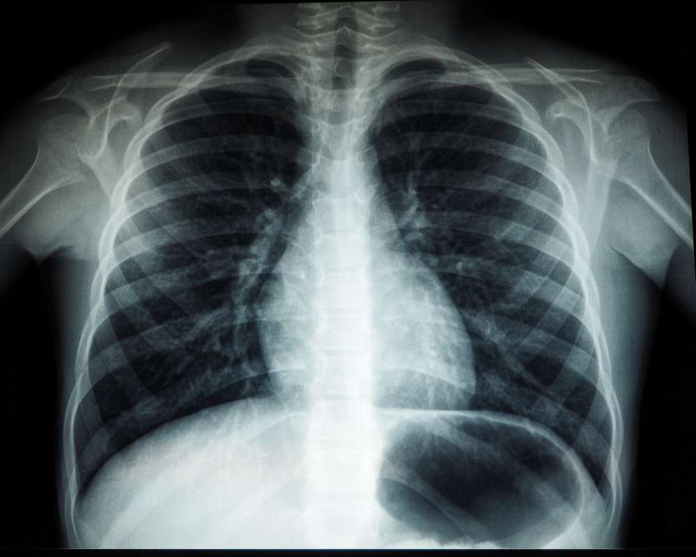
A modern chest X-ray machine is a marvel of precision engineering. The X-ray tube generates a controlled beam of photons directed toward the patient’s chest. On the opposite side, the digital detector captures the photons that pass through. Dense materials, such as bone or metal (like a pacemaker), absorb more photons and appear white. Less dense areas, such as air-filled lungs, allow most photons to pass through and appear black. The “gray” areas represent the heart, blood vessels, and fluid. The technology’s ability to differentiate these subtle gradients of gray is what determines the diagnostic quality of the image.
Visualizing the Interior: What the Data Reveals
A chest X-ray provides a comprehensive “snapshot” of the thoracic cavity. By manipulating the contrast and brightness of the digital image, technologists can highlight specific systems within the chest.
Lung Health and Respiratory Diagnostics
The primary focus of most chest X-rays is the lungs. Because lungs are filled with air, they provide a stark contrast to the surrounding tissue. The technology will show:
- Hyperinflation: Common in COPD or emphysema, where the lungs appear darker and larger than normal.
- Infiltrates and Opacities: When air is replaced by fluid, pus, or blood (as seen in pneumonia or pulmonary edema), the digital image shows cloudy, white “patches.”
- Pneumothorax: A collapsed lung is identified when the digital sensor detects air in the pleural space, showing a distinct lack of lung markings (the fine white lines representing blood vessels) in a specific area.
Cardiac Outlines and Vascular Imaging
The chest X-ray is a fundamental tool for monitoring the heart’s “silhouette.” The technology reveals the size and shape of the cardiac shadow. An enlarged heart (cardiomegaly) is easily detected by calculating the cardio-thoracic ratio on the digital workstation. Furthermore, the tech shows the “vascular pedicle”—the collection of large vessels (aorta and vena cava) entering and exiting the heart. Distortions in these shapes can indicate aortic aneurysms or pulmonary hypertension.
Skeletal Structures and Soft Tissue Density
Beyond the organs, a chest X-ray provides a high-resolution view of the rib cage, spine, and clavicles. Digital post-processing allows radiologists to “bone-suppress” the image—a software technique that digitally removes the ribs from the view to provide an unobstructed look at the lung tissue beneath. Conversely, the “bone-enhanced” view can highlight subtle stress fractures or metastatic lesions within the skeletal frame that might be missed on a standard analog print.
The Intersection of AI and Chest Radiography
Perhaps the most exciting trend in medical technology is the integration of Artificial Intelligence (AI) and Machine Learning (ML) into the interpretation of chest X-rays. What an X-ray “shows” is now being augmented by algorithms that can see beyond human visual perception.
Computer-Aided Detection (CAD) Systems
CAD software acts as a second set of eyes for the radiologist. These AI tools are trained on millions of previous X-rays to identify patterns associated with specific pathologies. For example, AI can highlight a tiny pulmonary nodule—the possible early sign of lung cancer—that might be obscured by a rib. By flagging these “areas of interest,” the technology reduces human error and ensures that the most critical findings are addressed immediately.
Predictive Analytics in Emergency Radiology
In high-volume hospital environments, AI-enabled X-ray tech is used for “triage.” When an X-ray is taken, the software instantly scans the digital file for life-threatening conditions like a tension pneumothorax or a misplaced tracheal tube. If the algorithm detects a critical issue, it can automatically move that patient’s image to the top of the radiologist’s reading queue. This tech-driven prioritization is transforming emergency care, ensuring that what the X-ray shows is acted upon within minutes.
Security and Data Management in Medical Imaging
As chest X-rays have transitioned into digital files, the technology surrounding their storage and transmission has become a critical pillar of digital security and IT infrastructure.
DICOM Standards and Interoperability
Chest X-rays are not stored as simple JPEGs. They use a specialized format called DICOM (Digital Imaging and Communications in Medicine). This protocol ensures that an image taken on a GE machine in New York can be read perfectly on a Siemens workstation in London. DICOM files contain not just the image, but a “header” of metadata including patient demographics, radiation dose, and equipment calibration settings. This interoperability is what allows for the seamless “Brand” experience of modern healthcare networks.
Protecting Patient Data in the Cloud
With the rise of Picture Archiving and Communication Systems (PACS), chest X-rays are now frequently stored in the cloud. This brings the tech world’s focus on cybersecurity into the radiology department. Encryption at rest and in transit is mandatory to protect sensitive health information (PHI). Modern imaging centers utilize blockchain technology and advanced firewalls to ensure that the “what” of a chest X-ray—your internal health data—remains private and accessible only to authorized personnel.
The Future of Diagnostic Imaging Tech
The trajectory of chest X-ray technology points toward even greater portability, lower radiation, and higher resolution.
Portable X-Ray Devices and Remote Diagnostics
We are seeing a trend toward “Point-of-Care” (POC) imaging. Modern portable X-ray units are now small enough to be wheeled into a patient’s home or a remote battlefield. These devices utilize high-sensitivity sensors that require a fraction of the radiation dose used ten years ago. These devices are often IoT-enabled (Internet of Things), wirelessly transmitting images to a central hub for instant consultation. This democratizes diagnostic tech, bringing the power of the chest X-ray to underserved populations.

Quantum Sensing and Higher Resolution
Looking further ahead, the development of quantum sensors may soon allow for even more detailed chest imaging. These sensors could potentially detect the “phase shift” of X-rays as they pass through tissue, not just the absorption. This would provide a level of detail currently only available in CT scans, but with the speed and low cost of a standard X-ray. It would revolutionize what we can “see”—moving from broad outlines of organs to the microscopic textures of lung tissue.
In conclusion, a chest X-ray shows far more than just “bones and lungs.” In the modern tech landscape, it is a sophisticated data product of advanced physics, high-speed digital sensors, and intelligent software. Whether it is being analyzed by a human expert or a machine-learning algorithm, the chest X-ray remains a cornerstone of technology-driven medicine, providing a window into the human body that is clearer, safer, and more accessible than ever before.
aViewFromTheCave is a participant in the Amazon Services LLC Associates Program, an affiliate advertising program designed to provide a means for sites to earn advertising fees by advertising and linking to Amazon.com. Amazon, the Amazon logo, AmazonSupply, and the AmazonSupply logo are trademarks of Amazon.com, Inc. or its affiliates. As an Amazon Associate we earn affiliate commissions from qualifying purchases.