In the realm of modern diagnostics, the sonogram—or echocardiogram—stands as a pinnacle of non-invasive medical engineering. While patients often view it as a simple “look” at their heart, the technology underlying these images is a complex orchestration of high-frequency acoustics, sophisticated signal processing, and increasingly, artificial intelligence. Understanding what a sonogram of the heart shows requires a deep dive into the technological stack that captures the invisible and translates it into actionable data.
The Mechanics of Sound: How Ultrasonic Hardware Visualizes Cardiac Structure
At its core, a cardiac sonogram is a feat of physics and hardware engineering. The process begins with a transducer, a sophisticated piece of hardware that acts as both a speaker and a microphone. This device utilizes the piezoelectric effect to generate ultrasound waves—frequencies far beyond the range of human hearing.

Piezoelectric Transducers and Wave Propagation
The hardware inside a sonogram probe contains piezoelectric crystals. When an electric current is applied, these crystals vibrate, sending high-frequency sound waves into the thoracic cavity. As these waves encounter different densities—such as the muscular wall of the myocardium, the fluid density of blood, or the fibrous tissue of heart valves—they bounce back. The tech must be incredibly precise; modern transducers can pulse and listen thousands of times per second, ensuring that the resulting image reflects the heart’s movement in real-time without significant latency.
High-Frequency Resolution in Modern Probes
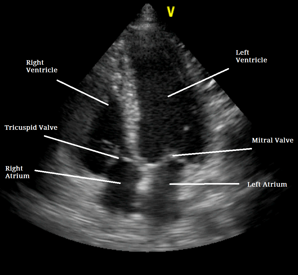
The evolution of sonogram tech has seen a massive shift toward high-frequency resolution. Older systems struggled with “noise” or artifacts caused by bone or lung tissue interference. Contemporary probes utilize “matrix array” technology, allowing for thousands of individual elements to fire in sequence. This creates a high-definition “slice” of the heart, showing the thickness of the ventricular walls and the delicate, parachute-like motion of the mitral valves with millimeter-level accuracy.
Software Processing: Translating Echoes into Live 3D Models
What the clinician sees on the screen is not a raw recording of sound, but a highly processed digital reconstruction. The software layer of a sonogram machine is responsible for interpreting the delay, intensity, and frequency shift of returning echoes to build a visual map.
Doppler Shift Algorithms and Hemodynamics
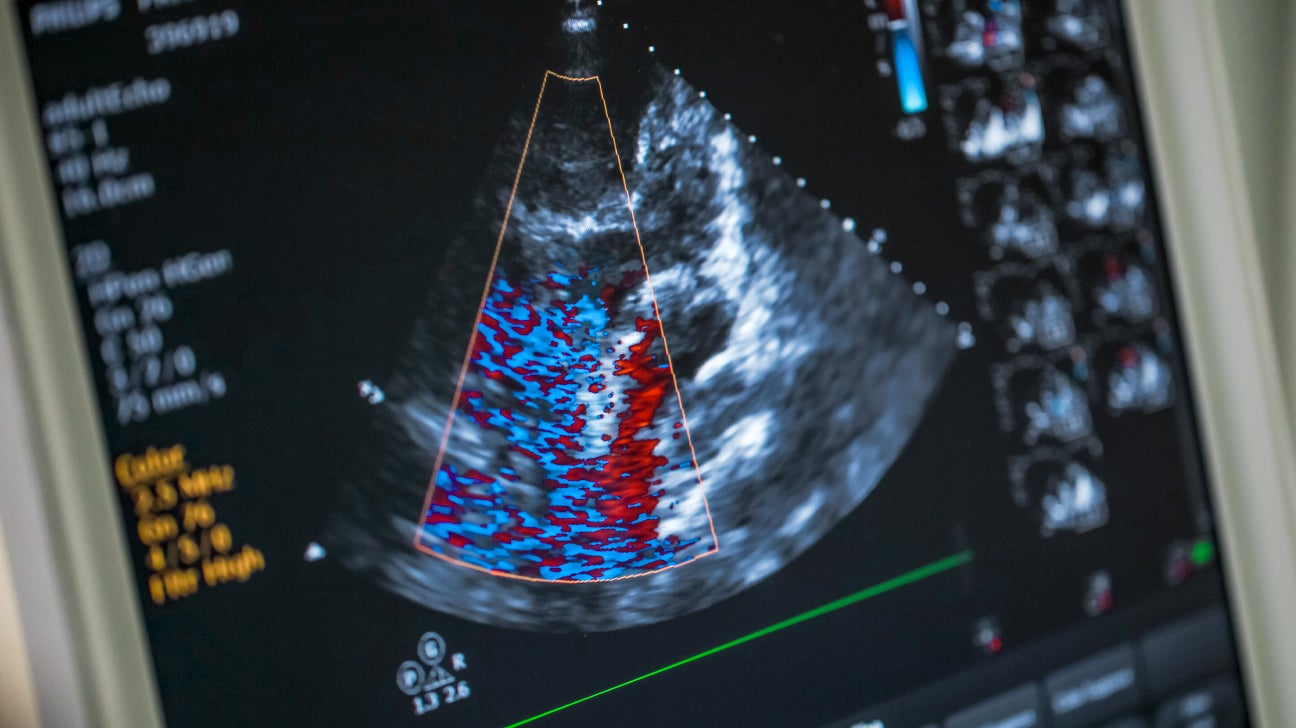
One of the most critical software features in a cardiac sonogram is Color Doppler imaging. By applying the Doppler effect—the same principle used in radar—the software calculates the velocity and direction of blood flow. On the monitor, this is visualized as a vibrant overlay: typically red for blood moving toward the probe and blue for blood moving away. These algorithms allow the tech to show turbulence or “regurgitation,” where blood leaks backward through a valve, identifying mechanical failures within the heart’s plumbing that a static image would miss.
Speckle-Tracking and Strain Imaging Technology
Modern cardiac software has moved beyond simple visual inspection to “Speckle-Tracking Echocardiography” (STE). This tech identifies natural acoustic markers (speckles) within the heart tissue and tracks them through the cardiac cycle. By analyzing how these speckles move relative to one another, the software calculates “strain”—a measure of how much the heart muscle is deforming and contracting. This provides a quantitative assessment of heart health, allowing the software to detect subtle dysfunction long before the human eye can see a change in heart rhythm or shape.
The AI Revolution: Enhancing Diagnostic Accuracy in Cardiac Scans
The most significant recent trend in sonogram technology is the integration of Artificial Intelligence (AI) and Machine Learning (ML). These tools are transforming the sonogram from a subjective visual tool into an objective diagnostic powerhouse.
Automated Chamber Quantification
In traditional sonography, a technician had to manually trace the edges of the heart’s chambers to calculate volumes and the “ejection fraction” (the percentage of blood pumped out with each beat). Today, AI-driven software performs automated chamber quantification. Using deep learning models trained on millions of previous scans, the tech can instantly identify the borders of the left ventricle and provide precise measurements. This reduces human error and ensures consistency across different hardware platforms and clinics.
Predictive Analytics for Early Heart Failure Detection
AI tools are now capable of identifying patterns in cardiac motion that are imperceptible to human observers. By analyzing the “digital signature” of a sonogram, AI can predict the onset of heart failure or structural deterioration years in advance. These predictive analytics transform what the sonogram shows from a “current state” report into a “future risk” assessment, shifting the focus of cardiac tech from reactive treatment to proactive management.
From Cart to Handheld: The Evolution of Portable Sonogram Tech
For decades, heart sonograms were performed using “cart-based” systems—large, heavy machines with massive processing power. However, the miniaturization of processors and the improvement of battery tech have led to a revolution in portability.
Point-of-Care Ultrasound (POCUS) and Smartphone Integration
The rise of Point-of-Care Ultrasound (POCUS) represents a major shift in the tech landscape. Companies have developed handheld transducers that plug directly into a smartphone or tablet. These devices leverage the high-resolution displays and mobile processing power of consumer tech to show detailed cardiac images at the bedside, in ambulances, or in rural clinics. This democratization of the tech means that the ability to “see” the heart is no longer confined to the imaging department of a major hospital.
Edge Computing in Medical Hardware
To support handheld sonography, manufacturers are utilizing “edge computing.” Instead of sending raw data to a central server for processing, the device itself handles the heavy lifting of image reconstruction. This requires specialized chips—Application-Specific Integrated Circuits (ASICs)—designed to handle the massive data throughput of ultrasound waves in real-time. This tech ensures that even a pocket-sized device can show a clear, jitter-free view of a beating heart.
Data Security and Interoperability in Modern Echocardiography
As sonogram technology becomes more digital and connected, the focus on the “backend” tech has intensified. A sonogram is no longer just a video file; it is a complex data packet that must be stored, shared, and secured.
Cloud-Based Storage and DICOM Standards
Modern sonogram machines are integrated into a hospital’s Digital Imaging and Communications in Medicine (DICOM) network. This protocol ensures that the high-resolution files generated by the sonogram are interoperable across different software platforms. Furthermore, many clinics are moving toward cloud-based storage, allowing a specialist in one city to review a “live” sonogram being performed in another. This connectivity relies on high-speed fiber optics and robust cloud infrastructure to handle the massive file sizes associated with 4D cardiac imaging.
Protecting Patient Privacy in the IoT Era
With the move to handheld and cloud-connected sonograms comes the challenge of digital security. Medical devices are now part of the Internet of Things (IoT), making them potential targets for cyberattacks. Manufacturers are building advanced encryption directly into the sonogram hardware and software. This ensures that the sensitive data—showing the most intimate mechanics of a patient’s life-sustaining organ—remains confidential while still being accessible to the authorized healthcare tech stack.
In conclusion, what a sonogram of the heart shows is a masterclass in technological integration. From the physical manipulation of sound waves via piezoelectric crystals to the AI-driven analysis of blood flow and muscle strain, the “echo” has evolved into a high-tech digital twin of the human heart. As hardware continues to shrink and software intelligence continues to grow, the sonogram will remain the most vital tool in the technological arsenal for understanding cardiac health.
aViewFromTheCave is a participant in the Amazon Services LLC Associates Program, an affiliate advertising program designed to provide a means for sites to earn advertising fees by advertising and linking to Amazon.com. Amazon, the Amazon logo, AmazonSupply, and the AmazonSupply logo are trademarks of Amazon.com, Inc. or its affiliates. As an Amazon Associate we earn affiliate commissions from qualifying purchases.