
The advent of advanced imaging technologies has revolutionized prenatal care, allowing unprecedented insights into early fetal development. While the primary goal of a 6-week ultrasound is clinical assessment, understanding the technological underpinnings of what is “seen” provides a richer appreciation of this diagnostic tool. At this nascent stage of gestation, the capabilities of modern ultrasound machines, their integrated software, and emerging AI applications are paramount in rendering detailed and interpretable images from what is still a microscopic entity.
The Evolution of Early Pregnancy Ultrasound Technology
The journey from rudimentary grayscale images to today’s high-resolution, nuanced scans represents decades of innovation in transducer design, signal processing, and display technologies. For a 6-week pregnancy, the precise capabilities of the ultrasound machine are critical in visualizing the minute structures indicative of viability and early developmental milestones.

From 2D to High-Resolution Imaging
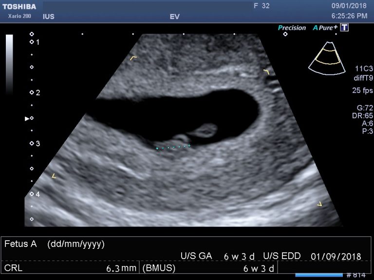
Early ultrasound machines produced relatively coarse 2D images, often making the identification of tiny structures challenging. Modern ultrasound systems, however, employ sophisticated beamforming techniques and broadband transducers that generate far clearer, higher-resolution 2D images. These advancements are particularly vital at 6 weeks gestation, where the embryo measures only a few millimeters. Improved spatial and contrast resolution allows for more definitive visualization of the gestational sac, yolk sac, and, most critically, the fetal pole and its nascent cardiac activity. Digital imaging processors have become exponentially more powerful, capable of handling vast amounts of raw data from multiple acoustic pulses and converting them into sharp, detailed images in real-time. This iterative refinement of image acquisition and reconstruction algorithms means that even subtle variations in tissue density can be rendered with greater clarity, aiding in the differentiation of early embryonic structures.
Transvaginal vs. Transabdominal Techniques
At 6 weeks, a transvaginal ultrasound (TVUS) is almost universally preferred due to its superior resolution and proximity to the uterus. Technologically, the TVUS transducer is specifically designed for internal use, featuring a higher frequency probe (typically 7-10 MHz, sometimes up to 12 MHz) compared to transabdominal probes. Higher frequencies allow for better resolution of superficial structures but have limited penetration depth. Since the uterus is relatively close to the transvaginal probe, this higher frequency can be effectively utilized to obtain exquisite detail of the very small gestational sac, yolk sac, and fetal pole. The focused beam and reduced interference from abdominal wall tissues and bowel gas contribute to clearer images. In contrast, a transabdominal approach at 6 weeks often yields less diagnostic information due to the distance and intervening tissues, making it more challenging to discern minute embryonic details with sufficient clarity, even with advanced signal processing. The technological design of the TVUS probe, optimized for close-range, high-frequency imaging, is thus central to successfully “seeing” what is expected at this early stage.
Decoding the Digital Image: Software and Algorithms
The raw data collected by ultrasound transducers is just the starting point. The true diagnostic power lies in the sophisticated software and algorithms that process, enhance, measure, and interpret this data. These digital tools transform acoustic signals into meaningful visual and quantitative information.
Image Processing and Enhancement
Once acoustic waves are converted into electrical signals, advanced image processing algorithms kick in. These algorithms perform various functions:
- Speckle Reduction: Ultrasound images inherently contain ‘speckle’ noise, which can obscure fine details. Sophisticated filters and algorithms (e.g., adaptive speckle reduction, compound imaging) are employed to reduce this noise without sacrificing true anatomical information, leading to smoother, clearer images.
- Harmonic Imaging: This technique utilizes the harmonic frequencies generated by tissue interactions with the ultrasound beam, rather than just the fundamental frequency. Harmonic images often have better contrast and reduced artifact levels, providing enhanced visualization of small structures like the yolk sac and fetal pole.
- Dynamic Range and Gain Control: Software allows operators to adjust the dynamic range (the number of shades of gray) and gain (amplification of signals) to optimize image contrast and brightness, highlighting specific structures within the uterine cavity. Automated optimization features use AI to suggest optimal settings based on tissue characteristics, further streamlining the imaging process.
Measurement and Interpretation Software
Critical to early pregnancy assessment are precise measurements, which modern ultrasound software facilitates with high accuracy.
- Gestational Sac Diameter (GSD): Software tools allow precise measurement of the gestational sac, helping to date the pregnancy and assess its growth. Automated calipers and measurement protocols ensure consistency.
- Yolk Sac Diameter: Similarly, the yolk sac’s size can be accurately measured, which is an important indicator of embryonic development.
- Crown-Rump Length (CRL): While often still very small at 6 weeks, software provides dedicated tools for measuring the CRL, the most accurate parameter for dating in the first trimester. Algorithms assist in identifying the longest axis of the fetal pole for accurate measurement.
- Heart Rate Calculation: The presence and rate of fetal cardiac activity are crucial. Advanced Doppler imaging and M-mode (motion mode) software allow for the visualization and precise calculation of the embryonic heart rate, displayed numerically and graphically. Automated detection algorithms can even flag potential bradycardia or tachycardia.
AI-Assisted Diagnostics in Early Pregnancy

The integration of Artificial Intelligence (AI) is beginning to transform how early pregnancy ultrasounds are interpreted. While still an evolving field, AI offers the potential for enhanced accuracy and efficiency.
- Automated Feature Detection: AI algorithms, trained on vast datasets of ultrasound images, can learn to automatically identify and delineate key structures such as the gestational sac, yolk sac, and fetal pole. This can reduce inter-observer variability and ensure all critical structures are assessed.
- Growth Curve Analysis: AI can compare current measurements (GSD, CRL) against established normative growth curves, flagging deviations that might warrant closer monitoring. This predictive capability moves beyond simple measurement to insightful analysis.
- Early Viability Assessment: Machine learning models are being developed to assist in assessing early pregnancy viability by analyzing a combination of parameters (size, growth rate, heart rate) and identifying patterns associated with successful pregnancies versus early pregnancy loss. These tools act as a second pair of “eyes,” providing decision support to clinicians.
- Quality Assurance: AI can evaluate the quality of acquired images, providing real-time feedback to sonographers on probe positioning and settings to ensure optimal diagnostic quality.
Digital Platforms and Patient Engagement
Beyond the immediate diagnostic session, digital technologies extend the utility and accessibility of ultrasound information, bridging the gap between clinical data and patient understanding.
Telemedicine and Remote Consultations
The secure transmission of ultrasound images and reports through telemedicine platforms allows for remote consultations with specialists, particularly beneficial in rural or underserved areas. High-resolution images can be encrypted and shared, enabling expert interpretation without the patient needing to travel. This digital infrastructure ensures continuity of care and access to specialized opinions, augmenting the local clinical assessment. Software platforms ensure HIPAA/GDPR compliance for patient data privacy.
Pregnancy Tracking Apps and Ultrasound Data Integration
A growing number of pregnancy tracking applications are designed to integrate with electronic health records (EHRs) or directly receive patient data. While raw ultrasound images are typically stored securely, summarized findings, measurements, and estimated due dates can be populated into these apps. This empowers expectant parents with personalized information, allowing them to track their pregnancy journey and understand the significance of measurements like CRL and heart rate in an accessible format. Some advanced apps might even display anonymized images or videos if the clinic’s system allows for secure, patient-controlled access.
Secure Digital Archiving and Sharing
Modern ultrasound machines are fully digital, saving images and video clips directly to PACS (Picture Archiving and Communication Systems) or EHRs. This digital archiving ensures that a complete, tamper-proof record of the 6-week ultrasound is available for future reference, comparative analysis, and potential sharing with other healthcare providers. Secure portals and encrypted email functions allow for safe dissemination of these digital assets, replacing outdated paper printouts and fostering a more integrated healthcare ecosystem.
Future Trends in Fetal Imaging Technology
The trajectory of ultrasound technology continues to advance, promising even more detailed insights and convenient access in the future.
Miniaturization and Portability
The trend towards smaller, more portable ultrasound devices, including handheld probes that connect to smartphones or tablets, is gaining momentum. While current handheld devices may not offer the same diagnostic power as high-end cart-based systems for detailed early pregnancy assessment, their ongoing technological refinement means they could increasingly play a role in initial screening or point-of-care verification. The software on these portable devices will need to be robust enough to process and interpret signals from miniaturized transducers accurately.
AI-Powered Predictive Analytics
Future AI applications will likely move beyond simple interpretation to predictive analytics. By integrating ultrasound data with other maternal health information, AI could potentially identify subtle patterns indicative of risk factors for early pregnancy complications even before clinical symptoms manifest. This could include more sophisticated analyses of embryonic growth patterns, vascular development, or even early markers for conditions like preeclampsia, using machine learning models trained on vast multimodal datasets. The 6-week ultrasound, enhanced by these intelligent systems, could thus become an even more powerful prognostic tool.
In summary, “what should you see” on a 6-week ultrasound is not merely a biological question but one deeply intertwined with the sophisticated technological apparatus that makes such early visualization possible. From the precision of transvaginal probes and high-resolution imaging to the intelligence embedded in software algorithms and the promise of AI, technology is continually refining our ability to observe, measure, and understand the earliest moments of life.
aViewFromTheCave is a participant in the Amazon Services LLC Associates Program, an affiliate advertising program designed to provide a means for sites to earn advertising fees by advertising and linking to Amazon.com. Amazon, the Amazon logo, AmazonSupply, and the AmazonSupply logo are trademarks of Amazon.com, Inc. or its affiliates. As an Amazon Associate we earn affiliate commissions from qualifying purchases.