
Nuclear cardiology represents a highly specialized branch of medical imaging that leverages advanced technology to assess heart health and function with remarkable precision. Far from being merely a diagnostic tool, it embodies a sophisticated interplay of physics, engineering, radiochemistry, and increasingly, data science and artificial intelligence. At its core, nuclear cardiology employs trace amounts of radioactive materials, known as radiopharmaceuticals, to visualize blood flow to the heart muscle, evaluate its pumping function, and detect areas of damage or disease. The technological underpinnings of this field are constantly evolving, pushing the boundaries of non-invasive cardiac assessment.
The Technological Core: Imaging Modalities and Principles
The diagnostic power of nuclear cardiology stems from its two primary imaging modalities: Single-Photon Emission Computed Tomography (SPECT) and Positron Emission Tomography (PET). Both are functional imaging techniques that provide insights into physiological processes rather than just anatomical structures, achieved through the detection of emitted radiation from tracers administered to the patient.
Single-Photon Emission Computed Tomography (SPECT)
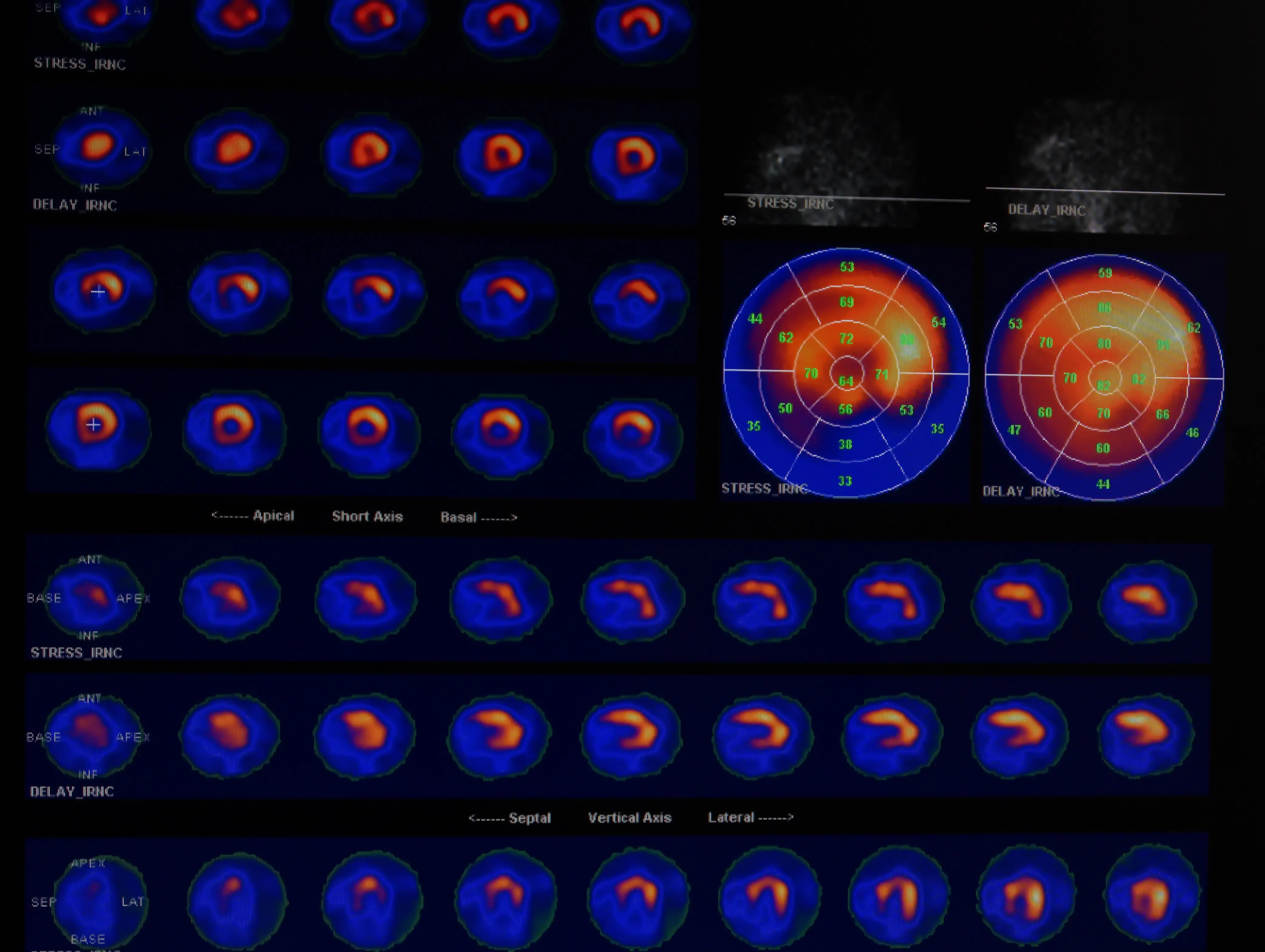
SPECT technology relies on radiopharmaceuticals that emit gamma rays. Once injected, these tracers localize in the heart muscle in proportion to blood flow. A SPECT scanner, equipped with gamma cameras, rotates around the patient, capturing multiple two-dimensional images from different angles. The technological marvel lies in the subsequent reconstruction process. Sophisticated algorithms process these planar projections to create a three-dimensional (3D) image of the heart. Key technological components of a SPECT system include highly sensitive scintillation detectors (often made of sodium iodide crystals) that convert gamma photons into light, and photomultiplier tubes that amplify these light signals into electrical impulses. Collimators—lead-based septa—are critical for directing gamma rays to the detectors, ensuring spatial resolution and image clarity by filtering out scattered photons. The evolution of SPECT has seen the introduction of solid-state cadmium zinc telluride (CZT) detectors, which offer superior energy resolution, faster acquisition times, and lower radiation doses compared to traditional Anger cameras.
Positron Emission Tomography (PET)
PET represents an even more advanced imaging technique, utilizing radiopharmaceuticals that emit positrons. When a positron encounters an electron in the body, they annihilate each other, producing two gamma rays that travel in opposite directions (at nearly 180 degrees). PET scanners are designed to detect these coincident events. The precise timing and spatial detection of these paired gamma rays allow highly accurate localization of the annihilation event, forming the basis for constructing detailed 3D images. PET tracers are typically cyclotron-produced radionuclides with very short half-lives, such as Fluorine-18 (F-18), Rubidium-82 (Rb-82), or Nitrogen-13 (N-13). The technological complexity of PET extends to the need for on-site or nearby cyclotrons for tracer production, and intricate detector systems (often using LSO or LYSO crystals) coupled with advanced electronics capable of picosecond timing resolution to accurately map the tracer distribution. This superior spatial and temporal resolution often makes PET the gold standard for quantifying myocardial blood flow and viability.
Radiopharmaceuticals: The Tracer Technology
The efficacy of nuclear cardiology fundamentally hinges on the design and production of radiopharmaceuticals. These aren’t just radioactive materials; they are molecular tools engineered to target specific biological pathways or structures within the heart. For SPECT, common tracers like Thallium-201 or Technetium-99m labeled compounds (e.g., sestamibi, tetrofosmin) are chosen for their ability to be taken up by viable myocardial cells in proportion to blood flow. PET tracers, such as F-18 fluorodeoxyglucose (FDG) for metabolic imaging or Rb-82 for perfusion, demonstrate higher sensitivity and allow for absolute quantification of physiological parameters. The development of new radiopharmaceuticals involves sophisticated radiochemistry, often leveraging automated synthesis modules, to ensure purity, efficacy, and safety, representing a continuous frontier in nuclear medicine technology.
The Hardware Evolution: From Detectors to Hybrid Systems
The physical hardware of nuclear cardiology scanners has undergone significant evolution, driven by the relentless pursuit of improved image quality, reduced scan times, and lower radiation exposure for patients.
Advancements in Detector Technology
Early gamma cameras relied on large, single sodium iodide (NaI) crystals. While effective, these designs had inherent limitations in terms of spatial resolution and count rate capability. Modern SPECT systems increasingly incorporate solid-state detectors, such as Cadmium Zinc Telluride (CZT). CZT detectors offer several key advantages: they directly convert gamma rays into electrical signals, eliminating the need for photomultiplier tubes and leading to higher energy resolution, better signal-to-noise ratio, and greater stability. This allows for smaller, more flexible detector heads, enabling closer patient positioning for improved image quality, and facilitating faster scans with lower administered doses. For PET, the move from Bismuth Germanate (BGO) crystals to Lutetium Oxyorthosilicate (LSO) and Lutetium Yttrium Oxyorthosilicate (LYSO) has dramatically improved light output and decay time, crucial for enhancing coincidence detection efficiency and overall image resolution.
The Rise of Hybrid Imaging: PET/CT and SPECT/CT
Perhaps the most transformative hardware innovation has been the integration of nuclear medicine scanners with anatomical imaging modalities, giving rise to hybrid systems like PET/CT and SPECT/CT. These systems combine the functional information from nuclear scans with the detailed anatomical context provided by a Computed Tomography (CT) scan. Technologically, this means housing two distinct imaging gantries within a single unit, controlled by a unified operating system. The CT component serves multiple crucial roles: it provides precise anatomical localization of tracer uptake, crucial for accurately interpreting the functional images; it’s used for attenuation correction, mathematically compensating for the absorption of photons by different tissues (bone, soft tissue) to improve the quantitative accuracy of the nuclear scan; and it can even be used for coronary artery calcium scoring or CT angiography in the same sitting. This technological synergy yields a more comprehensive diagnostic picture, allowing clinicians to correlate areas of functional impairment with specific anatomical structures, thereby enhancing diagnostic confidence and patient management.
Software and Data Science: Unlocking Diagnostic Precision
While the hardware captures the raw data, it is the sophisticated software and data science pipelines that transform this information into actionable diagnostic insights. The computational demands of nuclear cardiology are substantial, requiring advanced algorithms for image reconstruction, correction, and quantitative analysis.
Image Reconstruction and Processing Algorithms
The journey from raw detector counts to a 3D image is a complex computational one. Iterative reconstruction algorithms, such as Ordered Subset Expectation Maximization (OSEM), have largely superseded older filtered back-projection methods. These algorithms repeatedly model the physical process of photon emission and detection, comparing predicted data with actual measured data, and iteratively refining the image until a statistically optimal reconstruction is achieved. This results in images with reduced noise, improved contrast, and fewer artifacts. Furthermore, sophisticated software modules are employed for motion correction, compensating for patient movement during scans to prevent blurring, and for attenuation correction, which is critical for accurate quantification of myocardial perfusion.
Quantitative Analysis and Decision Support Systems
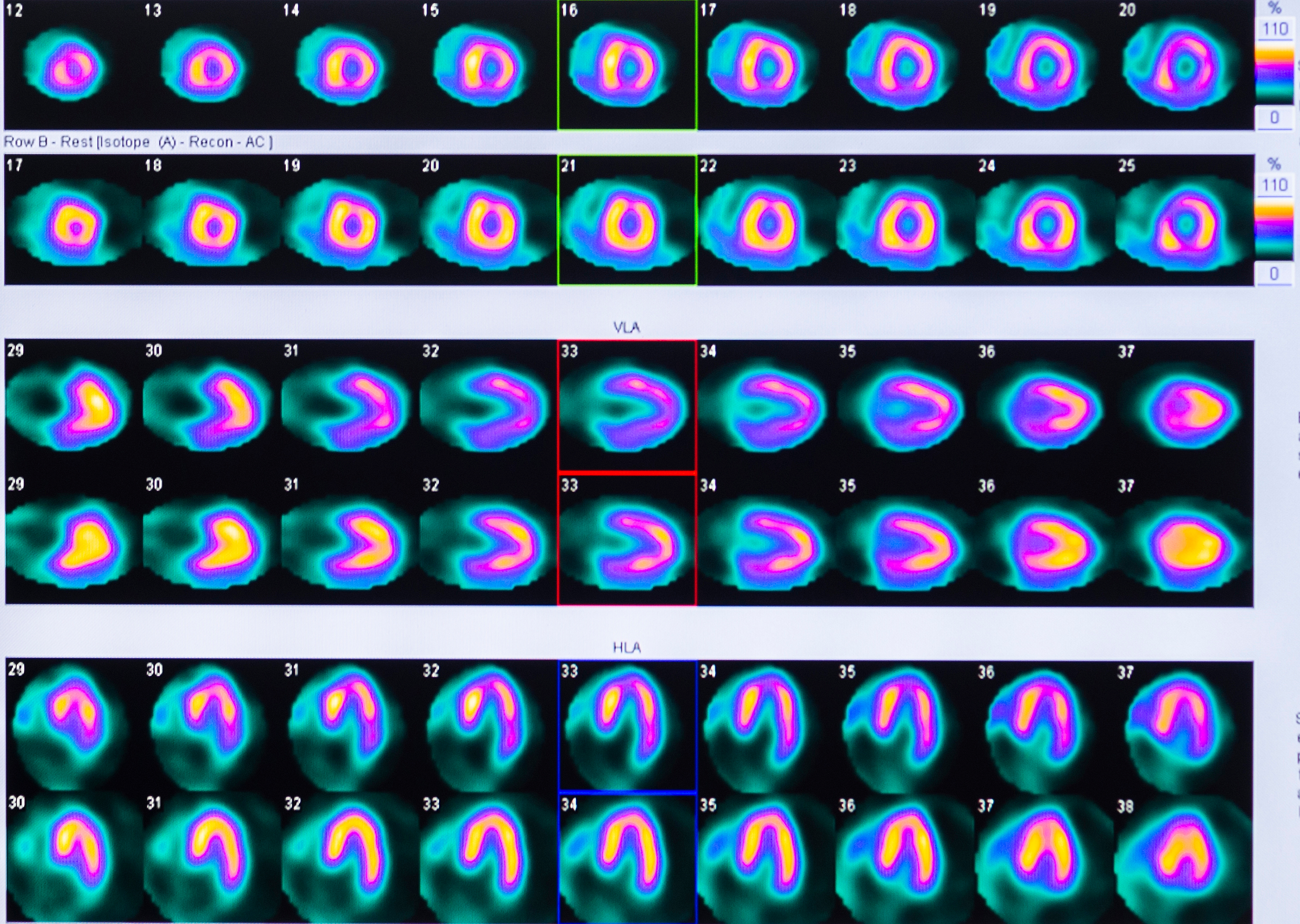
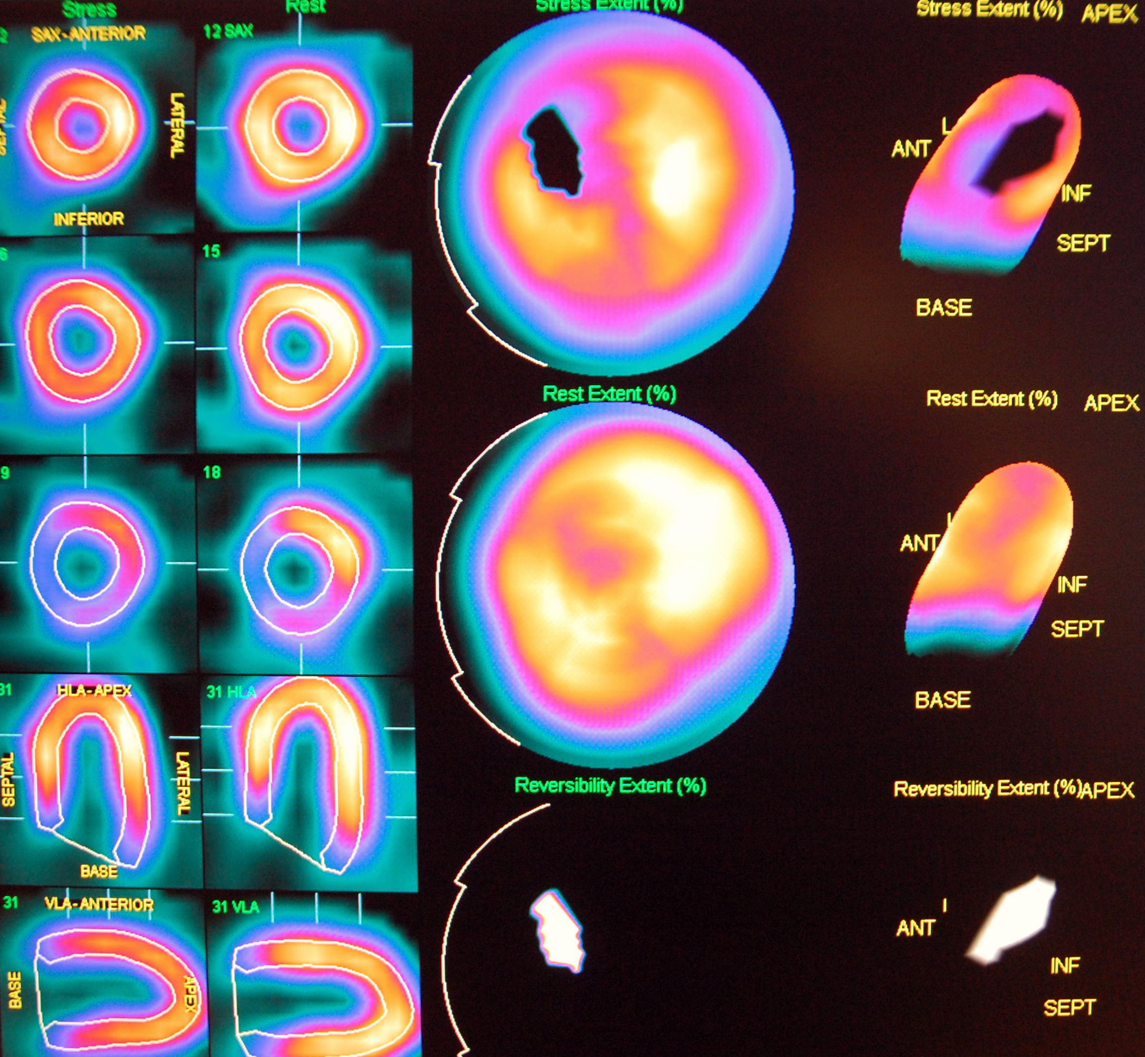
Beyond qualitative visual interpretation, modern nuclear cardiology heavily relies on quantitative analysis software. These tools automatically segment the heart, define regions of interest, and quantify parameters such as myocardial blood flow (MBF), myocardial flow reserve (MFR), and left ventricular ejection fraction (LVEF). Software packages can generate polar maps (bull’s-eye plots) that visually represent tracer distribution throughout the left ventricle, allowing for easy identification of perfusion defects and their severity. Decision support systems, integrated within these software platforms, can aid clinicians in interpreting complex data by providing standardized measurements and comparisons against normative databases, thereby enhancing reproducibility and consistency in diagnosis.
Digital Infrastructure and Data Management
The vast amount of image data generated by nuclear cardiology procedures necessitates robust digital infrastructure. Picture Archiving and Communication Systems (PACS) are central to storing, retrieving, and transmitting these images securely. Integration with Hospital Information Systems (HIS) and Radiology Information Systems (RIS) ensures a seamless workflow from patient scheduling to reporting. Secure cloud-based solutions are also emerging, offering scalable storage and remote access, which are crucial for multi-center studies, telemedicine applications, and facilitating collaborative diagnostic efforts. Data integrity, security, and privacy are paramount, leading to the implementation of advanced encryption and access control technologies.
Artificial Intelligence and Machine Learning: Revolutionizing Cardiac Diagnostics
The integration of Artificial Intelligence (AI) and Machine Learning (ML) is rapidly transforming nuclear cardiology, promising to enhance diagnostic accuracy, streamline workflows, and personalize patient care. AI’s capacity for pattern recognition and data analysis makes it uniquely suited to address some of the field’s long-standing challenges.
AI for Image Enhancement and Noise Reduction
One significant application of AI is in improving image quality. Deep learning algorithms can be trained on large datasets of nuclear cardiology images to denoise scans, reduce artifacts, and effectively reconstruct high-quality images from lower count rates or shorter acquisition times. This can potentially lead to significantly lower radiation doses for patients without compromising diagnostic utility, or enable faster scan protocols. AI models are also being developed to automatically optimize image acquisition parameters, ensuring consistent image quality across different patients and scanners.
Automated Quantification and Predictive Analytics
AI excels at automating repetitive and complex tasks. In nuclear cardiology, this includes automated segmentation of the left ventricle, quantification of perfusion defects, and calculation of ejection fraction. Beyond simple quantification, ML models are being developed for predictive analytics. By analyzing a multitude of features from nuclear scans, clinical data, and patient demographics, AI can help predict major adverse cardiac events, identify patients at high risk for certain conditions, or even predict a patient’s response to specific therapies. This moves the field towards more proactive and personalized medicine.
Streamlining Workflows and Personalized Medicine
AI tools can assist in various aspects of the workflow, from automated quality control of images to prioritizing studies for interpretation based on severity, thereby reducing the burden on clinicians and improving efficiency. Furthermore, by integrating data from nuclear scans with genetic information, electronic health records, and other ‘-omics’ data, AI can contribute to truly personalized medicine. It can help identify subtle patterns indicative of specific patient subgroups that may benefit most from particular interventions, tailoring treatment strategies based on an individual’s unique biological and physiological profile.
The Future Landscape: Innovation on the Horizon
The trajectory of nuclear cardiology technology points towards continued innovation, driven by the desire for greater precision, safety, and integration within the broader healthcare ecosystem.
Novel Tracers and Targeted Therapies
Research into new radiopharmaceuticals is ongoing, focusing on developing tracers that target specific molecular pathways involved in various cardiac diseases, beyond just perfusion and metabolism. This includes tracers for imaging inflammation, fibrosis, angiogenesis, and specific receptor expression. Such advancements promise to provide even more specific diagnostic information and pave the way for theranostics—the combination of diagnostic imaging with targeted radionuclide therapy—for cardiac conditions.
Ultra-Low Dose and Ultrafast Imaging Protocols
With advancements in detector technology, reconstruction algorithms, and AI, the trend towards ultra-low dose and ultrafast imaging protocols will continue. The goal is to minimize patient radiation exposure while maintaining or even improving diagnostic accuracy. This could involve dynamic imaging, where a series of rapid scans provide real-time information about tracer kinetics, offering deeper physiological insights.
Integrating Multi-Modal Data for Holistic Views
The future will see even greater integration of nuclear cardiology data with other imaging modalities (e.g., MRI, ultrasound) and clinical data. Advanced data fusion techniques and AI platforms will be crucial in synthesizing this disparate information into a holistic, comprehensive view of cardiac health, moving beyond isolated findings to a more integrated, predictive model of disease progression and patient management. This technological synergy aims to empower clinicians with unparalleled insights, ultimately enhancing diagnostic confidence and improving patient outcomes in the realm of cardiovascular health.
aViewFromTheCave is a participant in the Amazon Services LLC Associates Program, an affiliate advertising program designed to provide a means for sites to earn advertising fees by advertising and linking to Amazon.com. Amazon, the Amazon logo, AmazonSupply, and the AmazonSupply logo are trademarks of Amazon.com, Inc. or its affiliates. As an Amazon Associate we earn affiliate commissions from qualifying purchases.