
Dacryocystorhinostomy (DCR) is a specialized surgical procedure designed to restore the drainage of tears from the surface of the eye into the nose. When the nasolacrimal duct—the passageway responsible for carrying tears away from the eye—becomes obstructed, patients often experience chronic tearing, recurrent infections, and significant discomfort. While the term may sound intimidating to those unfamiliar with medical terminology, DCR is a refined intervention that effectively bridges the gap between the lacrimal sac and the nasal cavity. In the modern surgical landscape, DCR has evolved from an external, scar-prone incision method to sophisticated endoscopic techniques, making it a critical focus for those investigating the intersection of clinical precision and patient quality of life.

Understanding the Anatomy and Pathology of Lacrimal Obstruction
To grasp the necessity of a DCR procedure, one must first understand the lacrimal system. Tears are produced by the lacrimal glands and serve to lubricate, protect, and nourish the surface of the eye. Under normal physiological conditions, excess tears drain through small openings at the inner corners of the eyelids called puncta. From there, they travel through the canaliculi into the lacrimal sac, eventually exiting through the nasolacrimal duct into the nasal cavity.
The Mechanism of Epiphora
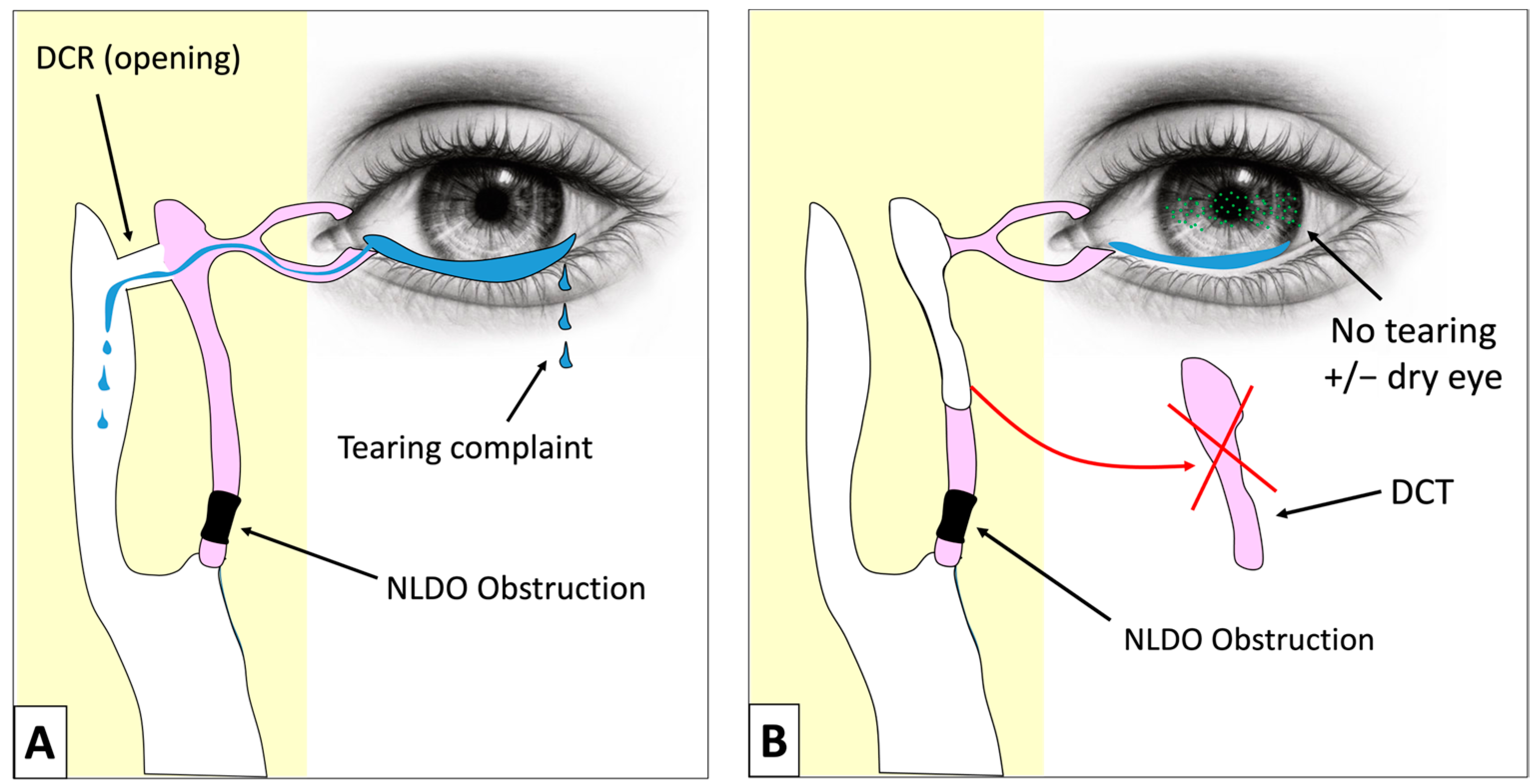
When this system experiences a blockage, a condition known as epiphora occurs. Epiphora is the constant overflow of tears onto the cheek, which can lead to blurred vision, skin irritation, and social embarrassment. Obstructions may result from congenital issues, aging, previous trauma, or chronic inflammation. If the blockage persists, the lacrimal sac can become a reservoir for stagnant fluid, creating an environment ripe for dacryocystitis—a painful infection characterized by redness, swelling, and purulent discharge.
Why Conservative Treatments Fail
In many cases, initial management of lacrimal obstruction involves conservative measures such as warm compresses, systemic antibiotics for acute infections, or lacrimal irrigation to attempt to clear the blockage. However, these methods are frequently temporary. When the anatomical obstruction is permanent or structural—often caused by narrow ducts or fibrotic changes—surgical intervention becomes the gold standard for resolution. Dacryocystorhinostomy serves to bypass the faulty duct entirely, creating a new, direct pathway for tear drainage.
The Evolution of Surgical Technique: External vs. Endoscopic DCR
Historically, the standard approach to this procedure was the external DCR. This involved a small skin incision on the side of the nose, through which the surgeon would expose the lacrimal sac and create an opening into the nasal bone. While effective, the external approach required careful suturing to prevent visible scarring and often involved a longer recovery period.
Endonasal DCR: The Modern Standard
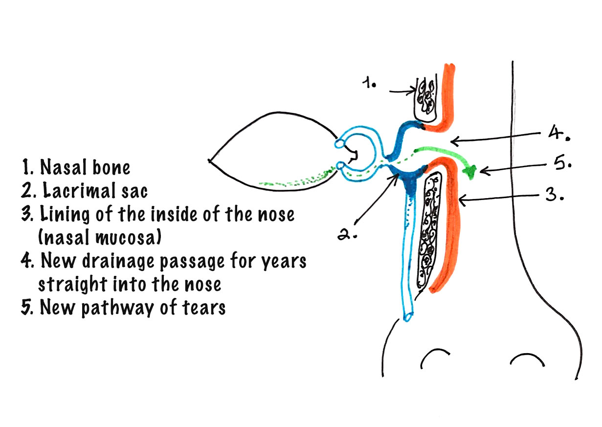
In the past two decades, the field of ophthalmology and rhinology has shifted significantly toward Endonasal Dacryocystorhinostomy (EnDCR). This minimally invasive technique utilizes high-definition endoscopes passed through the nostrils to perform the procedure from the inside. By visualizing the nasal cavity directly, surgeons can remove the bone overlying the lacrimal sac and create an ostium—a new drainage window—without making a single incision on the patient’s face.
Advantages of the Endoscopic Approach
The move toward endoscopic techniques represents a major leap forward in patient care. The primary benefits include the total absence of external scarring, reduced trauma to the surrounding tissues, and a significantly faster recovery time. Because the procedure is performed endonasally, there is less risk of damaging the medial canthal ligament, which is responsible for the anatomical stability of the eyelid. Furthermore, the ability to utilize advanced visualization tools allows surgeons to address concurrent nasal pathologies, such as a deviated septum or enlarged turbinates, which might otherwise impede successful drainage.
The Surgical Process and Post-Operative Recovery
Preparation for a DCR procedure typically involves a comprehensive evaluation to ensure that the tear drainage issue is indeed caused by a nasolacrimal duct obstruction rather than ocular surface disease or eyelid malposition. Once the diagnosis is confirmed, the patient undergoes the procedure under either local anesthesia with sedation or general anesthesia, depending on individual preference and surgical complexity.
Intraoperative Precision
During the procedure, the surgeon identifies the area of the nasal wall adjacent to the lacrimal sac. Using specialized micro-instruments, a small window of bone is removed. The wall of the lacrimal sac is then incised and sutured—or simply opened—to connect it directly to the nasal mucosa. In some cases, a small silicone stent may be temporarily placed in the new drainage channel to ensure it remains open during the initial healing phase. This stent is typically removed in the office after a few weeks.
Managing Expectations and Recovery
Post-operative care is relatively straightforward, though it requires strict adherence to surgeon guidelines to ensure the new opening heals correctly. Patients are often prescribed a combination of saline nasal rinses to keep the nasal cavity clean, as well as antibiotic or steroid eye drops to manage inflammation. While minor bruising or nasal congestion is expected in the first few days, most patients report a significant reduction in tearing within a short period. The long-term success rates for endoscopic DCR are impressively high, often exceeding 90% in primary cases, providing lasting relief to those who previously struggled with chronic tearing.
Assessing Long-Term Outcomes and Success Factors
The success of a dacryocystorhinostomy is measured not only by the immediate cessation of tearing but also by the long-term patency of the created drainage pathway. While the procedure is highly effective, several factors contribute to the ultimate outcome, including the patient’s baseline anatomy, the presence of systemic inflammatory conditions, and the surgeon’s expertise in endoscopic nasal surgery.
Factors Influencing Success
In complex cases—such as those involving extensive scar tissue from previous trauma or systemic autoimmune diseases—the body may attempt to close the new drainage window during the healing process. In these instances, surgeons may use mitomycin C, an anti-fibrotic agent, to reduce the likelihood of scar tissue formation at the site of the new opening. Additionally, careful follow-up is essential to monitor for the formation of “granulation tissue,” which can occasionally obstruct the new ostium.
Quality of Life Improvements
For the patient, the successful completion of a DCR procedure offers more than just the elimination of physical discomfort. Epiphora often affects a person’s daily life, impacting activities such as reading, driving, and even simple social interactions. The constant need to wipe the eyes can become a significant source of frustration. By restoring the natural flow of tears, DCR allows patients to resume these activities without the distraction and discomfort of blurred, watery vision.
The procedure stands as a testament to the advancements in modern medical technology, where the marriage of fiber-optic imaging and microsurgical techniques has transformed an invasive, scar-prone operation into a seamless, high-precision intervention. As the population continues to age, the demand for effective management of lacrimal obstruction will likely grow, further cementing Dacryocystorhinostomy as an essential and life-improving component of ophthalmic practice. Whether addressing a simple blockage or a more complex case of dacryocystitis, the refined approach to DCR continues to prioritize functional success and aesthetic preservation, setting the gold standard for restoring the delicate balance of the ocular surface.
aViewFromTheCave is a participant in the Amazon Services LLC Associates Program, an affiliate advertising program designed to provide a means for sites to earn advertising fees by advertising and linking to Amazon.com. Amazon, the Amazon logo, AmazonSupply, and the AmazonSupply logo are trademarks of Amazon.com, Inc. or its affiliates. As an Amazon Associate we earn affiliate commissions from qualifying purchases.