The term “buffy coat” may sound like something pulled from the wardrobe department of a period drama, but in the clinical laboratory, it represents a pivotal intersection of hematology, diagnostic technology, and biotechnology. As laboratory science evolves toward faster, more precise, and highly automated workflows, understanding the mechanics of blood fractionation has become essential for anyone involved in the diagnostic technology ecosystem. The buffy coat is not just a biological byproduct; it is a high-value concentrated source of material that fuels modern medical research and diagnostic innovation.
The Science of Fractionation: How Technology Separates Blood
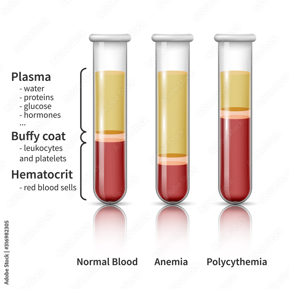
At its most fundamental level, the buffy coat is the thin, light-colored layer of white blood cells (leukocytes) and platelets that forms when anticoagulant-treated blood is centrifuged. When a blood sample is placed in a high-speed centrifuge, the varying densities of blood components cause them to separate into distinct layers.
The Layers of Centrifugation
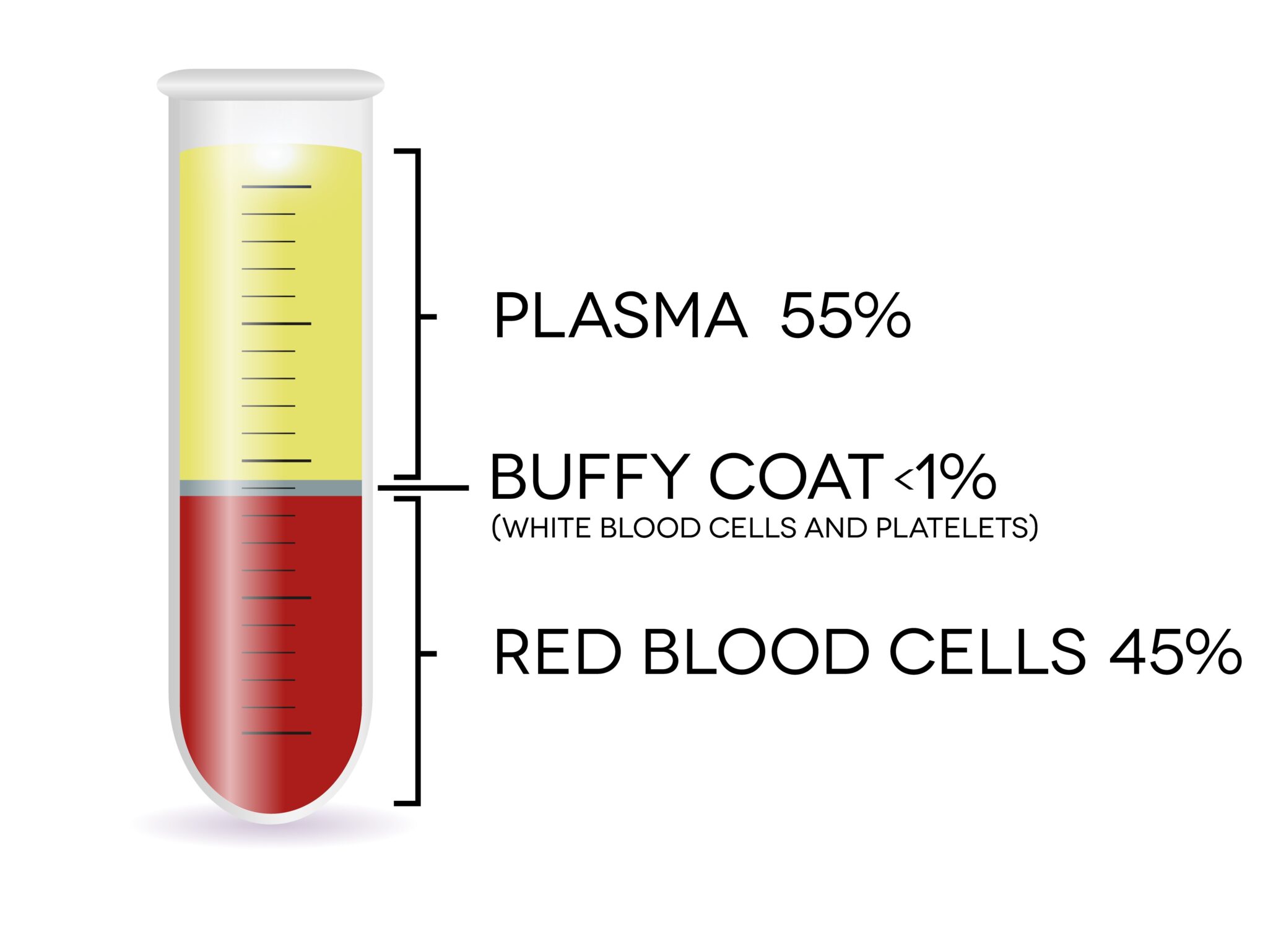
Under high centrifugal force, blood settles into three primary strata:
- Red Blood Cells (Erythrocytes): The heaviest component, these sink to the bottom of the tube, representing approximately 45% of the total volume.
- Plasma: The lightest component, this straw-colored liquid remains at the top, housing proteins, electrolytes, and clotting factors.
- The Buffy Coat: Sandwiched between the dense erythrocytes and the plasma, this thin, grayish-white layer contains the vast majority of the body’s white blood cells and platelets.
Automation in Laboratory Diagnostics
In modern clinical settings, the manual creation of a buffy coat is increasingly being replaced by automated diagnostic platforms. These systems utilize microfluidic chips and automated centrifugal analyzers to isolate the buffy coat with minimal human intervention. This shift is a significant trend in lab technology, as it reduces the risk of sample contamination and improves the reproducibility of results. By integrating sensors into the centrifugal housing, these machines can detect the precise boundary of the buffy coat layer, allowing for the automated aspiration of specific cell types for downstream analysis.
The Buffy Coat as a Diagnostic Goldmine
The importance of the buffy coat in diagnostic technology cannot be overstated. By concentrating white blood cells and platelets into a small volume, the buffy coat serves as a “purified” sample that simplifies downstream diagnostic applications. Instead of processing liters of whole blood, technicians can work with a concentrated volume where the target analytes—such as pathogens, rare cells, or genetic material—are significantly more accessible.
Molecular Diagnostics and DNA Extraction
One of the most common tech-driven applications of the buffy coat is in DNA extraction. Because red blood cells lack a nucleus, they provide no genetic information. Conversely, the white blood cells concentrated in the buffy coat are rich in genomic DNA. Genomic sequencing platforms, which require high-quality, high-yield DNA, rely on the buffy coat to provide the “input” for next-generation sequencing (NGS). By using automated robotic liquid handlers, laboratories can process multiple buffy coat samples simultaneously, drastically increasing throughput for population-wide genetic screening and personalized medicine projects.
Flow Cytometry and Cell Sorting
Flow cytometry is a cornerstone of modern diagnostic hardware. This technology involves passing cells in a fluid stream through an electronic detection apparatus. The buffy coat is the preferred sample type for these machines because it is essentially a “pre-enriched” population of leukocytes. This allows the flow cytometer to operate more efficiently, as the machine spends less time processing irrelevant red blood cells and can instead focus on identifying subtle cell markers associated with cancers like leukemia or lymphoma.

Biotechnological Applications and Future Trends
Beyond standard clinical diagnostics, the buffy coat has become a focal point for biotechnology firms developing new therapeutics. The ability to isolate specific cell populations—such as T-cells or stem cells—has opened the door to advanced therapies that were previously considered science fiction.
CAR-T Cell Therapy and Cell Engineering
Chimeric Antigen Receptor (CAR) T-cell therapy is a revolutionary approach to cancer treatment, and it relies heavily on the buffy coat. The process begins with leukapheresis, a procedure that collects the buffy coat from a patient’s blood. These collected T-cells are then genetically modified in a sterile manufacturing environment to better recognize and kill cancer cells. The tech industry has responded to this need by developing closed-system, automated bioreactors that handle these cells within the buffy coat without exposing them to the external environment. This ensures the integrity of the genetic modification process while scaling up production for clinical use.
The Rise of Point-of-Care (POC) Devices
One of the most exciting trends in diagnostic technology is the transition from massive, centralized laboratory equipment to palm-sized Point-of-Care (POC) devices. Engineers are now designing “lab-on-a-chip” systems that perform the separation of the buffy coat within a disposable cartridge. These devices utilize micro-channels and centrifugal forces induced by small, high-speed motors to isolate the buffy coat in minutes rather than hours. This is a game-changer for emergency medicine and resource-limited settings, where the rapid analysis of white blood cell counts can determine immediate intervention strategies for sepsis or acute infection.
Challenges and Future Technological Integration
While the buffy coat is a critical resource, the technology used to manage it faces ongoing challenges regarding standardization and data integration.
Standardization in a Digital Ecosystem
One of the primary hurdles in utilizing the buffy coat for AI-driven diagnostics is the lack of standardized collection protocols. Centrifugation speeds, durations, and tube geometries vary across laboratories, leading to inconsistent yields of cellular material. To address this, software developers are creating LIMS (Laboratory Information Management Systems) that integrate directly with centrifuge hardware to log environmental and physical parameters for every sample. This creates a data-rich trail, ensuring that the “quality” of a buffy coat sample is quantifiable and traceable, which is essential for AI algorithms trained on biological data.
AI and Image Recognition
The intersection of the buffy coat and AI is particularly promising in the realm of computer vision. High-resolution cameras installed within automated smear-making machines now take images of the buffy coat layers. AI models analyze these images to identify morphological abnormalities in the white blood cells before a human pathologist even glances at the slide. By digitizing the morphology of cells within the buffy coat, clinicians can achieve faster turn-around times for diagnosis, potentially flagging rare blood disorders or viral infections hours, or even days, faster than traditional manual review methods.
Sustainable Biobanking
As the volume of genetic and clinical research grows, so does the need for sustainable storage of biological samples. Automated cryo-storage systems, which manage the long-term preservation of buffy coat samples, represent a significant sector of the biotech infrastructure market. These systems use barcode-scanning robotics to retrieve specific samples from deep-freeze environments (-80°C), minimizing the risk of “thaw-cycles” that could degrade the cellular material. This level of technological precision ensures that the buffy coat remains a viable resource for longitudinal studies and future scientific discovery.
The buffy coat stands as a testament to how traditional hematology and modern diagnostic technology are inextricably linked. From the spinning centrifuge to the AI-driven molecular analyzer, the evolution of how we handle this small but potent layer of cells reflects the broader trajectory of medical technology: moving toward greater precision, higher levels of automation, and a deeper integration of digital data into the life sciences. As diagnostic platforms continue to shrink in size and expand in capability, the buffy coat will remain at the heart of our efforts to diagnose, treat, and understand human disease.
aViewFromTheCave is a participant in the Amazon Services LLC Associates Program, an affiliate advertising program designed to provide a means for sites to earn advertising fees by advertising and linking to Amazon.com. Amazon, the Amazon logo, AmazonSupply, and the AmazonSupply logo are trademarks of Amazon.com, Inc. or its affiliates. As an Amazon Associate we earn affiliate commissions from qualifying purchases.