
In today’s rapidly evolving technological landscape, understanding the tools and systems that underpin various industries is becoming increasingly crucial. While our focus on the website often centers on the latest tech trends, AI advancements, and digital security, it’s important to acknowledge that technology’s impact extends far beyond our screens and personal devices. One area where cutting-edge technology plays a vital role, with profound implications for human well-being, is medical imaging.
When it comes to diagnosing and understanding complex medical conditions, healthcare professionals rely on sophisticated imaging techniques. Among these, Magnetic Resonance Imaging (MRI) and Positron Emission Tomography (PET) scans are two prominent technologies, each offering a unique window into the human body. While both are powerful diagnostic tools, their underlying principles, the type of information they provide, and their applications differ significantly. This article aims to demystify these differences, exploring the technological foundations of MRI and PET scans, and highlighting why understanding these distinctions is as important as keeping up with the latest AI breakthroughs.
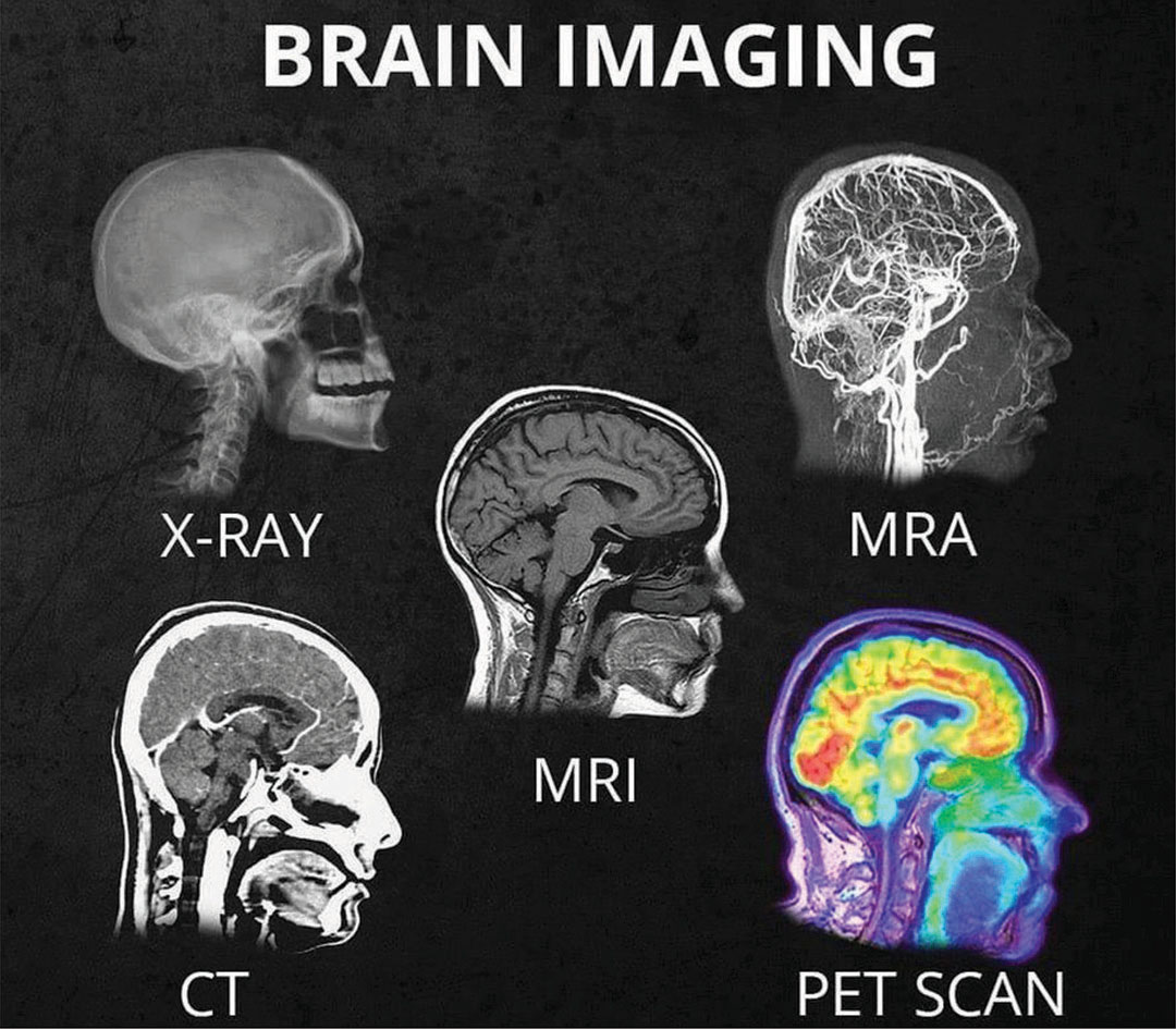
The Technological Foundation: How MRI and PET Scans Work
At their core, both MRI and PET scans are advanced imaging modalities that allow doctors to visualize internal body structures. However, the fundamental physics and the type of data they acquire are vastly different. This technological divergence dictates their respective strengths and weaknesses.
Magnetic Resonance Imaging (MRI): Leveraging Magnetism and Radio Waves
MRI technology is a marvel of applied physics, utilizing powerful magnetic fields and radio waves to generate highly detailed images of soft tissues, organs, and bones. The process starts with a strong magnetic field that aligns the protons within the water molecules of your body. Think of these protons as tiny magnets themselves. When you’re placed inside the MRI scanner, which is essentially a large, tube-shaped magnet, these protons naturally line up with the magnetic field.
Next, the MRI machine emits short bursts of radio waves. These radio waves knock the aligned protons out of their alignment. When the radio waves are turned off, the protons realign themselves with the magnetic field. As they realign, they release energy in the form of radio signals. The MRI scanner’s coils detect these signals, and a computer processes them to create cross-sectional images of the body. The strength and timing of these signals vary depending on the type of tissue the protons are in, allowing for differentiation between fat, water, muscle, and other structures.
The beauty of MRI lies in its ability to provide exquisite detail of anatomical structures. It excels at visualizing:
- Soft Tissues: This is where MRI truly shines. It can differentiate between various types of soft tissues with remarkable clarity, making it invaluable for imaging the brain, spinal cord, muscles, ligaments, tendons, and internal organs.
- Brain and Nervous System: MRI is the gold standard for detecting and characterizing tumors, strokes, multiple sclerosis, and other neurological conditions. Its ability to see subtle changes in brain tissue is unparalleled.
- Musculoskeletal System: It’s extensively used to diagnose injuries to joints, ligaments, tendons, and cartilage, providing crucial information for treatment planning.
- Abdominal and Pelvic Organs: MRI can offer detailed views of organs like the liver, kidneys, pancreas, and reproductive organs.
Crucially, MRI does not use ionizing radiation, making it a safe option for repeated imaging and for pregnant women (though precautions are still taken). The magnetic field strength and the precision of radio wave pulses are the core technological elements that enable its diagnostic power.
Positron Emission Tomography (PET): Visualizing Metabolic Activity
In contrast to MRI’s focus on anatomy, PET scans provide insights into the function and metabolic activity of cells and organs. PET technology works by tracking a radioactive tracer that is introduced into the body, typically injected into a vein. This tracer is usually a glucose molecule that has been tagged with a short-lived radioactive isotope.
Once injected, the tracer travels through the bloodstream and is absorbed by cells. Cells that are highly metabolically active, such as cancer cells or areas of active inflammation, will absorb more of the tracer. The radioactive isotope within the tracer emits positrons, which are tiny particles. When a positron encounters an electron in the body, they annihilate each other, producing two gamma rays that travel in opposite directions.
The PET scanner is equipped with detectors that capture these gamma rays. By analyzing the timing and direction of the detected gamma rays, the computer can reconstruct a three-dimensional image showing where the radioactive tracer has accumulated in the body. This accumulation directly correlates with areas of high metabolic activity.
The primary applications of PET scans include:
- Cancer Detection and Staging: PET is exceptionally useful for identifying cancerous tumors, determining their spread (staging), and assessing how well a tumor is responding to treatment. Cancer cells often have a higher metabolic rate than normal cells and thus take up more of the glucose-based tracer.
- Neurology: PET can help diagnose conditions like Alzheimer’s disease, Parkinson’s disease, and epilepsy by revealing patterns of altered brain metabolism. It can show areas of reduced activity in neurodegenerative diseases or areas of overactivity in epilepsy.
- Cardiology: PET can assess blood flow to the heart muscle, helping to identify areas of damage or reduced oxygen supply.
The key technological elements of PET are the production and administration of a radiotracer, the detection of gamma rays, and sophisticated reconstruction algorithms to create functional images. While PET scans do involve a small amount of radiation from the tracer, it is typically short-lived and considered safe.
Key Differences: Anatomy vs. Function
The fundamental distinction between MRI and PET scans can be distilled into their primary purpose: MRI images structure, while PET images function. This core difference leads to a cascade of other distinctions:
Information Provided
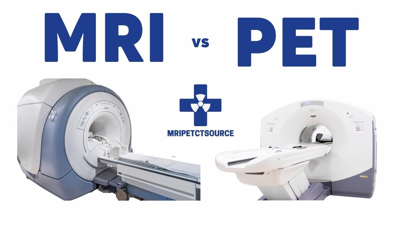
- MRI: Provides detailed anatomical images. It shows the size, shape, and position of organs and tissues, and can highlight structural abnormalities like tumors, cysts, inflammation, or tears. It’s like looking at a high-resolution blueprint of your body.
- PET: Provides metabolic or functional information. It reveals how actively cells are using energy (typically glucose). This allows for the detection of abnormal cellular activity, such as rapidly dividing cancer cells, before structural changes might be evident on other imaging. It’s like observing the energy consumption patterns within your body.
Imaging Focus
- MRI: Excels in visualizing soft tissues with excellent contrast. It’s particularly strong for the brain, spinal cord, muscles, and ligaments.
- PET: Focuses on areas of high metabolic activity. While it can show organs, its primary value is in detecting areas where cells are working harder or abnormally.
Radiation Exposure
- MRI: Uses magnetic fields and radio waves, and therefore does not involve ionizing radiation. This makes it a safer choice for patients requiring frequent imaging or those who are pregnant.
- PET: Uses a small amount of radioactive tracer, which emits ionizing radiation. While generally safe due to the short half-life of the isotopes, it is a consideration for certain patient populations or when repeated scans are necessary.
Procedure and Preparation
- MRI: Typically involves lying still on a table that slides into a large, tube-shaped scanner. The process can take anywhere from 15 minutes to over an hour, depending on the area being scanned. Patients may be asked to hold their breath at certain points. Some patients experience claustrophobia due to the confined space.
- PET: Requires an injection of the radioactive tracer, followed by a waiting period (usually 30-60 minutes) for the tracer to distribute throughout the body. The scanning process itself is usually shorter than an MRI, often lasting 20-40 minutes. Patients may be asked to fast before the scan.
Cost and Accessibility
While both are advanced imaging techniques, there can be variations in cost and availability. The complexity of the technology and the specialized personnel required for both MRI and PET scans generally make them more expensive than simpler imaging methods like X-rays or ultrasounds. However, the specific costs can vary significantly by region and healthcare provider.
When is an MRI Used? When is a PET Scan Used? Applications in Diagnosis and Treatment
The choice between an MRI and a PET scan, or even a combination of both, depends entirely on the suspected medical condition and the type of information needed for diagnosis and treatment.
MRI: Illuminating Structural Details
When a doctor suspects a problem with the physical structure of organs or tissues, MRI is often the preferred modality. For instance:
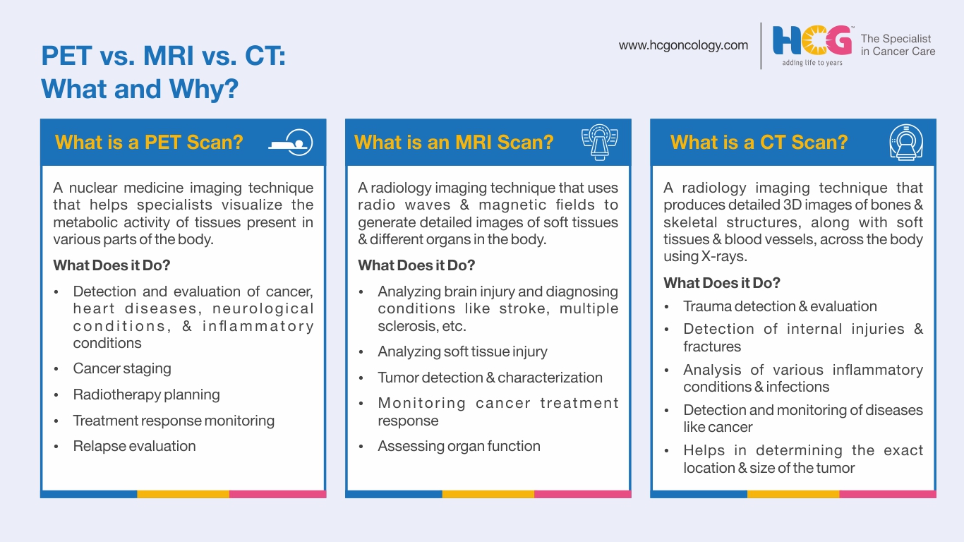
- Neurological Disorders: To diagnose brain tumors, assess the extent of a stroke, identify lesions in multiple sclerosis, or evaluate spinal cord injuries.
- Musculoskeletal Injuries: To pinpoint tears in ligaments or tendons, diagnose cartilage damage, or evaluate bone marrow abnormalities.
- Cancer Diagnosis (Initial): While PET is crucial for metabolic assessment, MRI can sometimes identify tumors by their structural characteristics, especially in organs like the breast, prostate, or liver, and help determine their size and invasion into surrounding tissues.
- Abdominal and Pelvic Issues: To examine conditions affecting the liver, kidneys, uterus, ovaries, or prostate.
PET: Revealing Metabolic Activity and Disease Progression
PET scans are invaluable when the focus is on cellular activity, disease progression, or treatment response. Common applications include:
- Oncology: This is where PET truly excels. It can detect cancer recurrence, stage cancers, differentiate between scar tissue and active tumor after treatment, and assess the effectiveness of chemotherapy or radiation therapy by observing changes in tumor metabolism.
- Neurology: To identify specific patterns of brain metabolism indicative of Alzheimer’s disease, evaluate the cause of seizures, or study brain function in research settings.
- Cardiology: To assess myocardial viability (whether heart muscle can be salvaged after a heart attack) or diagnose coronary artery disease.
- Infection and Inflammation: PET can sometimes be used to locate areas of infection or inflammation that may not be readily apparent with other imaging techniques.
The Power of Hybrid Imaging: PET-MRI
It’s also worth noting the emergence of hybrid imaging technologies, such as PET-MRI scanners. These combine the strengths of both modalities, allowing for simultaneous acquisition of detailed anatomical (MRI) and functional (PET) information in a single session. This can provide a more comprehensive picture, especially in complex cases like advanced cancer or neurological disorders, leading to more precise diagnoses and personalized treatment plans. This fusion of technologies represents a significant advancement in medical imaging, showcasing how the integration of different technological approaches can yield superior clinical insights.
Conclusion: Technology Empowering Healthcare
The differences between MRI and PET scans, rooted in their distinct technological principles, highlight the sophisticated advancements driving modern medicine. While MRI provides unparalleled anatomical detail, PET offers crucial insights into metabolic function. Understanding these distinctions is not just about demystifying medical procedures; it’s about appreciating how cutting-edge technology, from magnetic fields and radio waves to radioactive tracers and advanced computer algorithms, is continuously being harnessed to improve diagnosis, guide treatment, and ultimately, enhance patient outcomes. As we continue to explore the frontiers of technology on this website, it’s essential to remember its profound impact in fields like healthcare, where innovation translates directly into human well-being. The ongoing evolution of these imaging technologies promises even more precise and personalized approaches to medicine in the future.
aViewFromTheCave is a participant in the Amazon Services LLC Associates Program, an affiliate advertising program designed to provide a means for sites to earn advertising fees by advertising and linking to Amazon.com. Amazon, the Amazon logo, AmazonSupply, and the AmazonSupply logo are trademarks of Amazon.com, Inc. or its affiliates. As an Amazon Associate we earn affiliate commissions from qualifying purchases.