In the contemporary healthcare landscape, the term “lung infiltrates” describes the presence of substances denser than air—such as blood, pus, or edema—within the parenchyma of the lungs. While this is fundamentally a clinical observation, the methodology used to detect, analyze, and manage these findings has been entirely transformed by the technology sector. In the digital age, identifying what infiltrates are in the lungs is no longer a task relegated to a simple physical examination; it is a complex process driven by high-resolution imaging, artificial intelligence (AI), and sophisticated data analytics.

As medical technology continues to evolve, the distinction between “medicine” and “tech” blurs. Today, radiologists and pulmonologists rely on advanced software suites and hardware innovations to interpret the nuances of pulmonary health. This article explores the technological ecosystem surrounding the detection and diagnosis of lung infiltrates, highlighting the software, AI tools, and digital security measures that define modern respiratory care.
The Digital Evolution of Pulmonary Imaging
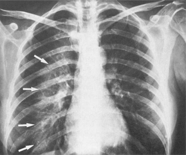
The journey of identifying lung infiltrates begins with the hardware and software used to visualize the interior of the human chest. The transition from traditional film-based X-rays to digital radiography (DR) marked the first major tech milestone in this field, allowing for immediate image processing and manipulation.
From Film to Digital Radiography (DR)
Digital radiography replaced physical film with sensitive plates that convert X-rays into digital signals. This technological shift allowed for “image enhancement,” a software-driven process where contrast and brightness can be adjusted post-capture to reveal subtle infiltrates that might have been invisible on traditional film. For technicians and clinicians, this means higher diagnostic accuracy and the ability to share files instantly across hospital networks via PACS (Picture Archiving and Communication Systems).
High-Resolution Computed Tomography (HRCT)
While a standard X-ray provides a two-dimensional view, HRCT utilizes complex algorithms to reconstruct cross-sectional “slices” of the lungs. The technology behind HRCT involves rapid-fire X-ray tubes and ultra-sensitive detectors that feed data into a computer. This software then performs back-projection algorithms to create a 3D map of the lungs. For identifying infiltrates—whether they are “ground-glass opacities” or dense consolidations—HRCT is the gold standard of pulmonary diagnostic tech, offering sub-millimeter resolution that human eyes alone could never achieve.
Point-of-Care Ultrasound (POCUS)
Miniaturization in hardware has led to the rise of POCUS. These are handheld devices that connect to smartphones or tablets, allowing clinicians to visualize lung infiltrates at the bedside. The tech here relies on sophisticated digital signal processing to filter out “noise” and produce clear images of the pleural line and lung tissue, proving that powerful diagnostic tools no longer require a dedicated imaging suite.
Artificial Intelligence: The New Frontier in Detecting Pulmonary Infiltrates
Perhaps the most significant tech trend in respiratory health is the integration of Artificial Intelligence and Machine Learning (ML). AI tools are no longer science fiction; they are active assistants in the radiology department, helping to categorize infiltrates with speed and precision that rivals senior clinicians.
Computer-Aided Diagnosis (CAD) Systems
CAD software uses deep learning models—specifically Convolutional Neural Networks (CNNs)—to analyze thousands of thoracic images. These models are trained on massive datasets of “normal” vs. “abnormal” lung scans. When a new scan is uploaded, the AI flags potential infiltrates, highlighting areas of concern for the radiologist. This “second pair of eyes” is crucial in high-volume environments where fatigue can lead to human error.
Automated Quantification and Trend Analysis
One of the most difficult tasks for a human is quantifying exactly how much of the lung is affected by an infiltrate. AI software can perform “segmentation,” where it digitally traces the boundaries of an infiltrate and calculates its volume. Furthermore, when a patient returns for a follow-up, the software can overlay the new scan on the old one, providing a pixel-by-pixel comparison to determine if the infiltrate is resolving or expanding. This level of data-driven tracking is a hallmark of modern health tech.
Predictive Analytics and Risk Stratification
Beyond just seeing the infiltrate, AI tools are now being used to predict the underlying cause. By analyzing the texture and distribution patterns of the infiltrates, machine learning algorithms can suggest whether the findings are more likely to be viral pneumonia, heart failure, or a chronic interstitial disease. This “decision support” tech helps prioritize urgent cases and streamlines the clinical workflow.
Telemedicine and Remote Diagnostic Ecosystems
The identification and monitoring of lung infiltrates have moved beyond the four walls of the hospital. Cloud computing and IoT (Internet of Things) connectivity have created a decentralized model of care, making pulmonary diagnostics more accessible than ever.
Cloud-Based Image Sharing and Teleradiology
The ability to move large DICOM (Digital Imaging and Communications in Medicine) files through the cloud has revolutionized how we treat lung conditions. Teleradiology platforms allow a scan taken in a rural clinic to be interpreted by a specialist in a major city within minutes. This tech ecosystem relies on high-speed fiber networks and optimized compression algorithms that maintain image quality while reducing file size for rapid transmission.
IoT and Wearable Tech for Monitoring
While a wearable device like a smartwatch cannot “see” a lung infiltrate, it can monitor the physiological effects caused by one. Advanced sensors now track peripheral oxygen saturation (SpO2), respiratory rate, and even lung sounds (via digital stethoscopes). This data is synced to mobile apps, providing a digital “dashboard” of a patient’s respiratory health. If an infiltrate causes a sudden drop in oxygen levels, the tech can trigger an automated alert to the healthcare provider.
The Rise of Digital Therapeutics (DTx)
In the recovery phase of treating lung infiltrates, software-based interventions known as Digital Therapeutics are gaining ground. These are FDA-cleared apps that guide patients through pulmonary rehabilitation exercises, using gamification and biofeedback to ensure compliance. This represents the shift from “tech as a diagnostic tool” to “tech as a treatment modality.”
Digital Security and Ethics in Medical Imaging
As we rely more heavily on software and the cloud to manage sensitive data regarding lung infiltrates, the importance of digital security cannot be overstated. Protecting patient privacy while maintaining data accessibility is a primary challenge for IT professionals in the healthcare sector.
Protecting DICOM Files and Patient Privacy
Medical images are highly sensitive data. Modern imaging software must comply with strict regulations like HIPAA (in the US) or GDPR (in Europe). This involves implementing end-to-end encryption for data both at rest and in transit. Tech companies are now focusing on “zero-trust” architectures, where every access request to a medical image must be verified, regardless of where it originates.
Blockchain for Medical Record Integrity
There is a growing movement toward using blockchain technology to manage medical records, including imaging reports for lung infiltrates. By creating a decentralized, immutable ledger of a patient’s diagnostic history, blockchain ensures that images cannot be tampered with or lost. It also gives patients more control over who can view their digital health data, using cryptographic keys to grant access.
Addressing Algorithmic Bias
From a tech-ethics perspective, the AI tools used to identify infiltrates must be scrutinized for bias. If an algorithm is trained primarily on data from one demographic, it may be less accurate for others. Leading tech developers are now focusing on “Explainable AI” (XAI), which allows clinicians to see why an AI flagged a specific area as an infiltrate. This transparency is essential for building trust in the digital tools that are increasingly making life-and-death decisions.

The Future: Integrating Tech into Every Breath
The question “what are infiltrates in lungs” is traditionally answered with a medical definition. However, in the modern era, the answer is inseparable from the technology used to find them. From the hardware that captures the image to the AI that interprets it, and the secure cloud networks that share it, the tech industry is the silent partner in every pulmonary diagnosis.
As we look forward, we can expect even greater integration. We may soon see “smart” inhalers that use AI to predict flare-ups before an infiltrate even forms, or augmented reality (AR) headsets that allow surgeons to see a 3D digital overlay of lung infiltrates during a biopsy. The synergy between software, hardware, and data security will continue to refine our understanding of respiratory health, proving that the future of medicine is, at its core, a technological journey.
aViewFromTheCave is a participant in the Amazon Services LLC Associates Program, an affiliate advertising program designed to provide a means for sites to earn advertising fees by advertising and linking to Amazon.com. Amazon, the Amazon logo, AmazonSupply, and the AmazonSupply logo are trademarks of Amazon.com, Inc. or its affiliates. As an Amazon Associate we earn affiliate commissions from qualifying purchases.