In an era defined by rapid technological advancements, where data is king and innovation constantly reshapes our understanding of the world, it’s easy to overlook the subtle yet profound revolutions happening within the realm of medicine. Among these, scintigraphy stands out as a powerful diagnostic tool, offering a unique window into the human body’s inner workings. Far from being a mere static image, scintigraphy provides dynamic, functional insights, revealing not just what an organ looks like, but how it’s actually performing. This article delves into the fascinating world of scintigraphy, exploring its scientific underpinnings, its technological evolution, its significant impact on healthcare economics and branding, and its promising future, viewed through the interconnected lenses of technology, branding, and money.
At its heart, scintigraphy is a non-invasive nuclear medicine imaging technique that uses small amounts of radioactive material, known as radiotracers, to diagnose and assess a variety of medical conditions. Unlike X-rays, CT scans, or MRI, which primarily focus on anatomical structures, scintigraphy excels at evaluating physiological processes and organ function. This capability makes it invaluable for detecting diseases in their early stages, sometimes even before structural changes become apparent, thereby offering critical opportunities for timely intervention and improved patient outcomes.
The Core Science: How Scintigraphy Works
To truly appreciate the power of scintigraphy, one must first grasp the elegant science behind it. The process is a testament to the sophisticated interplay of chemistry, physics, and advanced medical imaging. It begins with the introduction of a radiotracer into the patient’s body, typically through intravenous injection. These radiotracers are specially designed molecules that are chemically identical or similar to substances naturally used by the body, allowing them to selectively accumulate in specific organs, tissues, or cells based on metabolic activity, blood flow, or receptor binding.
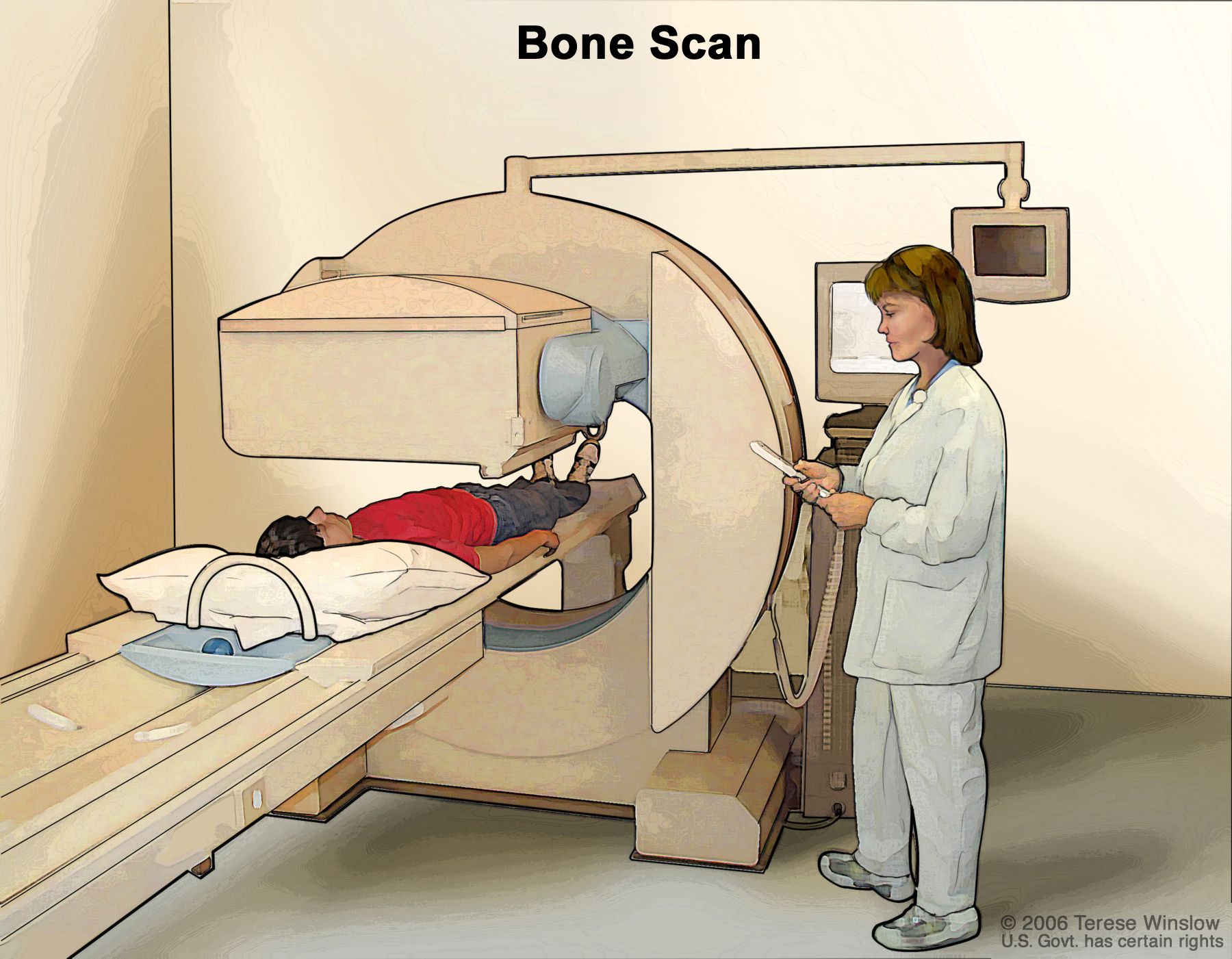
Once the radiotracer is administered, it begins its journey, circulating through the bloodstream and concentrating in target areas. For example, a bone scan uses a tracer that mimics calcium, accumulating in areas of active bone formation or repair. A cardiac scan might use a tracer that is absorbed by healthy heart muscle cells, allowing physicians to visualize blood flow and muscle function. As the tracer congregates, it emits gamma rays – a form of electromagnetic radiation. These gamma rays are then detected by a specialized external camera, known as a gamma camera.
The Radiotracer Revolution
The efficacy of scintigraphy hinges entirely on the radiotracer. This isn’t just a generic radioactive substance; it’s a meticulously engineered pharmaceutical. The development of new radiotracers is a continuous area of research and innovation within nuclear medicine, directly tying into technology trends. Advances in radiochemistry and molecular biology have led to an explosion in the variety and specificity of tracers available. From identifying cancerous cells based on their unique metabolic pathways to pinpointing areas of inflammation or infection, the precision of these tracers determines the diagnostic power of the scan.
The tech aspect here is multifaceted:
- Chemical Synthesis: Developing novel compounds that are both biologically active and capable of carrying a radioactive isotope.
- Isotope Production: Leveraging particle accelerators and nuclear reactors to produce the necessary radioactive isotopes with specific half-lives.
- Pharmacokinetics: Understanding how these tracers move through and are eliminated from the body, ensuring optimal imaging times and patient safety.
The ongoing “radiotracer revolution” is not just about expanding diagnostic capabilities; it’s about pushing the boundaries of non-invasive medicine, allowing for earlier, more accurate diagnoses with minimal patient discomfort.
Capturing the Invisible: The Gamma Camera
The second critical component of scintigraphy is the gamma camera, a marvel of modern engineering. This sophisticated “gadget” is designed to detect the subtle gamma rays emitted by the radiotracer within the body. When a gamma ray strikes the detector crystal (often made of sodium iodide), it creates a tiny flash of light. Photomultiplier tubes then convert these light flashes into electrical signals, which are amplified and processed by computer software.
The gamma camera doesn’t just record the presence of radiation; it maps its location and intensity. By acquiring data from various angles, the system can construct a two- or three-dimensional image of the distribution of the radiotracer. This allows physicians to visualize the functional activity within organs and tissues. Modern gamma cameras are often integrated with computed tomography (CT) scanners in what are known as SPECT/CT (Single-Photon Emission Computed Tomography/CT) systems. These hybrid machines provide both functional (scintigraphy) and anatomical (CT) information simultaneously, offering a more precise localization of metabolic activity and significantly enhancing diagnostic accuracy. The technological leap from early, cumbersome detectors to today’s highly sensitive, digitally integrated gamma cameras exemplifies the continuous drive for precision and efficiency in medical imaging.
Scintigraphy Through a Technological Lens
The journey of scintigraphy from its rudimentary beginnings to its current sophisticated state is a compelling narrative of technological evolution. Within the “Tech” domain, scintigraphy embodies several key trends, from AI integration to digital security, illustrating how cutting-edge innovation continually refines and empowers this diagnostic modality.
AI and Advanced Software: Revolutionizing Diagnosis
One of the most transformative technology trends impacting scintigraphy is the integration of Artificial Intelligence (AI) and advanced software. AI tools are rapidly changing how medical images are acquired, processed, interpreted, and managed.
- Image Reconstruction and Enhancement: AI algorithms can improve the quality of scintigraphy images, reducing noise, correcting for patient movement, and enhancing contrast, leading to clearer and more reliable diagnostic information. This means less need for repeat scans and more accurate initial diagnoses.
- Automated Interpretation and Anomaly Detection: AI-powered software can be trained on vast datasets of scintigraphy images to identify subtle patterns indicative of disease that might be missed by the human eye. Machine learning models can flag potential anomalies, assist in lesion detection, and even quantify tracer uptake in specific regions, standardizing measurements and improving diagnostic consistency.
- Workflow Optimization and Productivity: AI can automate routine tasks, such as image segmentation and report generation, freeing up nuclear medicine physicians to focus on complex cases. This boosts productivity within diagnostic departments, a critical factor for managing patient loads and reducing wait times.
- Predictive Analytics: Beyond diagnosis, AI can contribute to predictive analytics, helping to forecast disease progression or treatment response based on scintigraphy findings, moving towards a more personalized medicine approach.
This fusion of scintigraphy with AI represents a leap forward, transforming raw data into actionable insights and augmenting the capabilities of human experts.
The Evolution of Imaging Hardware: From Gamma Cameras to Hybrid Systems
The hardware underpinning scintigraphy has seen continuous evolution, driven by the demand for higher resolution, faster acquisition times, and greater diagnostic specificity. The “gadgets” of nuclear medicine are increasingly complex and powerful.
- Detector Technology: Advances in detector materials and designs have led to increased sensitivity and spatial resolution, allowing for the detection of smaller lesions and more precise localization of radiotracer uptake. Solid-state detectors, for instance, offer superior energy resolution compared to traditional scintillator crystals.
- SPECT/CT and PET/CT: The development and widespread adoption of hybrid imaging systems like SPECT/CT and PET/CT (Positron Emission Tomography/CT) are perhaps the most significant hardware innovations. These systems merge the functional information of scintigraphy (SPECT or PET) with the anatomical detail of CT in a single scan. This integration provides unparalleled diagnostic clarity, crucial for accurate staging of cancers, precise surgical planning, and detailed assessment of complex conditions. The ability to overlay functional data onto anatomical maps dramatically improves the accuracy of diagnosis and treatment monitoring.
- Patient Experience: Beyond diagnostic capability, hardware design also focuses on patient comfort and safety. From open gantry designs to faster scan times, engineers are constantly working to improve the patient experience, making these advanced procedures more accessible and less daunting.

Digital Security and Data Management in Nuclear Medicine
In an increasingly interconnected digital world, the robust “Digital Security” and meticulous data management of patient information generated by scintigraphy scans are paramount. Nuclear medicine departments deal with highly sensitive patient data, including personal health information (PHI) and detailed imaging results.
- PACS and EMR Integration: Picture Archiving and Communication Systems (PACS) and Electronic Medical Records (EMR) are central to managing scintigraphy data. These software systems store, retrieve, distribute, and display medical images and reports, ensuring they are readily available to healthcare providers while maintaining strict access controls.
- Cybersecurity Measures: Protecting this data from unauthorized access, breaches, and cyber threats is a critical concern. Healthcare organizations must implement stringent cybersecurity protocols, including encryption, multi-factor authentication, intrusion detection systems, and regular security audits, to comply with regulations like HIPAA (Health Insurance Portability and Accountability Act) and GDPR (General Data Protection Regulation).
- Data Integrity and Archiving: Ensuring the long-term integrity and reliable archiving of scintigraphy images and reports is essential for patient care, longitudinal studies, and legal compliance. Cloud-based solutions and robust backup strategies play a vital role in this aspect.
The technological sophistication extends beyond the scan itself to the entire ecosystem of data handling, underscoring the importance of a comprehensive digital strategy in modern healthcare.
The Business of Diagnostics: Scintigraphy’s Economic and Brand Impact
Beyond its scientific and technological marvels, scintigraphy significantly impacts the “Money” and “Brand” aspects of healthcare. For hospitals, clinics, and medical device manufacturers, investing in and offering scintigraphy services is a strategic decision with substantial financial implications and branding opportunities.
Investing in Innovation: The Financial Calculus for Healthcare Providers
From a “Business Finance” perspective, acquiring and operating a nuclear medicine department equipped for scintigraphy represents a significant investment.
- Capital Expenditure: The initial cost of gamma cameras, SPECT/CT machines, hot labs (for preparing radiotracers), and specialized software can run into millions of dollars. This necessitates careful financial planning, including capital budgeting and return on investment (ROI) analysis.
- Operational Costs: Ongoing expenses include the cost of radiotracers (which often have short half-lives and require just-in-time delivery), maintenance of sophisticated equipment, highly specialized staff salaries (nuclear medicine physicians, technologists, radiopharmacists), and regulatory compliance.
- Revenue Generation: Despite the costs, scintigraphy generates substantial revenue for healthcare providers through diagnostic services. Its unique ability to provide functional information often makes it indispensable for specific diagnoses, attracting patient referrals.
- Cost-Effectiveness: Scintigraphy can be highly cost-effective in the long run. By enabling early and accurate diagnosis, it can prevent more expensive and invasive procedures, reduce the need for prolonged treatments, and improve patient outcomes, ultimately lowering overall healthcare costs. This “Investing” in advanced diagnostics isn’t just about financial returns but about enhancing the quality and efficiency of care.
Brand Building Through Advanced Medical Imaging
For a healthcare institution, offering state-of-the-art scintigraphy services is a powerful “Brand Strategy” and “Corporate Identity” differentiator.
- Reputation for Excellence: A facility equipped with advanced nuclear medicine capabilities cultivates a reputation for cutting-edge technology, comprehensive diagnostic services, and medical innovation. This enhances its corporate identity and attracts both patients and top medical talent.
- Marketing and Differentiation: Hospitals can “Market” their advanced imaging capabilities, highlighting how scintigraphy offers superior diagnostic accuracy for conditions like heart disease, cancer, and neurological disorders. This differentiates them from competitors and positions them as leaders in specialized care.
- Patient Trust and Confidence: The availability of advanced, non-invasive diagnostic tools like scintigraphy instills confidence in patients and referring physicians. It signifies a commitment to providing the best possible care, thereby strengthening the hospital’s reputation.
- Case Studies: Showcasing successful diagnostic outcomes and improved patient journeys facilitated by scintigraphy can serve as compelling “Case Studies” in marketing materials, reinforcing the value proposition and building trust.
In a competitive healthcare landscape, the ability to provide sophisticated diagnostic tools like scintigraphy is not just a medical advantage, but a significant brand asset, shaping public perception and professional standing.
Navigating Patient Costs and Value: A Personal Finance Perspective
From a “Personal Finance” standpoint, patients often face questions regarding the cost of a scintigraphy scan and how it fits into their healthcare budget.
- Direct Costs: The cost of a scintigraphy procedure can vary widely depending on the type of scan, the facility, and geographical location. This typically includes charges for the radiotracer, the technical component of the scan, and the professional interpretation by a nuclear medicine physician.
- Insurance Coverage: Most scintigraphy procedures are covered by health insurance plans, given their diagnostic necessity. However, patients may still be responsible for deductibles, co-pays, or co-insurance, which can represent significant out-of-pocket expenses. Understanding one’s “Financial Tools” like insurance policies and health savings accounts is crucial.
- Value Proposition: While seemingly expensive, the “value” of scintigraphy often lies in its ability to provide early and accurate diagnoses, which can prevent more severe illness, avoid unnecessary invasive procedures, and lead to more effective treatment plans. Early detection of cancer or heart disease, for instance, can drastically alter prognosis and significantly reduce the overall long-term financial burden associated with advanced disease management. Educating patients on this long-term value is key to managing expectations and demonstrating the worth of the investment in their health.
The Future of Functional Imaging: Trends and Horizons
The trajectory of scintigraphy is one of continuous innovation, promising even more precise, personalized, and proactive healthcare. The “Tech” sphere will continue to drive these advancements, while “Money” and “Brand” considerations will shape their adoption and accessibility.
Emerging Radiotracers and Targeted Therapies
The frontier of scintigraphy is particularly exciting in the development of new radiotracers. Researchers are constantly designing “smarter” molecules that can target specific disease markers with even greater sensitivity.
- Theranostics: A major trend is “theranostics,” which combines diagnostics and therapeutics into a single agent. A theranostic radiotracer can first be used for diagnostic imaging (scintigraphy) to pinpoint diseased cells (e.g., metastatic cancer). Once localized, a higher dose of a therapeutic radionuclide (often attached to the same targeting molecule) can then be administered to specifically treat those cells, minimizing damage to healthy tissue. This highly personalized approach is revolutionizing cancer treatment, offering a blend of precision medicine and targeted therapy.
- Molecular Imaging: Future tracers will delve deeper into molecular pathways, allowing for the visualization of gene expression, receptor activity, and other cellular processes, offering unprecedented insights into disease pathogenesis.
The Promise of Precision Medicine
Scintigraphy is a cornerstone of “precision medicine,” tailoring medical treatment to the individual characteristics of each patient. As technology advances, scintigraphy will play an even greater role in:
- Personalized Treatment Selection: Helping physicians choose the most effective therapies based on a patient’s unique molecular profile and how their disease interacts with specific tracers.
- Monitoring Treatment Response: Precisely tracking the effectiveness of therapies in real-time, allowing for rapid adjustments to treatment plans.
- Disease Prevention and Risk Assessment: Potentially identifying individuals at high risk for certain diseases even before symptoms appear, enabling proactive interventions.
These advancements underscore how scintigraphy is evolving from a purely diagnostic tool to an integral part of a comprehensive, individualized healthcare strategy, further intertwining with digital security as more sophisticated data is generated and processed.
In conclusion, scintigraphy is far more than just a medical procedure; it is a dynamic field at the intersection of cutting-edge technology, strategic healthcare branding, and complex economic considerations. From the intricate science of radiotracers and gamma cameras to the transformative power of AI and hybrid imaging, its technological prowess is undeniable. Its role in building a hospital’s reputation for excellence and its impact on the financial landscape of healthcare—both for providers and patients—are profound. As we look to the future, scintigraphy promises to continue its evolution, offering even more precise diagnoses and paving the way for truly personalized medicine, ensuring it remains an indispensable tool in our quest for healthier lives.
aViewFromTheCave is a participant in the Amazon Services LLC Associates Program, an affiliate advertising program designed to provide a means for sites to earn advertising fees by advertising and linking to Amazon.com. Amazon, the Amazon logo, AmazonSupply, and the AmazonSupply logo are trademarks of Amazon.com, Inc. or its affiliates. As an Amazon Associate we earn affiliate commissions from qualifying purchases.