The question “what does a fatty tumor look like on a dog” has traditionally been answered in the sterile environment of a veterinary clinic through manual palpation and physical observation. However, as we move further into the decade, the intersection of veterinary medicine and advanced technology is redefining how we visualize, diagnose, and monitor these common growths. Known medically as lipomas, fatty tumors are benign masses of fat cells. While they are usually non-threatening, the technological tools used to identify them are becoming increasingly sophisticated, moving away from “wait and see” approaches toward high-resolution digital analysis.
The Evolution of Veterinary Diagnostics: From Manual Palpation to Digital Precision
For decades, the standard procedure for identifying a fatty tumor involved a veterinarian feeling a lump and performing a Fine Needle Aspirate (FNA) to look at cells under a microscope. While effective, this method is subjective and prone to human error. Today, the tech industry is transforming this process through high-resolution imaging and digital documentation.
The Limitations of the Physical Exam
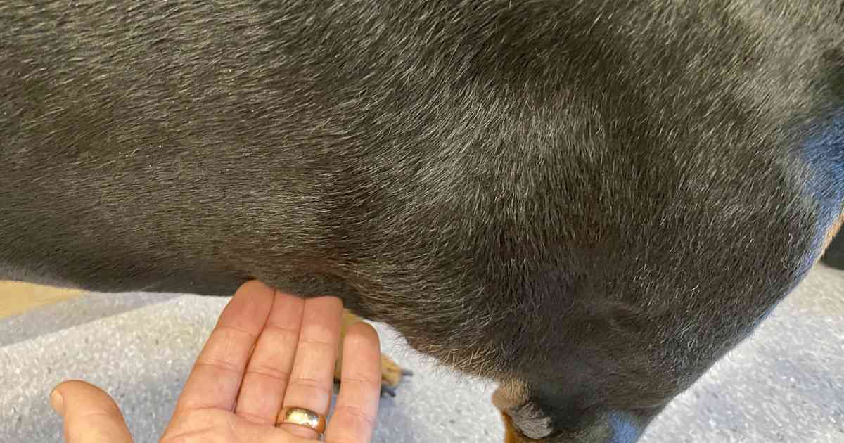
Manual palpation is the “analog” version of diagnostics. While a vet can describe a fatty tumor as soft, mobile, and located just under the skin, these descriptions are qualitative. In the realm of modern “PetTech,” qualitative data is being replaced by quantitative data. Technology allows us to measure the exact density, volume, and vascularity of a mass, providing a much clearer picture of whether a lump is a simple lipoma or something more subcutaneous and complex.
Integrating High-Resolution Digital Imaging
Modern veterinary practices are increasingly adopting advanced imaging software that integrates with ultrasound and CT hardware. When a dog presents with a potential fatty tumor, digital ultrasound tech can now render 3D visualizations of the mass. This software analyzes the “echo-texture” of the lump. Because fat has a specific density compared to muscle or cancerous tissue, ultrasound software can provide a high-probability digital signature of a lipoma long before a needle ever touches the skin.
Artificial Intelligence and Pattern Recognition in Lipoma Identification
The most significant leap in answering what a fatty tumor looks like is the application of Artificial Intelligence (AI) and Machine Learning (ML). By feeding millions of images of canine skin masses into neural networks, tech companies are developing diagnostic tools that can identify lipomas with startling accuracy.
Machine Learning Algorithms for Soft Tissue Classification
AI tools are now capable of analyzing “Visual Data Points.” When a digital image or a microscopic slide of a fatty tumor is uploaded to a cloud-based AI platform, the algorithm compares the cellular patterns against a massive database of both benign and malignant tumors. This “Computer Vision” technology looks for specific markers—such as the uniformity of adipocytes (fat cells)—that the human eye might miss. This reduces the “diagnostic lag” and provides pet owners with immediate, tech-backed reassurance.
Computer-Aided Diagnosis (CAD) in Veterinary Clinics
CAD systems are becoming a staple in modern animal hospitals. These software suites act as a “second pair of eyes” for radiologists. When viewing an X-ray or MRI of a dog’s torso, the CAD software highlights areas of concern. For fatty tumors, which can sometimes be deep-seated (infiltrative lipomas), CAD technology is essential. It can differentiate between the fatty tissue of the tumor and the surrounding muscle fibers by analyzing Hounsfield units—a tech-based measurement of radiodensity—ensuring that the “look” of the tumor is interpreted through data rather than just sight.
The Role of Telemedicine and Mobile Tech in Pet Health Monitoring
As smartphone technology improves, the ability to monitor a dog’s fatty tumor has moved into the hands of the owner via specialized apps and mobile peripherals. The “look” of a tumor can now be tracked over time using digital logs, creating a chronological data set that is far more useful than a one-time photo.
App-Based Triage: Bridging the Gap for Pet Owners
Several startups have launched apps that use the smartphone camera to perform preliminary assessments of skin lumps. While these are not replacements for professional veterinary care, they use AI-driven image analysis to tell an owner if a lump “looks” like a typical lipoma. The user takes a photo, often with a calibration tool (like a coin for scale), and the app’s software analyzes the borders, color, and texture. This use of “Edge Computing”—where the processing happens directly on the device—is a prime example of how tech is democratizing pet health.
Smart Wearables and Early Detection Metrics
The next frontier in identifying and monitoring fatty tumors lies in wearable technology. Smart collars equipped with bio-sensors can now monitor changes in a dog’s gait or local body temperature. If a fatty tumor begins to grow in a location that restricts movement—such as the axilla (armpit)—the software detects the subtle change in the dog’s activity “fingerprint.” This “Internet of Medical Things” (IoMT) for pets ensures that even if a tumor doesn’t “look” different yet, its impact on the dog’s physiology is recorded and flagged digitally.
Future Trends: Non-Invasive Diagnostic Tech and 3D Modeling
The future of identifying fatty tumors on dogs lies in even more advanced, non-invasive gadgets and software-driven models that eliminate the need for traditional biopsies.
The Rise of Bio-Sensing and Optical Coherence Tomography (OCT)
OCT is a technology originally used in human ophthalmology that is now finding its way into veterinary dermatology. It uses light waves to take cross-section pictures of the skin. This allows a veterinarian to see the “look” of a fatty tumor beneath the surface in microscopic detail without making an incision. The software converts light reflections into a high-definition map of the tissue layers, identifying the classic “honeycomb” structure of fat cells that defines a lipoma.
3D Mapping for Surgical Planning and Monitoring
For larger fatty tumors that require removal, 3D printing and modeling software are becoming invaluable. By taking data from a CT scan, surgeons can create a digital 3D model of the tumor. This allows them to see exactly how the fatty mass interacts with nearby nerves and blood vessels. In the “Tech” niche, this is referred to as a “Digital Twin.” By manipulating the digital twin of the dog’s anatomy, the veterinary surgeon can plan the most efficient route for removal, minimizing the “digital footprint” of the surgery and improving recovery times.
Digital Security and Data Integrity in Veterinary Tech
As we lean more heavily on AI, cloud diagnostics, and mobile apps to identify what a fatty tumor looks like, the importance of digital security cannot be overstated. The transition from physical paper charts to digital health records (EHR) brings both opportunities and risks.
Protecting Pet Health Data
Pet owners are often unaware that their dog’s diagnostic images and health metrics are valuable data points. Tech platforms that host AI diagnostic tools must employ robust encryption to ensure that this biometric data is not compromised. As veterinary clinics move toward cloud-based practice management software, the integration of blockchain technology is being explored to create immutable records of a pet’s health history. This ensures that the “digital history” of a fatty tumor—from its first appearance on an ultrasound to its eventual removal—is secure and accessible only to authorized providers.

The Ethics of AI in Diagnostics
The tech industry is also grappling with the “Black Box” problem of AI in veterinary medicine. If an algorithm identifies a lump as a fatty tumor, developers must ensure the software is transparent about why it reached that conclusion. “Explainable AI” (XAI) is a burgeoning field within pet tech that aims to make these digital decisions understandable to human veterinarians, ensuring that technology remains a tool for enhancement rather than a replacement for professional judgment.
In conclusion, the question of what a fatty tumor looks like on a dog is no longer just a matter of sight and touch. Through the lens of modern technology—ranging from AI-driven image recognition to 3D surgical modeling and secure digital health records—we are gaining a deeper, more accurate understanding of these common growths. As these tools continue to evolve, they promise a future where pet health is more proactive, data-driven, and non-invasive than ever before.
aViewFromTheCave is a participant in the Amazon Services LLC Associates Program, an affiliate advertising program designed to provide a means for sites to earn advertising fees by advertising and linking to Amazon.com. Amazon, the Amazon logo, AmazonSupply, and the AmazonSupply logo are trademarks of Amazon.com, Inc. or its affiliates. As an Amazon Associate we earn affiliate commissions from qualifying purchases.