
The seemingly simple act of analyzing urine sediment is a cornerstone of modern diagnostics, offering a window into the intricate workings of the human body. Far from being a rudimentary visual inspection, the examination of urine sediment is a sophisticated process deeply intertwined with technological advancements. From the fundamental principles of microscopy to the cutting-edge capabilities of automated analyzers, technology plays an indispensable role in identifying, quantifying, and interpreting the various cellular and non-cellular components suspended within urine. This article delves into the technological underpinnings of urine sediment analysis, exploring the key components and the advanced tools that allow us to glean invaluable health information.

The Microscopic Battlefield: Technologies for Visualizing Sediment
At its core, urine sediment analysis relies on the ability to visualize microscopic elements. The journey from a urine sample to diagnostic insight begins with technologies that facilitate this crucial step.
Enhanced Microscopy: Beyond the Basic Light Microscope
While the light microscope remains a fundamental tool, its capabilities have been significantly enhanced over the years. Modern clinical laboratories utilize high-quality phase-contrast microscopy and differential interference contrast (DIC) microscopy. These advanced optical techniques manipulate light waves to increase the contrast of transparent or nearly transparent specimens, such as cells and crystals, without the need for staining. This allows for better visualization of subtle morphological details, improving the accuracy of identifying and differentiating various elements like red blood cells, white blood cells, and epithelial cells.
The adoption of digital imaging and microscopy systems has further revolutionized this field. High-resolution cameras coupled with advanced software allow for the capture, storage, and real-time display of microscopic images. This technology facilitates easier documentation, sharing of findings among pathologists and clinicians, and the potential for remote analysis. Furthermore, sophisticated image analysis software is increasingly being integrated, capable of automatically detecting and classifying certain elements within the field of view, thereby reducing subjective variability and increasing throughput.
Automation in Microscopy: Streamlining the Process
The sheer volume of urine samples processed in clinical settings necessitates automation. Automated urine analyzers combine sample preparation, centrifugation, and microscopic analysis into a single integrated system. These instruments employ sophisticated robotics and fluidics to handle samples precisely, ensuring consistent centrifugation speeds and volumes, critical for reproducible sediment preparation.
Following centrifugation, automated microscopy modules within these systems utilize high-magnification objectives and advanced imaging sensors. These modules are equipped with powerful algorithms that can differentiate between various cellular types (e.g., neutrophils, lymphocytes, red blood cells), detect casts, and identify other significant findings like crystals and bacteria. The ability of these systems to perform preliminary classifications and provide quantitative data significantly reduces the workload for laboratory technicians, allowing them to focus on more complex or ambiguous cases. The integration of artificial intelligence (AI) and machine learning (ML) algorithms within these automated systems is a significant technological leap. These AI-powered systems are trained on vast datasets of annotated urine sediment images, enabling them to achieve high levels of accuracy in identifying and categorizing sediment components, often surpassing human capabilities in speed and consistency for routine analyses.
Key Sediment Components and Their Technological Detection
The diagnostic value of urine sediment lies in the identification of its various constituents. Technology is instrumental in not only visualizing these components but also in providing quantitative data that aids in diagnosis.
Cellular Elements: The Body’s Microscopic Messengers
-
Red Blood Cells (RBCs): The presence of RBCs (hematuria) can indicate a wide range of conditions, from minor urinary tract infections to serious kidney diseases. Automated analyzers employ light scattering techniques and impedance principles to count and differentiate RBCs from other cells. Advanced systems can also employ fluorescence flow cytometry for highly accurate enumeration and characterization of RBCs, particularly in complex cases.
-
White Blood Cells (WBCs): Elevated WBC counts (pyuria) are a strong indicator of inflammation or infection within the urinary tract. Similar to RBCs, automated analyzers use light scattering and impedance methods for quantification. Cytochemical staining integrated into automated platforms can further differentiate WBC types (e.g., neutrophils, eosinophils) based on their characteristic enzymatic activity, providing more specific diagnostic clues.
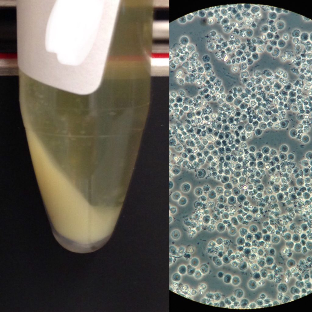
- Epithelial Cells: Various types of epithelial cells line the urinary tract. The presence of squamous epithelial cells is generally normal, while renal tubular epithelial cells can indicate tubular damage. Morphological analysis by automated microscopy, aided by AI algorithms, is crucial for distinguishing between these cell types and assessing their morphology for signs of abnormality.
Non-Cellular Components: Clues from the Urinary Matrix
-
Casts: These cylindrical structures formed in the renal tubules are highly diagnostic. Different types of casts (e.g., hyaline, granular, waxy, red blood cell, white blood cell casts) provide specific insights into kidney function and pathology. Automated microscopy systems excel at identifying casts due to their characteristic shapes and refractile properties. Sophisticated image processing algorithms are essential for accurate classification of cast types, often distinguishing between them based on subtle differences in their internal structure and refractivity.
-
Crystals: Various types of crystals can precipitate in urine, depending on pH and concentration. Some are benign (e.g., calcium oxalate), while others can indicate metabolic disorders or kidney stone formation (e.g., uric acid, cystine). Automated visual identification systems are programmed to recognize the characteristic shapes and birefringence of different crystal types. Advanced spectroscopic techniques, though less common in routine analysis, hold potential for rapid and specific crystal identification.
-
Bacteria and Other Microorganisms: The presence of bacteria can signal a urinary tract infection. Automated microscopy can detect and even semi-quantify bacteria, often aided by AI algorithms trained to recognize bacterial morphology. Rapid diagnostic tests, often employing immunoassay principles or molecular detection methods, are increasingly integrated into laboratory workflows to quickly identify specific pathogens, complementing sediment analysis.
Advanced Technologies Enhancing Diagnostic Accuracy
Beyond standard microscopy and automated analysis, emerging technologies are continuously pushing the boundaries of urine sediment diagnostics, offering greater precision and broader insights.
Flow Cytometry: Precision Cell Counting and Characterization
Flow cytometry, a powerful cell analysis technology, is increasingly being applied to urine sediment. This technology allows for the rapid, high-throughput, and highly accurate enumeration and characterization of cellular elements, including RBCs, WBCs, and even renal tubular cells. By interrogating individual cells as they pass through a laser beam, flow cytometry can measure their size, internal complexity (granularity), and the presence of specific surface markers (through immunofluorescence). This level of detail surpasses traditional microscopy and can be invaluable in diagnosing hematuria of unknown origin, assessing inflammatory responses, and identifying early signs of renal damage.
Spectroscopy and Imaging Techniques: Unlocking Deeper Molecular Insights
While currently more in the realm of research and specialized applications, spectroscopic techniques like Raman spectroscopy and infrared spectroscopy hold promise for the future of urine sediment analysis. These methods can provide information about the molecular composition of sediment elements, potentially identifying specific proteins, lipids, or crystalline structures without extensive sample preparation. Coupled with advanced digital imaging and pattern recognition algorithms, these techniques could offer highly specific and non-invasive diagnostic capabilities. Furthermore, advancements in multispectral imaging can capture images across a range of wavelengths, providing more detailed information about the optical properties of sediment components, thus enhancing their identification and characterization.
The Role of Data Analytics and AI in Interpreting Complex Findings
The sheer volume of data generated by modern automated urine analyzers necessitates sophisticated data processing and interpretation. Artificial intelligence (AI) and machine learning (ML) algorithms are playing an increasingly crucial role. These technologies are trained to recognize subtle patterns and correlations within the sediment findings that might be missed by human observers. AI can assist in:
- Differential diagnosis: By analyzing a combination of sediment components and correlating them with clinical data, AI can suggest potential diagnoses.
- Prognostic assessment: AI models can potentially predict the likelihood of disease progression or treatment response based on the characteristics of the urine sediment.
- Quality control: AI can continuously monitor the performance of automated analyzers and flag any deviations from expected results.
- Personalized medicine: In the future, AI-powered analysis of urine sediment, integrated with other patient data, could contribute to more personalized diagnostic and treatment strategies.
In conclusion, the analysis of urine sediment is a prime example of how technology permeates even the most fundamental aspects of healthcare. From the fundamental optics of microscopy to the sophisticated algorithms of AI, technology is not merely an aid but an integral part of understanding what lies within urine sediment and, consequently, unlocking vital insights into human health. The ongoing evolution of these technologies promises even more precise, efficient, and informative diagnostic capabilities in the years to come.
aViewFromTheCave is a participant in the Amazon Services LLC Associates Program, an affiliate advertising program designed to provide a means for sites to earn advertising fees by advertising and linking to Amazon.com. Amazon, the Amazon logo, AmazonSupply, and the AmazonSupply logo are trademarks of Amazon.com, Inc. or its affiliates. As an Amazon Associate we earn affiliate commissions from qualifying purchases.