
The realm of medicine is a complex tapestry woven with specialized terminology, acronyms, and abbreviations that can often leave patients and even some healthcare professionals feeling a bit lost. Among these, “IOP” is a term that frequently surfaces, particularly in discussions surrounding eye health. While its meaning might seem straightforward to those within ophthalmology, for the uninitiated, understanding “IOP” is crucial for comprehending diagnoses, treatment plans, and the overall trajectory of eye care. This article delves into the meaning of IOP in medical terms, exploring its significance, the conditions it relates to, and its vital role in maintaining ocular health.

Understanding Intraocular Pressure (IOP): The Foundation of Ocular Health
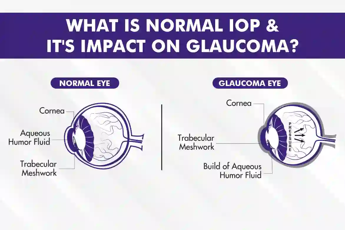
At its core, “IOP” stands for Intraocular Pressure. This term refers to the fluid pressure inside the eye. Imagine the eye as a somewhat rigid, spherical balloon filled with a liquid. This liquid, known as aqueous humor, is constantly produced and drained within the eye. The pressure exerted by this fluid against the inner walls of the eye is what we call intraocular pressure. Maintaining this pressure within a healthy range is paramount for the eye to function correctly and for the delicate structures within it, such as the optic nerve and retina, to remain undamaged.
The Aqueous Humor: The Eye’s Internal Hydraulics
The aqueous humor is a clear, watery fluid that fills the anterior and posterior chambers of the eye, the spaces in front of and behind the iris, respectively. Its primary functions are:
- Nutrient Supply: It nourishes the avascular structures of the eye, namely the cornea and the lens, which do not have their own blood vessels.
- Waste Removal: It helps to remove metabolic waste products from these structures.
- Maintaining Shape: Crucially, it contributes to the structural integrity and shape of the eyeball, providing the necessary rigidity to allow for clear vision.
The production of aqueous humor occurs in the ciliary body, a ring-shaped tissue inside the eye. It then flows from the posterior chamber, through the pupil, into the anterior chamber, and finally drains out of the eye through the trabecular meshwork, a sponge-like tissue located at the angle where the iris meets the cornea. This continuous cycle of production and drainage maintains a delicate balance, and disruptions to this balance are what lead to abnormal IOP.
Normal IOP Ranges: A Vital Equilibrium
The normal range for intraocular pressure is generally considered to be between 10 and 21 millimeters of mercury (mmHg). However, it’s important to understand that this is a general guideline, and individual variations can exist. Factors such as age, race, and even the time of day can slightly influence IOP readings. A healthcare professional will consider these factors when interpreting IOP measurements.
- “Normal Tension” Glaucoma: Some individuals may have optic nerve damage and visual field loss characteristic of glaucoma, yet their IOP readings consistently fall within the “normal” range. This condition, known as normal-tension glaucoma, highlights that simply having an IOP within the normal range doesn’t guarantee the absence of eye disease.
- Ocular Hypertension: Conversely, some individuals may have elevated IOP readings without any detectable optic nerve damage or vision loss. This condition is termed ocular hypertension. While not glaucoma itself, it signifies an increased risk of developing glaucoma, and therefore requires close monitoring.
The measurement of IOP is typically performed using an instrument called a tonometer. There are several types of tonometers, with the most common being Goldmann applanation tonometry, which is considered the gold standard, and non-contact tonometry (air puff test), which is quicker and less invasive.
The Significance of Elevated IOP: The Glaucoma Connection
The most critical implication of elevated intraocular pressure is its strong association with glaucoma. Glaucoma is a group of eye diseases that damage the optic nerve, which connects the eye to the brain. This damage is often caused by abnormally high IOP, which puts pressure on the optic nerve fibers. If left untreated, glaucoma can lead to irreversible vision loss and even blindness.
Glaucoma: A Silent Thief of Sight
Glaucoma is often referred to as the “silent thief of sight” because it typically progresses without noticeable symptoms in its early stages. Peripheral vision is usually affected first, and individuals may not realize they are losing vision until the disease has advanced significantly and central vision is compromised. This is why regular eye examinations, including IOP measurement, are so crucial, especially for individuals at higher risk.
Risk factors for developing glaucoma include:

- Age: The risk of glaucoma increases with age, particularly after 40.
- Family History: A genetic predisposition plays a significant role. If a close family member has glaucoma, your risk is substantially higher.
- Race: Certain racial groups, such as African Americans, are at a higher risk of developing primary open-angle glaucoma, the most common type.
- Medical Conditions: Conditions like diabetes, high blood pressure, and heart disease can increase the risk.
- Eye Conditions: Thin corneas, severe myopia (nearsightedness), and previous eye injuries can also be contributing factors.
Mechanisms of IOP Elevation in Glaucoma
In the most common form of glaucoma, primary open-angle glaucoma (POAG), the drainage angle of the eye (where the trabecular meshwork is located) appears normal, but the outflow of aqueous humor is somehow impeded. The exact reasons for this blockage are not fully understood but are believed to involve changes in the trabecular meshwork itself.
In angle-closure glaucoma, the iris bulges forward, narrowing or blocking the drainage angle. This can happen suddenly (acute angle-closure glaucoma), leading to a rapid and severe increase in IOP, intense eye pain, redness, blurred vision, and halos around lights, or it can be a more gradual process (chronic angle-closure glaucoma).
Managing IOP: Treatments and Monitoring
The primary goal in managing elevated IOP is to lower it to a level that prevents or slows further damage to the optic nerve. This is achieved through various treatment strategies, often involving a combination of medication and, in some cases, surgical interventions. Close monitoring of IOP is essential to assess the effectiveness of treatment and to detect any progression of the disease.
Medical Management: Medications to Lower IOP
Ophthalmic medications, commonly in the form of eye drops, are the first line of treatment for most patients with elevated IOP. These medications work by either reducing the production of aqueous humor or increasing its outflow from the eye. There are several classes of glaucoma medications, each with its own mechanism of action and potential side effects:
- Prostaglandin Analogs: These are often the first choice as they are highly effective and generally well-tolerated. Examples include latanoprost, travoprost, and bimatoprost. They work by increasing the uveoscleral outflow of aqueous humor.
- Beta-Blockers: Medications like timolol reduce the production of aqueous humor. They were a mainstay of treatment for many years but are now often used in combination with other agents.
- Alpha-Adrenergic Agonists: These medications can both decrease aqueous humor production and increase outflow. Brimonidine is a common example.
- Carbonic Anhydrase Inhibitors: These drugs reduce aqueous humor production. They can be administered as eye drops (dorzolamide, brinzolamide) or orally (acetazolamide), though oral forms are typically reserved for more severe cases due to systemic side effects.
- Miotic Agents: These constrict the pupil and increase outflow through the trabecular meshwork. Pilocarpine is an example, but they are less commonly used today due to side effects like blurred vision and headaches.
It’s crucial for patients to take their eye drops exactly as prescribed by their doctor. Missing doses or using them incorrectly can lead to fluctuations in IOP, potentially undermining treatment effectiveness.
Surgical and Laser Interventions: When Medications Aren’t Enough
When medications fail to adequately control IOP, or if a patient cannot tolerate them, surgical or laser procedures may be recommended. These interventions aim to create new pathways for aqueous humor to drain from the eye.
- Laser Trabeculoplasty: This procedure uses a laser to improve the drainage of aqueous humor through the trabecular meshwork. It is typically performed for open-angle glaucoma.
- Trabeculectomy: This is a more invasive surgical procedure that creates a new drainage channel for the aqueous humor, often by making a flap in the sclera (the white part of the eye).
- Glaucoma Drainage Devices (Tube Shunts): These involve surgically implanting a small tube into the eye to divert aqueous humor to a reservoir placed under the conjunctiva.
- Minimally Invasive Glaucoma Surgery (MIGS): A growing area of glaucoma treatment, MIGS procedures involve a variety of techniques using microscopic instruments to enhance aqueous humor outflow. They are generally less invasive than traditional surgeries and are often performed in conjunction with cataract surgery.
Ongoing Monitoring: The Key to Long-Term Eye Health
Regardless of the treatment employed, regular monitoring is indispensable. This involves:
- IOP Measurements: Frequent checks to ensure IOP remains within the target range.
- Optic Nerve Examination: Visual inspection of the optic nerve by an ophthalmologist.
- Visual Field Testing: Assessing peripheral vision to detect any loss.
- Optical Coherence Tomography (OCT): A non-invasive imaging test that provides detailed cross-sectional images of the retina and optic nerve, allowing for precise measurement of nerve fiber layer thickness and detection of subtle changes over time.
By diligently adhering to treatment plans and attending all scheduled follow-up appointments, individuals diagnosed with conditions related to IOP can significantly improve their chances of preserving their vision. Understanding what IOP means in medical terms is the first step towards actively participating in one’s own eye care journey.
aViewFromTheCave is a participant in the Amazon Services LLC Associates Program, an affiliate advertising program designed to provide a means for sites to earn advertising fees by advertising and linking to Amazon.com. Amazon, the Amazon logo, AmazonSupply, and the AmazonSupply logo are trademarks of Amazon.com, Inc. or its affiliates. As an Amazon Associate we earn affiliate commissions from qualifying purchases.