
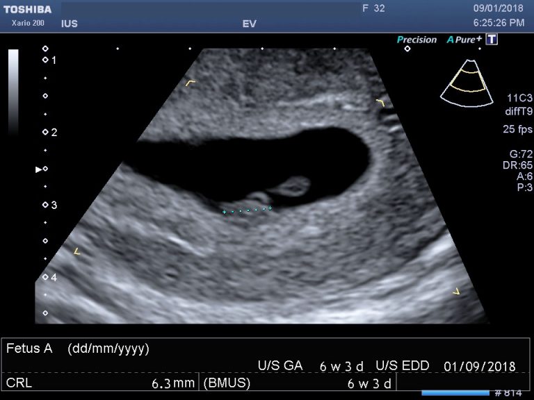
The image is often described by patients as a “flickering grain of rice” or a small, glowing smudge against a sea of static. However, to the engineer, the data scientist, and the medical technologist, a 6-week sonogram represents a pinnacle of acoustic physics and digital signal processing. While the human eye sees a modest blur, the technology behind the screen is performing millions of calculations per second to visualize a biological entity no larger than a sweet pea.
To understand what a 6-week sonogram “looks like” from a tech perspective, we must look beyond the grainy black-and-white monitor. We must look at the sophisticated hardware, the AI-driven software enhancements, and the cloud-integrated infrastructure that allow us to peer into the microscopic beginnings of human development.
The Evolution of Medical Imaging: From Analog Shadows to High-Resolution Data
The visual output of a 6-week sonogram is limited by the laws of physics, yet modern technology has pushed these boundaries further than ever before. At six weeks, an embryo is approximately 5 to 9 millimeters long. Capturing this requires specialized hardware capable of high-frequency output and sensitive data reception.
Transvaginal Transducers: The Hardware of Precision
At the six-week mark, a standard abdominal probe often lacks the resolution to provide a clear image due to the depth of the tissue and the minute size of the subject. The technological solution is the transvaginal transducer. This device utilizes high-frequency sound waves (typically between 5 MHz and 10 MHz).
The technology relies on the piezoelectric effect—the ability of certain materials to generate an electric charge in response to applied mechanical stress. Inside the probe, quartz crystals or ceramic elements vibrate at high speeds, sending out ultrasonic pulses. Because these high frequencies have shorter wavelengths, they provide superior axial resolution, allowing the software to distinguish between two structures that are very close together—such as the yolk sac and the nascent fetal pole.
Signal Processing and Pixel Density in Early Gestation
What the user sees on the screen is not a “photo” but a reconstructed data map. Once the sound waves bounce off the embryonic structures, the transducer captures the returning echoes. This raw analog data is then converted into digital signals.
Advanced Digital Beamformers (DBF) are the “brains” of the modern sonogram machine. They process the timing and phase of the returning signals to focus the image digitally. At six weeks, the challenge is “noise.” The surrounding maternal tissue creates acoustic clutter. Modern software uses “Harmonic Imaging” technology, which listens for the “overtones” of the sound waves rather than just the fundamental frequency. This tech results in a crisper image with higher contrast, making that tiny 6-week flickering heart move from a blurry gray shadow to a distinct, measurable digital event.
AI-Assisted Diagnostics: Deciphering the “Fetal Pole” via Algorithms
In the past, the quality of a 6-week sonogram depended entirely on the steady hand and sharp eye of the sonographer. Today, Artificial Intelligence and Machine Learning (ML) are integrated into the software suites of high-end ultrasound machines (like those produced by GE Healthcare or Philips), drastically changing how we interpret the “look” of the image.
Automated Measurements and Crown-Rump Length (CRL) Accuracy
One of the most critical data points at six weeks is the Crown-Rump Length (CRL). This measurement determines the gestational age and the estimated due date. Historically, a technician would manually place “calipers” on the screen—a process prone to human error.
Modern sonography tech utilizes AI-driven auto-measurement tools. These algorithms have been trained on millions of previous scans to identify the specific anatomical markers of a 6-week embryo. The software can automatically detect the boundaries of the fetal pole and provide a CRL measurement with sub-millimeter precision. This reduces variability between different machines and operators, ensuring that the “look” of the sonogram is backed by consistent, verifiable data.
Pattern Recognition in Early Cardiac Activity Detection
Perhaps the most significant technological feat at the 6-week mark is the detection of the primitive heartbeat. At this stage, the heart is not a fully formed organ but a tube of rhythmic cells. The “flicker” seen on the screen is often hard to distinguish from the mother’s own pulse or general electronic interference.

AI pattern recognition software can now isolate these rhythmic movements. By utilizing “Color Doppler” or “Power Doppler” technology, the software translates the frequency shift of the returning sound waves (the Doppler effect) into visual colors—usually red and blue—to represent blood flow. This allows the technology to “see” a heartbeat that might be invisible to the naked eye in a standard 2D grayscale mode.
Cloud Integration and Telehealth: Sharing the First “Digital Footprint”
The “look” of a 6-week sonogram has moved beyond the thermal-printed paper strip. It is now a high-fidelity digital asset, part of a larger ecosystem of Health IT and data management.
Secure Portals and DICOM Standards
Medical images are stored in a specific format known as DICOM (Digital Imaging and Communications in Medicine). This is not a simple JPEG; it contains layers of metadata including patient history, equipment settings, and calibrated measurement data.
In a modern tech-forward clinic, the 6-week sonogram is instantly uploaded to a secure, HIPAA-compliant cloud server. This allows for “Picture Archiving and Communication Systems” (PACS) integration, where a specialist in a different city can review the high-resolution files in real-time. For the patient, this means the 6-week image is accessible via a secure mobile app, allowing the “first look” to be shared digitally with family members through encrypted links.
The Rise of App-Based Monitoring and Remote Consultations
As telehealth tech matures, we are seeing the emergence of “Point-of-Care Ultrasound” (POCUS). While 6-week scans are still primarily done in-clinic, the software used to view them is becoming increasingly mobile. Advanced software-as-a-service (SaaS) platforms now allow physicians to view sonogram streams on tablets or smartphones without losing resolution. This mobility ensures that if a technical anomaly is spotted at the 6-week mark, data can be ported to a maternal-fetal medicine specialist for an immediate second opinion, leveraging the power of high-speed 5G networks and cloud computing.
The Future of Sonography: 3D Rendering and Portable POCUS
While the standard 6-week sonogram is a 2D “slice,” we are on the precipice of a shift toward 3D and 4D rendering even at these incredibly early stages. The technology is moving from massive, stationary consoles to handheld, silicon-chip-based devices.
Miniature Circuits and Smartphone Compatibility
The most disruptive trend in sonogram tech is the “Ultrasound-on-a-Chip.” Companies like Butterfly Network have replaced traditional piezoelectric crystals with Thousands of Micro-machined Ultrasound Transducers (MUTs) etched onto a single silicon chip.
This technology allows a probe to connect directly to an iPhone or iPad. At six weeks, this means the “look” of the sonogram can be generated by the processing power of a smartphone’s GPU. While currently more common in emergency rooms for quick assessments, the miniaturization of this tech suggests a future where early-stage monitoring could be more frequent and accessible, providing a continuous stream of data rather than a single “snapshot” at a scheduled appointment.
Enhanced Visualization: Moving Beyond the Grainy 2D Image
The future of the 6-week sonogram “look” involves Virtual Reality (VR) and Augmented Reality (AR). Tech firms are experimenting with taking the 2D slices of a 6-week scan and using “volumetric rendering” to create a 3D model.
Instead of looking at a flat, gray screen, a clinician wearing an AR headset could see a three-dimensional representation of the gestational sac floating in space. This would allow for a much more intuitive understanding of the embryo’s placement and the health of the surrounding yolk sac. By applying “Cinematic Rendering” algorithms—the same tech used in Hollywood CGI—software can simulate how light would bounce off the embryo, creating an image that looks less like a sonar map and more like a high-definition photograph of a life-in-progress.

Conclusion: A Masterpiece of Modern Engineering
When we ask “what does a 6-week sonogram look like,” we are really asking how well our current technology can translate the invisible into the visible. The answer is a testament to the synergy between hardware and software.
The “look” is defined by the frequency of the transducer, the efficiency of the AI algorithms, the security of the cloud storage, and the processing power of the digital beamformer. As we continue to integrate machine learning and miniaturized hardware into the field of medical imaging, the 6-week sonogram will only become clearer, more data-rich, and more accessible. We are no longer just looking at a “grain of rice”; we are looking at the frontier of digital health technology.
aViewFromTheCave is a participant in the Amazon Services LLC Associates Program, an affiliate advertising program designed to provide a means for sites to earn advertising fees by advertising and linking to Amazon.com. Amazon, the Amazon logo, AmazonSupply, and the AmazonSupply logo are trademarks of Amazon.com, Inc. or its affiliates. As an Amazon Associate we earn affiliate commissions from qualifying purchases.