
In an era defined by rapid technological advancement, our ability to peer into the complexities of the human body has become nothing short of revolutionary. Among the suite of diagnostic tools at a physician’s disposal, the Chest Computed Tomography (CT) scan stands out as a powerful, non-invasive imaging technique that offers an unparalleled detailed view of the structures within the chest cavity. Far beyond what a conventional X-ray can achieve, a CT scan provides cross-sectional images, often referred to as “slices,” which a sophisticated computer then reassembles into incredibly detailed 2D or even 3D representations. This article delves into the specifics of what a chest CT scan reveals, exploring the intricate technology that underpins this diagnostic marvel and touching upon its broader implications across technology, finance, and even brand perception in modern healthcare.

The Core Technology Behind Chest CT Scans: A Marvel of Modern Engineering
At its heart, a chest CT scan is a testament to the convergence of advanced physics, powerful computing, and innovative software design. It represents a significant leap from traditional radiography, offering a level of detail crucial for accurate diagnosis. Understanding the foundational technology helps appreciate the depth of information a CT scan can provide.
How CT Scans Work: Beyond X-Rays with Digital Precision
While a CT scan utilizes X-rays, its methodology is vastly different from a standard chest X-ray. Instead of a single beam, a CT scanner features an X-ray tube that rotates around the patient’s body, emitting numerous narrow beams. On the opposite side of the patient, a corresponding array of detectors captures the X-rays that pass through. As the X-ray tube and detectors rotate, they collect data from various angles around the body. Each “slice” or cross-section is generated by the computer processing this vast amount of data. Different tissues (bone, soft tissue, air, fluid) absorb X-rays to varying degrees, allowing the computer to assign distinct numerical values and display them as shades of gray, forming a highly detailed image. This process is inherently digital, relying on sophisticated algorithms and substantial computing power to transform raw data into clinically meaningful images. The speed and precision with which these digital images are acquired and processed are continuously evolving, driven by the relentless pace of technological innovation.
Advancements in Imaging Technology: Driving Efficiency and Accuracy
The journey of CT technology has been marked by continuous innovation, transforming what was once a slow, high-dose procedure into a rapid, more patient-friendly diagnostic tool. Modern CT scanners, often termed “multi-slice” or “multi-detector” CT (MDCT) scanners, can acquire multiple slices simultaneously during a single rotation. This significantly reduces scan time, minimizes motion artifacts, and allows for much finer anatomical detail. Spiral CT, where the patient moves continuously through the scanner while the X-ray tube rotates, further enhances efficiency and enables comprehensive volumetric data acquisition.
These technological trends extend to dose reduction techniques, where sophisticated software algorithms optimize X-ray exposure while maintaining image quality. Iterative reconstruction techniques, for instance, have revolutionized dose management, allowing for significantly lower radiation exposure compared to older methods. The development of dual-energy CT, which uses two different X-ray energy levels, provides additional material characterization, helping differentiate between various tissue types, such as uric acid stones from calcium stones, or to better characterize plaques. These advancements underscore how technology trends directly translate into better patient outcomes and safer diagnostic practices.
Software and AI in Image Interpretation: The Future of Diagnostics
Perhaps one of the most exciting integrations of “Tech” in modern CT diagnostics is the role of advanced software and artificial intelligence (AI) tools. Radiologists, while indispensable, are increasingly supported by AI-powered applications that enhance efficiency and accuracy. Computer-Aided Detection (CAD) software, for example, can flag potential abnormalities like lung nodules, assisting radiologists by highlighting areas that warrant closer inspection.
Machine learning algorithms are being trained on vast datasets of CT scans to identify subtle patterns indicative of disease that might be imperceptible to the human eye. AI can aid in quantifying disease progression, such as measuring emphysema severity or liver fat content, providing objective and reproducible metrics. Beyond detection, AI is improving image quality by reducing noise, correcting for patient motion, and even generating synthetic images from lower-dose scans. Furthermore, AI tools are streamlining workflow by automating image registration, segmentation, and even report generation, boosting productivity in busy radiology departments. This integration of AI tools and specialized software is not just an enhancement; it’s a fundamental shift in how diagnostic imaging is perceived, processed, and ultimately utilized for patient care.
What a Chest CT Scan Can Reveal: A Diagnostic Compass
With its intricate technological foundation, a chest CT scan serves as an invaluable diagnostic compass, guiding clinicians through a myriad of conditions affecting the thoracic cavity. It offers a comprehensive view of the lungs, heart, major blood vessels, lymph nodes, and surrounding structures, often pinpointing issues that would remain hidden with other imaging modalities.
Detecting Lung Conditions: From Infections to Malignancies
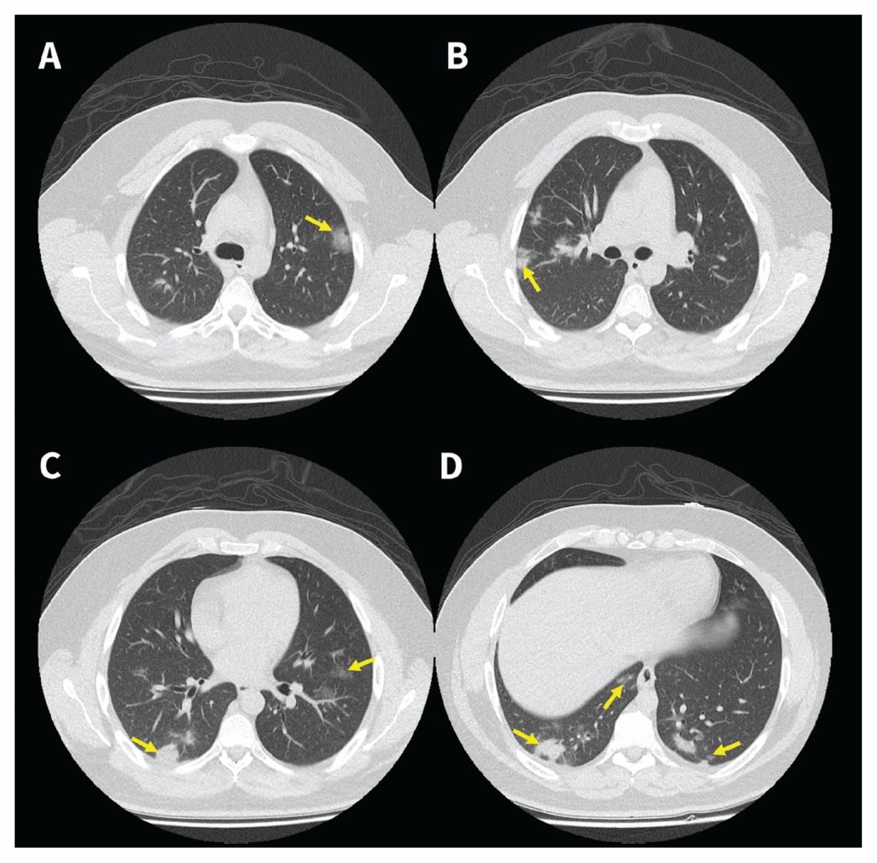
The primary utility of a chest CT scan often lies in its ability to thoroughly evaluate the lungs. It can detect and characterize a wide range of pulmonary conditions with remarkable precision:
- Infections: Conditions like pneumonia, bronchitis, and tuberculosis often present with characteristic patterns on a CT scan, showing areas of consolidation, ground-glass opacities, or cavitation.
- Inflammatory and Chronic Lung Diseases: Emphysema, a form of COPD, can be accurately quantified, showing areas of destroyed lung tissue. Interstitial lung diseases like pulmonary fibrosis, characterized by scarring of lung tissue, are vividly displayed, allowing for staging and monitoring. Bronchiectasis, involving widening of the airways, is also clearly visible.
- Lung Nodules and Tumors: A CT scan is highly effective in detecting small lung nodules, which may be benign or indicative of early-stage lung cancer. It provides detailed information on their size, shape, location, and characteristics, aiding in differentiation and guiding further management, such as biopsy or follow-up surveillance. It is the gold standard for lung cancer screening in high-risk individuals.
Assessing Cardiovascular Structures: Beyond the Heart Muscle
While a chest CT is not primarily a cardiac function study, it provides crucial information about the large blood vessels within the chest and surrounding cardiac structures:
- Aorta and Pulmonary Arteries: It can identify aneurysms (bulges), dissections (tears), and stenoses (narrowing) of the aorta and pulmonary arteries. Pulmonary embolism, a life-threatening condition involving blood clots in the lung arteries, is often definitively diagnosed with a CT pulmonary angiogram (CTPA), a specialized CT scan using contrast.
- Pericardium and Pleura: Fluid accumulation around the heart (pericardial effusion) or lungs (pleural effusion) can be clearly visualized and quantified. Thickening of the pleura, indicative of conditions like asbestos exposure, is also readily apparent.
- Coronary Artery Calcification: While not a dedicated coronary angiogram, a non-contrast chest CT can often reveal calcification in the coronary arteries, an indicator of atherosclerotic plaque burden and increased risk for heart disease.
Identifying Other Thoracic Issues: A Holistic View
Beyond the lungs and major vessels, a chest CT scan provides critical insights into other components of the thoracic cavity:
- Lymph Nodes: Enlarged lymph nodes (lymphadenopathy) in the mediastinum (the space between the lungs) or hilar regions often indicate infection, inflammation, or malignancy. The precise location and size of these nodes are critical for staging cancers like lymphoma or lung cancer.
- Mediastinal Masses: Abnormal growths or masses within the mediastinum, which could be benign cysts, thymomas, or other tumors, are accurately localized and characterized.
- Bones and Soft Tissues: Fractures of the ribs, sternum, or vertebrae within the chest can be clearly identified, especially in trauma cases. It also provides a view of surrounding soft tissues, aiding in the detection of abscesses or other soft tissue abnormalities.
Trauma and Emergency Diagnostics: Speed in Critical Situations
In emergency settings, the speed and comprehensive nature of a chest CT scan are invaluable. For patients experiencing trauma, such as car accidents or falls, a CT scan can rapidly detect life-threatening injuries like pneumothorax (collapsed lung), hemothorax (blood in the chest cavity), aortic injury, or multiple rib fractures, guiding immediate intervention and saving lives. Its ability to quickly produce detailed images makes it a cornerstone of modern emergency medicine protocols.
The Broader Impact: Data, Dollars, and Digital Health
The deployment and utilization of sophisticated diagnostic tools like chest CT scans extend far beyond the immediate medical diagnosis, intertwining with broader aspects of technology, financial management, and even the strategic branding of healthcare providers.
Digital Security and Data Management: Safeguarding Patient Information
With every CT scan, an immense volume of digital data is generated—images, patient demographics, scan parameters, and diagnostic reports. The secure handling and storage of this sensitive information are paramount, directly relating to the “Digital Security” aspect of our website topics. Healthcare institutions invest heavily in Picture Archiving and Communication Systems (PACS) and Electronic Health Records (EHR) systems to manage this data securely. These systems must comply with stringent regulations like HIPAA (Health Insurance Portability and Accountability Act) to protect patient privacy. Robust cybersecurity measures are essential to prevent data breaches, ransomware attacks, and unauthorized access, ensuring the integrity and confidentiality of medical images and patient information. The infrastructure required to store, transmit, and retrieve these large imaging files securely is a complex challenge that drives continuous innovation in digital security and data management platforms within healthcare tech.
The Financial Aspect of Diagnostic Imaging: Investment and Value
From a “Money” perspective, the implementation and operation of CT scanning services involve substantial financial considerations. A modern multi-slice CT scanner can cost anywhere from hundreds of thousands to several million dollars, representing a significant capital investment for hospitals and imaging centers. Beyond the initial purchase, there are ongoing costs for maintenance, software licenses, highly trained personnel (radiologists, technologists, physicists), electricity, and consumables.
For patients, the cost of a chest CT scan can vary widely depending on location, whether contrast is used, and insurance coverage, often ranging from a few hundred to several thousand dollars. Understanding these costs is crucial for personal finance and healthcare budgeting. From a business finance perspective, healthcare providers must carefully manage these expenses against reimbursement rates to ensure profitability and sustainability of services. Furthermore, the financial tools used for managing inventory, billing, and insurance claims for such high-value services are complex and require specialized software. The economic impact also extends to research and development in new imaging technologies, which require massive investment but promise future returns in improved health outcomes and efficiency.
Bridging the Gap: Telemedicine and Remote Diagnostics
The digital nature of CT images has profoundly impacted healthcare delivery through “Technology Trends” like telemedicine and remote diagnostics. CT scans, once performed and read exclusively in person, can now be transmitted securely over networks to radiologists located anywhere in the world. This capability is particularly vital for rural hospitals or clinics that may not have immediate access to a specialized radiologist. “Apps” and secure communication platforms enable rapid sharing and interpretation of critical images, significantly reducing diagnostic turnaround times and improving patient access to expert opinions. This rise of teleradiology enhances productivity for radiologists, allows for load balancing across different time zones, and ultimately brings high-quality diagnostic services to underserved populations. It exemplifies how digital innovation can overcome geographical barriers in healthcare.
The “Brand” of Advanced Healthcare: Reputation and Patient Trust
For hospitals and healthcare systems, offering state-of-the-art diagnostic imaging services, including the latest CT scanning technology, is a critical component of their “Brand Strategy” and “Corporate Identity.” A facility equipped with advanced, low-dose CT scanners that leverage AI for enhanced diagnosis projects an image of cutting-edge care, expertise, and patient safety. This contributes significantly to its “Reputation” and builds “Patient Trust.”
Marketing efforts often highlight investments in such technology to differentiate from competitors, attracting patients and skilled medical professionals alike. Case studies showcasing successful diagnoses through advanced CT scans can be powerful tools in demonstrating clinical excellence. In today’s competitive healthcare landscape, the ability to provide accurate, efficient, and technologically advanced diagnostics directly impacts a healthcare provider’s brand perception and its standing within the community. It signifies a commitment to leveraging the best available technology for the well-being of its patients.
Conclusion
A chest CT scan is a formidable diagnostic instrument, capable of revealing an astonishing array of conditions within the thoracic cavity, from subtle infections to life-threatening cancers and vascular emergencies. Its power is not solely in the physics of X-rays but in the sophisticated “Tech” ecosystem that supports it: the advanced hardware that acquires data rapidly and precisely, the intricate “Software” and “AI Tools” that process and interpret images, and the robust digital infrastructure that ensures “Digital Security” and seamless data management. Beyond the immediate medical application, the realm of chest CT scans profoundly intersects with “Money” – from the significant investments required for equipment and staffing to the financial implications for patients and the business models of healthcare providers. Moreover, the quality and accessibility of these services play a pivotal role in shaping a healthcare organization’s “Brand,” influencing its reputation and attracting patient trust. As technology continues to evolve, the capabilities of CT scans, augmented by AI and integrated into broader digital health platforms, will undoubtedly continue to expand, offering ever more precise insights into human health and further cementing their role as an indispensable tool in modern medicine.
aViewFromTheCave is a participant in the Amazon Services LLC Associates Program, an affiliate advertising program designed to provide a means for sites to earn advertising fees by advertising and linking to Amazon.com. Amazon, the Amazon logo, AmazonSupply, and the AmazonSupply logo are trademarks of Amazon.com, Inc. or its affiliates. As an Amazon Associate we earn affiliate commissions from qualifying purchases.