
The human body is an exquisite, complex machine, with an intricate network of vessels keeping it alive. Among these, the vertebral arteries play a crucial, yet often overlooked, role, supplying vital blood flow to the brainstem, cerebellum, and posterior cerebrum – areas fundamental for balance, coordination, and numerous life-sustaining functions. Traditionally, understanding such deep-seated anatomical structures relied on manual examination and rudimentary imaging.
However, in the 21st century, the very question “What is the vertebral artery?” has evolved beyond a simple anatomical definition. It now encompasses how cutting-edge technology allows us to visualize, diagnose, intervene, and even predict the health of this critical vessel. Modern innovation is not just observing the vertebral artery but actively transforming our understanding and management of cerebrovascular health. This article explores the profound impact of technology – from advanced imaging and AI diagnostics to robotic surgery and predictive modeling – on our approach to this vital component of the human circulatory system, illustrating an unprecedented convergence of biology and silicon.
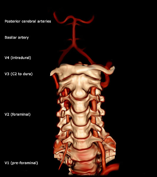
The Intersection of Anatomy and Innovation: Visualizing the Vertebral Artery
For centuries, the internal intricacies of the human body remained largely a mystery. The vertebral artery, nestled within the cervical spine and ascending into the brain, was particularly challenging to assess without invasive methods. Today, technology has revolutionized our ability to peer inside, offering unparalleled clarity that transforms diagnostic capabilities and therapeutic planning.
Advanced Imaging Modalities: Seeing the Unseen
Modern medicine leverages a sophisticated arsenal of imaging techniques to provide detailed insights into the vertebral artery’s structure, blood flow, and potential pathologies. These modalities have dramatically enhanced early detection and precise characterization of cerebrovascular conditions.
Magnetic Resonance Imaging (MRI) and Magnetic Resonance Angiography (MRA): Utilizing powerful magnetic fields and radio waves, MRI provides detailed soft tissue images, while MRA specifically visualizes blood vessels. These non-ionizing techniques are invaluable for detecting stenoses (narrowing), aneurysms (bulges), and dissections (tears) within the vertebral arteries. Advanced MRA sequences can even assess blood flow dynamics, offering crucial functional information. Its safety profile makes it suitable for repeated follow-up studies.
Computed Tomography Angiography (CTA): Employing X-rays and contrast material, CTA generates rapid, high-resolution cross-sectional images of the vertebral arteries. It excels in visualizing arterial wall characteristics, calcifications, and the relationship between the artery and surrounding bone structures. CTA’s speed is particularly advantageous in acute settings, such as stroke diagnosis, where swift assessment of arterial patency is critical for treatment decisions. Three-dimensional reconstructions further aid in surgical planning.
Digital Subtraction Angiography (DSA): Considered the gold standard for dynamic, real-time visualization of blood flow, DSA is an invasive technique involving catheter insertion and contrast dye. It offers the highest spatial resolution, allowing interventional specialists to precisely pinpoint blockages or malformations. DSA is often performed concurrently with endovascular treatments, providing immediate feedback on interventions and detailed insights into collateral circulation.
These advanced imaging tools have collectively transformed the diagnostic landscape, enabling earlier and more accurate detection of vertebral artery conditions, significantly improving patient outcomes by guiding timely interventions.
AI-Powered Diagnostics: Enhancing Accuracy and Speed
Beyond generating images, technology is augmenting our ability to interpret them. Artificial intelligence (AI) and machine learning (ML) are rapidly integrating into radiology workflows, promising to enhance diagnostic accuracy, reduce human error, and accelerate the identification of critical findings related to the vertebral artery.
Automated Anomaly Detection: AI algorithms, trained on vast datasets of medical images, can identify subtle abnormalities in vertebral artery morphology. These include early signs of atherosclerosis, small dissections, or minute aneurysms that might be missed by the human eye, particularly during demanding shifts. AI acts as a “second pair of eyes,” improving the sensitivity and consistency of detection.
Quantitative Analysis and Risk Stratification: AI can perform precise, quantitative measurements of arterial diameter, plaque volume, and blood flow characteristics with unmatched consistency. This allows for superior monitoring of disease progression and better risk stratification for conditions like vertebrobasilar insufficiency or stroke. For example, AI can analyze hemodynamic flow patterns to predict the likelihood of an aneurysm rupture or the clinical significance of a stenosis.
Workflow Optimization: AI-driven tools streamline image processing, prioritize urgent cases by flagging critical findings, and assist in generating preliminary reports. This not only speeds up diagnosis but also frees up radiologists’ time to focus on complex cases requiring nuanced human interpretation, thereby optimizing the entire diagnostic pipeline for conditions affecting the vertebral artery. The synergy between advanced imaging and AI creates a diagnostic ecosystem that is more precise, efficient, and proactive.
Navigating the Neural Network: Technology in Vertebral Artery Interventions
Once a pathology of the vertebral artery is identified, technology again steps forward, offering increasingly sophisticated and minimally invasive approaches to treatment. The goal is to restore blood flow, repair damage, or prevent future complications with the highest possible precision and the least possible trauma to the patient.
Robotic-Assisted Surgery: Precision at its Peak
Robotic surgical systems represent a significant advancement in complex vascular procedures, providing surgeons with enhanced dexterity, tremor filtration, and superior visualization. These capabilities are critical when operating on delicate structures like the vertebral artery, given its proximity to the spinal cord and brainstem.
Enhanced Dexterity and Control: Robotic platforms translate a surgeon’s hand movements into smaller, more precise movements of miniaturized instruments. This allows for intricate maneuvers in confined spaces, invaluable for procedures such as vertebral artery bypass, decompression, or the repair of small aneurysms, significantly minimizing collateral damage to surrounding neural tissue.
3D Visualization and Magnification: Robotic systems offer highly magnified, high-definition 3D views of the operative field. This immersive visual experience enhances depth perception and clarifies the complex anatomy around the vertebral artery, facilitating more accurate dissection and suturing. Surgeons can manipulate the viewpoint and zoom, achieving an unparalleled level of detail.
Reduced Invasiveness and Faster Recovery: By enabling smaller incisions and more precise movements, robotic-assisted surgery reduces trauma. This often leads to less post-operative pain, reduced blood loss, shorter hospital stays, and quicker recovery times compared to traditional open surgeries, making previously daunting procedures safer and more accessible for patients.
Minimally Invasive Techniques: Reducing Risk and Recovery
Beyond fully robotic systems, a broad spectrum of minimally invasive, catheter-based techniques has emerged, allowing access to the vertebral artery through small punctures rather than large incisions. These endovascular approaches are now central to many cerebrovascular interventions.
Catheter-Based Interventions: Using catheters guided through blood vessels from a remote entry point (e.g., the groin or wrist), interventional radiologists and neurosurgeons can access the vertebral artery directly. Techniques include:
- Angioplasty and Stenting: To treat vertebral artery stenosis, a balloon-tipped catheter opens the narrowed vessel, and a stent is deployed to maintain patency, ensuring continuous blood flow.
- Coiling and Flow Diversion for Aneurysms: For vertebral artery aneurysms, tiny platinum coils are deployed inside to block blood flow, preventing rupture. Flow diverters, mesh-like stents, redirect blood flow away from the aneurysm.
- Embolization: Used for arteriovenous malformations (AVMs) or fistulas, embolic agents block abnormal connections, preventing hemorrhage or “steal” phenomena.
Image-Guided Navigation: These procedures are performed under real-time imaging guidance, primarily DSA, often augmented by fusion imaging that overlays pre-operative CT or MRI data onto live fluoroscopy. This allows precise navigation of catheters and instruments through the intricate vascular tree, significantly enhancing safety and efficacy. Minimally invasive techniques have dramatically reduced morbidity and mortality, offering effective treatment options to a wider range of patients.
Digital Twins and Predictive Modeling: Simulating Vascular Health
Looking beyond immediate diagnosis and intervention, technology is enabling a proactive and personalized approach to vascular health. The concept of “digital twins” and advanced predictive modeling is poised to revolutionize how we understand and manage the long-term health of critical vessels like the vertebral artery.
Personalized Medicine through Digital Representations
A “digital twin” is a virtual replica of a patient’s individual vascular system, including their vertebral arteries. This highly detailed computer model is based on unique anatomical scans (MRI, CT), physiological data (blood pressure, heart rate, blood rheology), and genetic information.
Individualized Risk Assessment: With a digital twin, physicians can simulate various patient-specific scenarios. For example, they can model the effects of different blood pressure levels on a vertebral artery with known plaque buildup, or predict the growth trajectory of a small aneurysm. This allows for highly individualized risk assessment, moving beyond generalized population-level statistics.

Pre-Surgical Simulation and Optimization: Surgeons can use a digital twin to practice complex procedures on a virtual replica of their patient’s vertebral artery before actual surgery. This allows them to optimize surgical approaches, anticipate challenges, and refine techniques, potentially reducing operative time and improving outcomes. It functions as a personalized surgical simulator.
Drug and Treatment Pathway Testing: Digital twins can simulate the impact of different medications or lifestyle changes on vertebral artery health, predicting how a patient might respond to a particular drug regimen for hypertension or cholesterol management, without the need for trial-and-error in the real patient. This enables truly personalized medicine, optimizing treatment pathways before they are even initiated.
Forecasting Outcomes and Proactive Care
Predictive modeling, often powered by machine learning, analyzes large datasets (anonymized patient records, imaging studies, genetic markers) to identify patterns and forecast future health outcomes related to the vertebral artery.
Early Warning Systems: By analyzing various factors, predictive models can identify individuals at higher risk for developing vertebral artery stenosis, dissection, or stroke. This enables targeted screening and preventive interventions before symptoms manifest, allowing for proactive health management.
Prognosis and Treatment Response Prediction: For patients already diagnosed with a vertebral artery condition, predictive models can estimate the likelihood of disease progression, the success rate of various treatments, or the long-term prognosis. This data empowers both clinicians and patients to make more informed decisions about treatment strategies and lifestyle modifications.
Resource Allocation and Public Health: On a broader scale, predictive analytics can help healthcare systems anticipate demand for specific treatments, allocate resources more effectively, and design public health campaigns aimed at reducing risk factors for cerebrovascular diseases affecting the vertebral arteries. These technologies promise a future where healthcare is not just reactive but profoundly proactive, tailored precisely to the individual.
Wearable Technology and Remote Monitoring: A Proactive Approach
The power of technology extends beyond the clinical setting, integrating into our daily lives through wearable devices and remote monitoring systems. These innovations are shifting the paradigm from episodic care to continuous, real-time health surveillance, offering unprecedented opportunities for early detection and personalized management of conditions that could affect the vertebral artery.
Early Detection and Continuous Oversight
Wearable devices, once primarily for fitness tracking, are evolving into sophisticated medical-grade sensors capable of gathering vital physiological data that can hint at underlying vascular health issues.
Continuous Blood Pressure Monitoring: Advanced wearable sensors offer continuous, cuffless blood pressure monitoring. Persistent fluctuations or consistently elevated blood pressure, key risk factors for vertebral artery atherosclerosis or dissection, can be flagged early, prompting timely medical intervention.
Heart Rate Variability (HRV) and Arrhythmia Detection: Smartwatches and patches can monitor heart rate and detect irregular heart rhythms, such as atrial fibrillation, a major risk factor for stroke, including those originating from emboli traveling to the vertebral artery. Early detection of such arrhythmias allows for prompt anticoagulation therapy, significantly reducing stroke risk.
Sleep Apnea Detection: Wearables can monitor breathing patterns during sleep, identifying sleep apnea, a condition linked to hypertension and increased risk of cardiovascular disease. Addressing sleep apnea indirectly contributes to better vertebral artery health by reducing systemic stress.
Remote Patient Monitoring Platforms: Beyond personal wearables, dedicated remote patient monitoring (RPM) systems allow healthcare providers to continuously track vital signs, symptoms, and medication adherence for patients at high risk for cerebrovascular conditions. This continuous oversight enables prompt intervention if a patient’s condition deteriorates, potentially preventing acute events related to the vertebral artery.
Empowering Patients in Their Own Health Journey
These technological advancements are not merely tools for clinicians; they are powerful instruments for patient empowerment, fostering a more engaged and informed approach to personal health management.
Increased Health Awareness: By providing real-time data on their physiological metrics, patients gain a deeper understanding of their body’s responses to lifestyle choices, medications, and stress. This awareness can motivate positive behavioral changes, such as improved diet, regular exercise, and stress reduction, all of which benefit vascular health.
Proactive Engagement with Healthcare Providers: Patients can share their continuously collected data with their doctors, leading to more informed consultations. Instead of relying on intermittent readings, clinicians have a richer, more comprehensive dataset to guide diagnoses and treatment adjustments, making care for vertebral artery-related concerns more personalized and effective.
Early Symptom Recognition: In some cases, wearable technology might provide early indicators of subtle physiological changes that precede more overt symptoms of conditions affecting the vertebral artery, such as transient ischemic attacks (TIAs) or pre-stroke warnings. This can prompt patients to seek medical attention sooner, potentially averting severe neurological damage. The integration of wearable technology and remote monitoring is transforming passive patients into active participants, extending the reach of care beyond the clinic walls.
The Future Landscape: Merging Biology and Silicon
As we look ahead, the trajectory of technological advancement promises even more profound transformations in our understanding and interaction with the vertebral artery. The ultimate frontier involves a seamless integration of biological systems with engineered solutions, pushing the boundaries of what’s possible in medicine.
Nanotechnology and Targeted Therapies
Nanotechnology, the manipulation of matter on an atomic and molecular scale, holds immense promise for highly precise interventions within the intricate vascular network.
Nanobots for Plaque Removal: Imagine microscopic robots, or nanobots, designed to patrol the bloodstream, detect atherosclerotic plaques within the vertebral artery, and precisely remove or dissolve them without damaging healthy tissue. This could offer a non-invasive, highly targeted treatment for stenosis, preventing strokes and improving blood flow.
Targeted Drug Delivery: Nanoparticles can be engineered to deliver therapeutic agents directly to diseased areas of the vertebral artery, such as areas of inflammation or nascent aneurysms. This precision delivery minimizes systemic side effects, allowing for higher drug concentrations where needed and improving treatment efficacy for conditions like vasculitis or a fragile arterial wall.
Biosensors for Early Disease Markers: Nanoscale biosensors could be deployed within the bloodstream or embedded in arterial walls to continuously monitor for ultra-early molecular markers of disease, such as specific proteins associated with aneurysm formation or plaque instability. This would allow for intervention at an incredibly early stage, long before structural changes are detectable by current imaging.

Brain-Computer Interfaces and Beyond
While seemingly distant from the vertebral artery itself, advancements in brain-computer interfaces (BCIs) and regenerative medicine represent the ultimate convergence of tech and biology, with profound implications for managing the outcomes of vertebral artery pathologies.
Rehabilitation Post-Stroke: For patients who suffer a stroke due to vertebral artery occlusion or hemorrhage, BCIs could revolutionize rehabilitation. By allowing patients to control prosthetic limbs or communicate directly with devices using their thoughts, BCIs can help regain lost function and improve quality of life, effectively bypassing damaged neural pathways.
Neuroprosthetics and Functional Restoration: In cases of severe neurological damage resulting from vertebral artery events, neuroprosthetics powered by BCIs could offer unprecedented levels of functional restoration, such as restoring motor control or sensory perception, by directly interfacing with the brain’s remaining healthy networks.
Vascular Tissue Engineering and Regeneration: Beyond repair, the ultimate goal is regeneration. Advances in tissue engineering, stem cell therapy, and 3D bioprinting hold the potential to create patient-specific vertebral artery segments or even entire vessels to replace damaged ones. Imagine custom-printed arteries that seamlessly integrate into the body, solving complex vascular repair challenges and offering entirely new solutions for congenital defects or extensive damage.
The journey of understanding “what is the vertebral artery” has transformed from a purely anatomical inquiry into a dynamic exploration of how advanced technology can diagnose, treat, and even prevent its pathologies, ultimately enhancing human health and longevity. The fusion of biological insights with technological prowess is not just shaping the future of medicine; it is fundamentally redefining the human experience in the face of physiological challenges.
aViewFromTheCave is a participant in the Amazon Services LLC Associates Program, an affiliate advertising program designed to provide a means for sites to earn advertising fees by advertising and linking to Amazon.com. Amazon, the Amazon logo, AmazonSupply, and the AmazonSupply logo are trademarks of Amazon.com, Inc. or its affiliates. As an Amazon Associate we earn affiliate commissions from qualifying purchases.