
The term “gross anatomy” traditionally evokes images of medical students huddled around cadavers, meticulously dissecting tissues to understand the macroscopic structures of the human body. Historically, gross anatomy was the study of what can be seen with the naked eye. However, in the 21st century, the definition of gross anatomy has undergone a technological revolution. Today, gross anatomy is as much about pixels, polygons, and processing power as it is about physical biology.
As we move deeper into the era of digital health and “HealthTech,” the technology used to visualize and interact with anatomical structures has transformed the field from a static, physical discipline into a dynamic, data-driven science. This article explores how technology is redefining gross anatomy, the software tools driving this change, and the future of structural visualization.
The Digital Twin: Reimagining Gross Anatomy through 3D Modeling
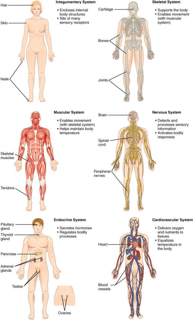
The most significant technological shift in the study of gross anatomy is the transition from two-dimensional textbook illustrations to three-dimensional digital twins. A digital twin is a virtual representation of a physical object—in this case, the human body—that serves as its real-time digital counterpart.
The Rise of Volumetric Rendering and DICOM Data
At the heart of modern digital anatomy is the processing of DICOM (Digital Imaging and Communications in Medicine) files. Technologies such as Computed Tomography (CT) and Magnetic Resonance Imaging (MRI) generate massive datasets that represent the “gross” structures of a patient. Advanced software now allows these 2D slices to be reconstructed into 3D volumetric models.
Engineers and software developers have created rendering engines that can differentiate between bone density, vascular pathways, and organ tissues, allowing a user to “dissect” a digital model layer by layer. This tech-first approach allows for a non-destructive exploration of human structures, where a mistake can be undone with a simple “Ctrl+Z,” a luxury not afforded in traditional laboratories.
High-Fidelity Anatomical Software Platforms
Platforms like BioDigital and Complete Anatomy have revolutionized how professionals interact with gross anatomy. These are not merely digital books; they are sophisticated cloud-based engines that utilize WebGL and high-end graphics processing to render millions of polygons in real-time. By leveraging cloud computing, these tools make high-fidelity anatomical models accessible on mobile devices and tablets, democratizing medical knowledge that was once locked behind expensive laboratory doors. This shift represents the “Software-as-a-Service” (SaaS) model entering the medical education space, providing continuous updates and collaborative tools for users worldwide.
Immersive Reality: AR, VR, and the Spatial Computing Frontier
If 3D modeling provided the map, then Extended Reality (XR)—comprising Virtual Reality (VR) and Augmented Reality (AR)—provides the territory. The integration of spatial computing into gross anatomy has fundamentally changed the user’s relationship with physical space.
Breaking Physical Constraints with Virtual Reality
Virtual Reality allows medical professionals and students to enter a completely simulated environment where they can interact with gross anatomical structures at any scale. Through headsets like the Meta Quest 3 or the Apple Vision Pro, a user can stand “inside” a human heart or expand a nervous system to the size of a room.
From a tech perspective, this involves complex spatial mapping and low-latency tracking. To make the experience immersive, developers must solve the “vergence-accommodation conflict” to prevent motion sickness during long study sessions. These VR environments allow for “procedural repetition,” where a user can practice a surgical approach to a specific anatomical structure hundreds of times before ever touching a physical tool.
Augmented Reality and Intraoperative Navigation
While VR replaces the world, Augmented Reality (AR) enhances it. In the context of gross anatomy, AR overlays digital anatomical maps directly onto a physical subject or a patient. This is particularly transformative in the field of “Image-Guided Surgery.”
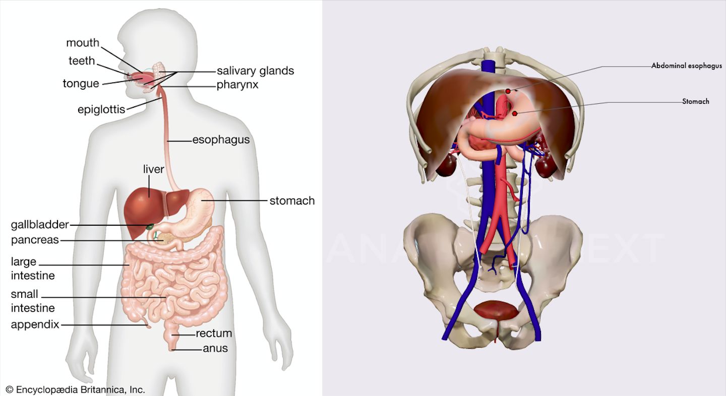
Using AR glasses, a surgeon can see the “gross anatomy” (the underlying veins, tumors, or bone structures) projected onto the patient’s skin in real-time. This tech relies on sophisticated registration algorithms that align the digital model with the physical body with sub-millimeter accuracy. By turning the human body into a transparent interface, AR technology reduces the risk of accidental damage to vital structures during complex procedures.
AI and Machine Learning: Automating Structural Identification
The sheer volume of anatomical data generated by modern medical imaging is too vast for human manual processing alone. This is where Artificial Intelligence (AI) and Machine Learning (ML) play a pivotal role in modern gross anatomy.
Computer Vision and Automated Segmentation
One of the most labor-intensive aspects of gross anatomy in a digital context is “segmentation”—the process of labeling every pixel in a scan to identify specific organs or tissues. Modern AI models, specifically Convolutional Neural Networks (CNNs), are now capable of automated segmentation.
These AI tools can scan a full-body CT in seconds and accurately identify the gross anatomy of the liver, lungs, kidneys, and skeletal system. This automation allows for “Precision Anatomy,” where software can identify minute structural variations between individuals that might be missed by the human eye. This tech-driven insight is crucial for personalized medicine, as it allows for the customization of implants and surgical plans based on an individual’s unique gross anatomical layout.
Predictive Modeling and Synthetic Data
Beyond just identifying what is there, AI is being used to predict how gross anatomical structures will change over time. Using generative adversarial networks (GANs), researchers can simulate the progression of muscular atrophy or the growth of a tumor within a specific anatomical region. Furthermore, AI is used to create “synthetic” anatomical datasets. These are artificially generated models that provide a diverse range of anatomical variations (different ages, ethnicities, and pathologies) without compromising the privacy of real patients. This ensures that the tech used for training is robust and inclusive.
The Cloud-Based Anatomy Lab: Connectivity and Cybersecurity
As gross anatomy becomes more digital, the infrastructure supporting it has moved from local servers to the cloud. This transition has significant implications for how anatomical data is stored, shared, and protected.
SaaS Architectures and Global Collaboration
The “Cloud-Based Anatomy Lab” allows for real-time collaboration across continents. A specialist in New York can interact with the same 3D anatomical model as a student in Nairobi, using “multi-user sync” technology. This requires high-bandwidth data transfer and robust back-end architecture to ensure that the “gross anatomy” being viewed is consistent and synchronized for all parties. These platforms often use a subscription-based model, ensuring that the software is constantly updated with the latest medical discoveries and technological patches.
The Ethics and Security of Anatomical Data
With the digitizing of gross anatomy comes the significant challenge of cybersecurity. Anatomical data is highly sensitive “Biometric Data.” As we create more detailed digital twins, the risk of “anatomical identity theft” increases.
Tech companies in the anatomy space are now prioritizing end-to-end encryption and blockchain-based verification to ensure that the structural data of individuals remains private. Moreover, the ethical use of digital cadavers—essentially 3D scans of deceased individuals—is a burgeoning field of “Tech Ethics.” Ensuring that digital remains are treated with the same respect as physical remains requires strict access controls and digital rights management (DRM) within the software.
Conclusion: The Future of the Human Map
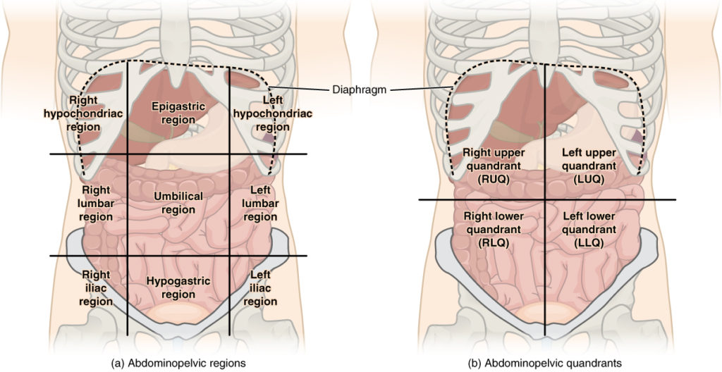
The question of “what is gross anatomy” no longer has a purely biological answer. In the modern world, it is a sophisticated intersection of medicine and high technology. From the 3D rendering of complex organ systems to the AI-driven analysis of structural data, technology has turned the human body into a navigable, data-rich landscape.
As we look toward the future, the integration of haptic feedback (technology that simulates the sense of touch) and real-time biometric syncing will further blur the lines between the physical and the digital. Gross anatomy is no longer a static discipline confined to the basement of a medical school; it is a cutting-edge tech frontier that is fundamentally changing how we understand, visualize, and interact with the human machine. Through the lens of technology, the “gross” structures of our bodies have never been more clearly defined, more accessible, or more profound.
aViewFromTheCave is a participant in the Amazon Services LLC Associates Program, an affiliate advertising program designed to provide a means for sites to earn advertising fees by advertising and linking to Amazon.com. Amazon, the Amazon logo, AmazonSupply, and the AmazonSupply logo are trademarks of Amazon.com, Inc. or its affiliates. As an Amazon Associate we earn affiliate commissions from qualifying purchases.