In the landscape of modern preventive medicine, few topics have been as significantly transformed by technological innovation as the study of breast density. For decades, the phrase “dense breasts” was a source of clinical frustration—a biological variable that made traditional screening less effective. However, we are currently witnessing a paradigm shift. What it means to have dense breasts today is fundamentally different from what it meant twenty years ago, thanks to the integration of high-resolution imaging, artificial intelligence (AI), and automated quantification software.
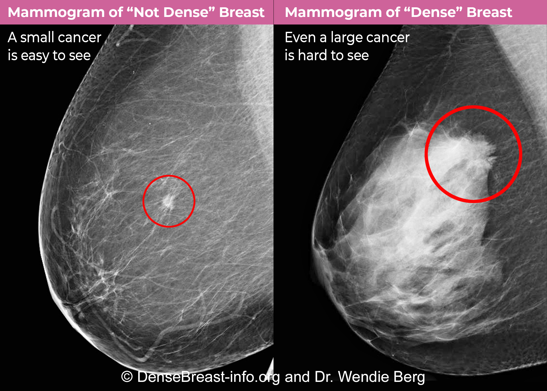
To understand the technological implications of breast density, one must first understand the technical challenge. Breast density is a measure of the ratio between glandular and fibrous tissue versus fatty tissue. On a standard mammogram, fatty tissue appears dark, while both dense tissue and cancerous tumors appear white. This creates a “camouflage effect,” a classic signal-to-noise problem where the “noise” (dense tissue) obscures the “signal” (potential malignancies).
As we move deeper into the era of Health-Tech, the industry is moving away from subjective visual assessments and toward data-driven, multi-modal diagnostic ecosystems.
The Physics of Density: Why Traditional Mammography Faces a “White-out” Problem
The evolution of breast density diagnostics began with the transition from analog film to digital mammography. However, even with digital sensors, the fundamental limitation of 2D X-ray imaging remains a challenge for dense tissue.
Digital Mammography vs. Analog Foundations
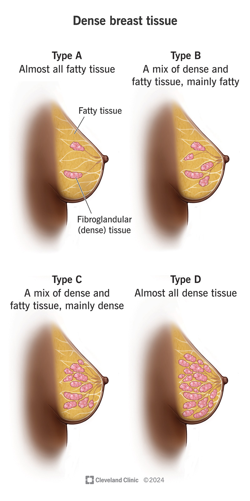
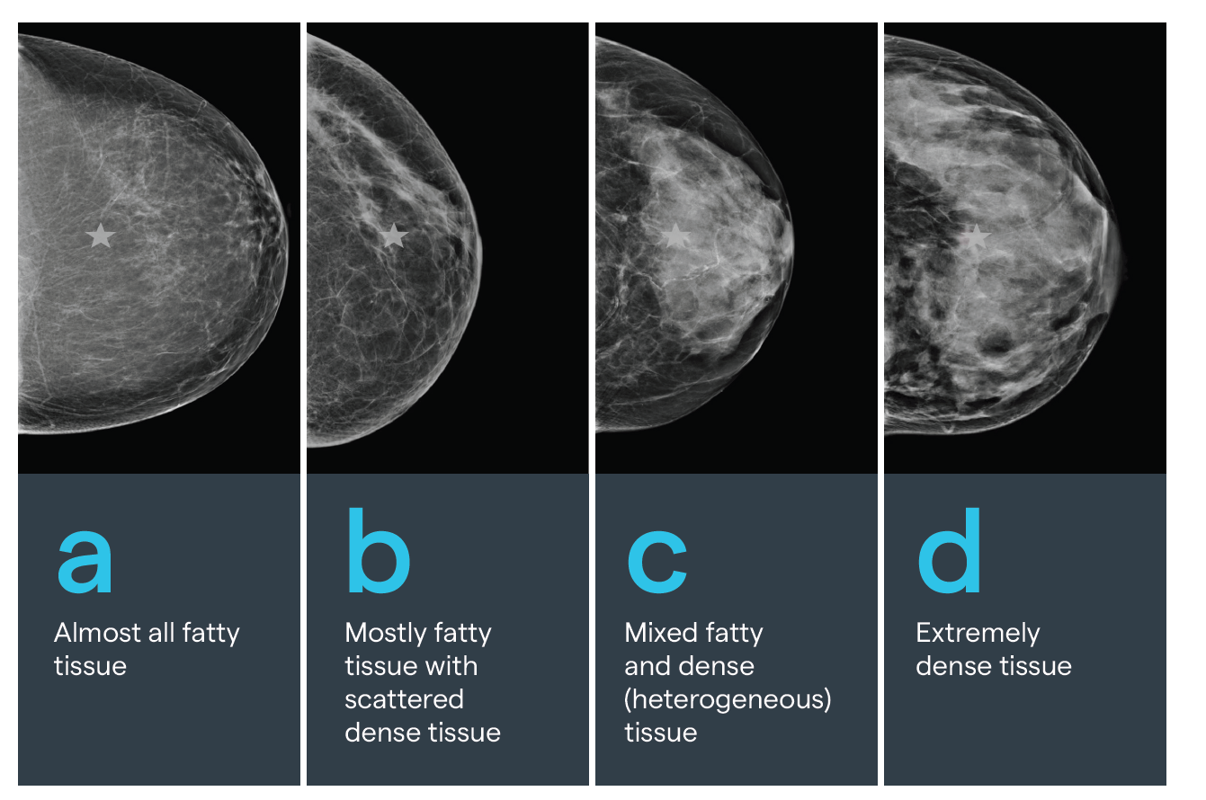
In the analog era, radiologists relied on physical film, which had a limited dynamic range. The move to Full-Field Digital Mammography (FFDM) was the first major tech milestone. FFDM allows for post-processing—software can adjust contrast and brightness to peer through layers of tissue. Despite this, FFDM still collapses a three-dimensional organ into a two-dimensional image. For a patient with Category C or D density (the highest levels on the BI-RADS scale), the 2D image often results in a “white-out,” where the sensitivity of the test drops significantly.
The Signal-to-Noise Ratio in Dense Tissue
From an engineering perspective, dense breast tissue represents a high-attenuation medium. X-ray photons are absorbed or scattered more frequently by fibrous tissue than by fat. This reduces the signal-to-noise ratio (SNR). When the SNR is low, the probability of detection (Pd) decreases while the probability of a false alarm (Pfa) increases. Tech developers have addressed this by developing higher-kvp (kilovoltage peak) systems and more sensitive cesium iodide (CsI) detectors, but hardware alone was not the solution. The solution required a change in imaging geometry.
Revolutionary Imaging Modalities: Beyond the Standard X-Ray
To solve the camouflage effect, the medical technology sector has introduced supplemental screening tools that leverage different physical properties—such as sound waves, magnetic resonance, and metabolic activity—to bypass the density barrier.
Digital Breast Tomosynthesis (3D Mammography)
Digital Breast Tomosynthesis (DBT) is arguably the most significant advancement in hardware in the last decade. Instead of taking a single static image, the X-ray tube moves in an arc over the breast, capturing multiple “slices.” Sophisticated reconstruction algorithms then compile these images into a 3D volume. This allows radiologists to scroll through the breast tissue millimeter by millimeter. By eliminating the “overlap” of tissue, DBT increases cancer detection rates in dense breasts by approximately 30-40% compared to 2D mammography alone.
Automated Breast Ultrasound (ABUS) Systems
While hand-held ultrasound has long been used for follow-ups, it is operator-dependent and time-consuming. The tech industry responded with Automated Breast Ultrasound (ABUS). These systems use large-format transducers and automated scanning arms to capture consistent, reproducible high-frequency sound wave data of the entire breast. Because ultrasound reacts differently to dense tissue (which is sonolucent) than to solid masses, it provides a vital secondary data layer that X-rays might miss.
Molecular Breast Imaging (MBI) and Contrast Enhancement
For patients where density makes anatomical imaging nearly impossible, functional imaging offers a tech-heavy alternative. Molecular Breast Imaging (MBI) involves injecting a short-lived radioactive tracer that is absorbed by cells with high metabolic activity (like tumors). Unlike a mammogram, which looks at the structure of the breast, MBI looks at the behavior of the cells. This “functional” approach effectively renders density irrelevant, as the dense tissue does not take up the tracer, allowing tumors to “glow” on the digital sensor.
The Role of Artificial Intelligence and Machine Learning in Classification
The most significant current trend in the “dense breast” space is not a new camera or sensor, but the software used to interpret the data. Artificial Intelligence (AI) and Machine Learning (ML) are now being deployed to standardize density assessment and improve diagnostic accuracy.
Quantifying Density with Algorithmic Precision
Historically, breast density was assessed subjectively by a radiologist, leading to significant inter-observer variability. Today, FDA-cleared software platforms like Volpara or Quantra use machine learning to calculate the volumetric density of the breast. These tools analyze the raw X-ray data (the “for processing” images) to provide a precise percentage of fibroglandular tissue. This eliminates human bias and ensures that patients are correctly categorized, which is crucial for triggering insurance coverage for supplemental screening.
Computer-Aided Detection (CAD) 2.0
Early CAD systems were notorious for “crying wolf,” marking every speck of calcium and creating “alert fatigue” for doctors. Modern AI, powered by Deep Learning and Neural Networks, is far more sophisticated. These algorithms are trained on millions of images to recognize the subtle architectural distortions that dense tissue often hides. By acting as a “second set of eyes,” AI helps radiologists prioritize high-risk cases and reduces the rate of false negatives in dense-tissue screenings.
Predictive Modeling and Risk Assessment Software
Having dense breasts is not just a screening challenge; it is an independent risk factor for developing breast cancer. New software integrations take density data and combine it with genomic data, family history, and lifestyle factors to create a personalized risk score (such as the Tyrer-Cuzick model). This move toward “Precision Medicine” allows clinics to tailor their tech stacks—determining exactly which patients need an MRI vs. an ultrasound—based on a calculated data profile.
Data Security and the Integration of Tele-Radiology
The move toward high-resolution 3D imaging and AI analysis has created a massive data problem. A single DBT exam can be 20 times the size of a standard 2D mammogram. Managing this data requires a robust tech infrastructure focused on storage, interoperability, and security.
Cloud-Based Image Sharing and Interoperability
In the past, patients had to carry physical CDs of their images from one clinic to another. Modern Health-Tech utilizes Picture Archiving and Communication Systems (PACS) integrated with cloud-based VNA (Vendor Neutral Archives). This allows for seamless transfer of high-resolution “heavy” files across networks. For a patient with dense breasts, this interoperability is vital; it allows for “temporal comparison,” where AI can compare this year’s 3D volume to last year’s to detect the most minute changes in tissue architecture.
Protecting Sensitive Biometric and Genomic Data
As we integrate more AI and cloud-based diagnostics, cybersecurity becomes a paramount concern. Health-Tech firms are now employing end-to-end encryption and blockchain-based audit trails to ensure that a patient’s diagnostic images and density scores are protected. This is especially critical as density data is increasingly linked to genetic testing results, creating a comprehensive but highly sensitive digital health identity.
Conclusion: The Convergence of Tech and Health
What does it mean to have dense breasts in the current technological era? It means you are at the center of one of the most advanced intersections of hardware and software in modern medicine. We are moving away from the era of “one size fits all” screening and into an era defined by precision diagnostics.
The challenge of dense tissue is being systematically dismantled by 3D reconstruction, automated ultrasound, and AI-driven quantification. As these technologies become more accessible and integrated into standard clinical workflows, the “camouflage effect” that once hindered early detection is being replaced by a transparent, data-rich view of breast health. For the tech industry, the mission is clear: continue refining the algorithms and imaging sensors until density is no longer a barrier, but simply another data point in a successful, proactive health strategy.
aViewFromTheCave is a participant in the Amazon Services LLC Associates Program, an affiliate advertising program designed to provide a means for sites to earn advertising fees by advertising and linking to Amazon.com. Amazon, the Amazon logo, AmazonSupply, and the AmazonSupply logo are trademarks of Amazon.com, Inc. or its affiliates. As an Amazon Associate we earn affiliate commissions from qualifying purchases.