
The human heart is a marvel of biological engineering, a tireless pump that sustains life. At its core are four intricate valves, each playing a critical role in directing blood flow and ensuring efficiency. Among these, the semilunar valves stand out for their unique structure and crucial function. While often discussed in the realm of biology and medicine, understanding semilunar valves takes on a new dimension when viewed through the lens of technological innovation. From advanced diagnostic imaging to the development of sophisticated prosthetic replacements, technology is increasingly intertwined with our understanding and treatment of these vital heart components.
The Anatomy and Physiology of Semilunar Valves
To appreciate the technological implications, it’s essential to grasp the fundamental nature of semilunar valves. These valves are not merely passive flaps; they are dynamic structures meticulously designed for optimal performance, a design that engineers and material scientists strive to replicate and enhance.
Structure: Three Cusps, One Purpose
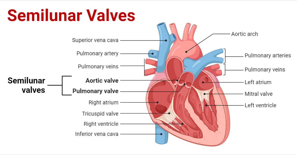
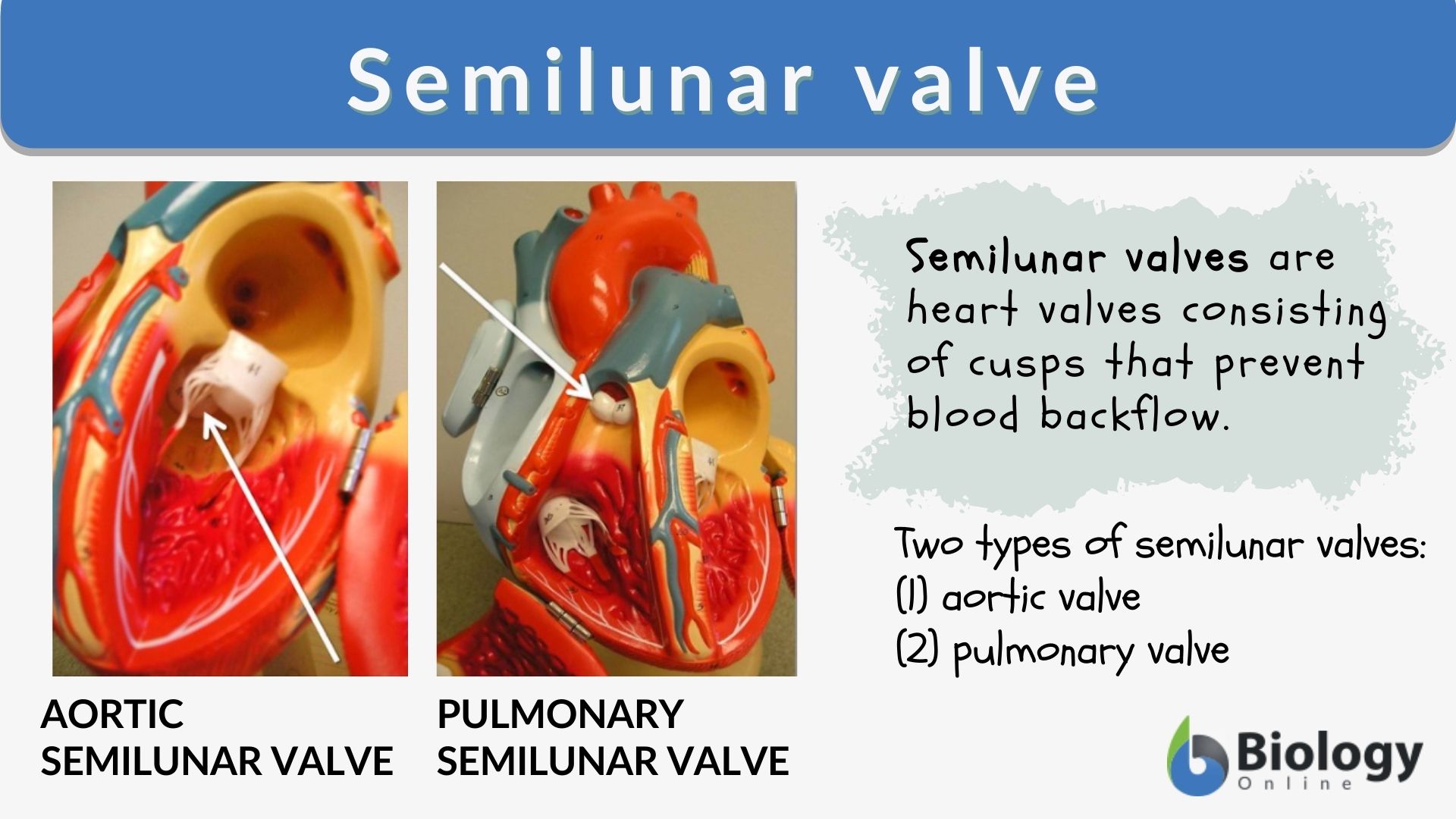
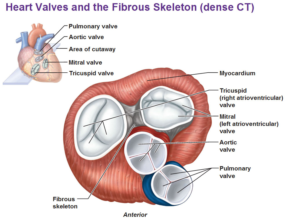
The defining characteristic of semilunar valves is their morphology. Unlike the atrioventricular valves (mitral and tricuspid), which possess two or three leaflets, respectively, the semilunar valves are composed of three crescent-shaped, or “half-moon” shaped, cusps. This distinctive structure is key to their function.
- Aortic Valve: Located between the left ventricle and the aorta, the aortic valve has three cusps. These cusps are robust, designed to withstand the high pressures generated by the powerful left ventricle as it pumps oxygenated blood to the entire body. When closed, the cusps coapt, forming a tight seal that prevents blood from flowing back into the left ventricle during diastole (relaxation).
- Pulmonary Valve: Situated between the right ventricle and the pulmonary artery, the pulmonary valve also features three cusps. Its role is to ensure that deoxygenated blood pumped by the right ventricle flows efficiently to the lungs for oxygenation and does not return to the ventricle. While also critical, the pressures it manages are generally lower than those handled by the aortic valve.
The fibrous tissue forming these cusps is highly specialized, providing both flexibility and strength. Each cusp is attached to a ring of connective tissue called the annulus, which provides structural support. The arrangement of these cusps is such that when the ventricles contract, the increased pressure within them forces the cusps open, allowing blood to flow forward. As the ventricles relax, the pressure gradient reverses, and the blood that has moved into the aorta and pulmonary artery exerts pressure back on the cusps, pushing them together to close the valve.
Function: The Unidirectional Flow Regulator
The primary function of semilunar valves is to act as one-way gates, ensuring that blood flows in the correct direction through the circulatory system. This unidirectional flow is fundamental to the efficiency of the heart as a pump.
- Ejection Phase: During ventricular contraction (systole), the pressure inside the ventricles rises. When this pressure exceeds the pressure in the aorta and pulmonary artery, the semilunar valves are forced open. This allows the blood to be ejected from the ventricles into these major arteries. The opening of the semilunar valves marks the beginning of the ejection phase of the cardiac cycle.
- Diastolic Phase: As the ventricles begin to relax (diastole), the pressure within them falls. When the ventricular pressure drops below the pressure in the aorta and pulmonary artery, the blood in these arteries starts to flow backward. This backward flow fills the cusps, pushing them together and closing the semilunar valves. This closure prevents regurgitation, ensuring that the blood remains in the aorta and pulmonary artery, ready for the next cycle. The closure of the semilunar valves also contributes to the second heart sound, often described as “dub.”
The seamless and synchronized action of these valves, driven by pressure gradients, is a testament to the body’s sophisticated design. However, like any mechanical system, they are susceptible to dysfunction, which is where technological intervention becomes paramount.
Technological Innovations in Understanding and Treating Semilunar Valve Dysfunction
The limitations of the biological system necessitate technological advancements for diagnosis, treatment, and even prevention of semilunar valve diseases. The field of cardiovascular technology has seen remarkable progress, offering new hope and improved outcomes for patients.
Advanced Imaging and Diagnostics: Seeing the Unseen
The ability to visualize the heart and its valves in real-time and with unprecedented detail is a cornerstone of modern cardiology. Technology has transformed our understanding of valve structure and function, enabling earlier and more accurate diagnoses.
- Echocardiography: This ultrasound-based imaging technique is indispensable. 2D echocardiography provides cross-sectional images, allowing assessment of valve structure, motion, and leaflet thickness. 3D and 4D echocardiography offer even more comprehensive views, enabling detailed reconstruction of valve anatomy and dynamics, crucial for evaluating complex valve pathologies like prolapse or regurgitation. Doppler echocardiography measures blood flow velocities across the valves, quantifying the severity of stenosis (narrowing) or regurgitation (leakage). Technologically, this involves sophisticated transducer design, advanced signal processing algorithms, and powerful computing to render these detailed images and measurements.
- Cardiac Magnetic Resonance Imaging (CMR): CMR provides high-resolution anatomical images of the heart and great vessels, offering excellent soft tissue contrast. It is particularly useful for assessing the size and function of the ventricles, the integrity of the aortic root, and the presence of any associated cardiac abnormalities. CMR can also quantify blood flow and regurgitant volumes with high accuracy, complementing echocardiography. The underlying technology involves powerful magnetic fields, radiofrequency pulses, and complex pulse sequences to generate detailed images.
- Computed Tomography (CT) Angiography: For specific indications, cardiac CT can provide detailed anatomical information about the aortic root and valve, especially when planning interventions like transcatheter aortic valve implantation (TAVI). It excels at visualizing calcification and assessing the dimensions of the aortic annulus. Advanced reconstruction algorithms and radiation dose reduction techniques are key technological aspects here.
These imaging modalities, powered by continuous advancements in hardware and software, allow clinicians to not only diagnose problems with semilunar valves but also to precisely assess their severity and plan the most effective course of treatment.
Minimally Invasive Interventions and Prosthetic Valves: Engineering a Solution
When semilunar valve dysfunction becomes significant, medical intervention is often required. The development of minimally invasive surgical techniques and advanced prosthetic valves represents a triumph of biomedical engineering.
- Transcatheter Aortic Valve Implantation (TAVI): This groundbreaking procedure has revolutionized the treatment of severe aortic stenosis, particularly in patients who are high-risk for traditional open-heart surgery. TAVI involves delivering a collapsible prosthetic aortic valve via a catheter, typically inserted through a major artery in the leg or chest. The valve is then deployed in the native aortic valve’s position, expanding to open and immediately restoring proper blood flow. This technology relies on advanced catheter design, steerable delivery systems, and sophisticated prosthetic valve designs, often featuring self-expanding or balloon-expandable frames made from nitinol alloys.
- Transcatheter Pulmonary Valve Implantation (TPVI): Similar to TAVI, TPVI is used to treat pulmonary valve dysfunction, particularly in patients with congenital heart defects who may have undergone previous surgeries. Devices like the Melody valve allow for catheter-based replacement of dysfunctional pulmonary valves, avoiding the need for repeat open-heart surgery.
- Mechanical and Bioprosthetic Valves: For traditional open-heart surgery, a range of prosthetic valves are available. Mechanical valves are typically made from durable materials like pyrolytic carbon and are designed to last a lifetime. However, they require patients to take lifelong anticoagulant medication due to the risk of clot formation. Bioprosthetic valves, made from animal tissue (porcine or bovine pericardium) mounted on a frame, offer a more natural function and often do not require lifelong anticoagulation. However, they have a limited lifespan and may need replacement after 10-20 years. The ongoing technological challenge is to create prosthetic valves that offer the durability of mechanical valves with the hemodynamic performance and low thrombogenicity of biological tissues. This involves advancements in material science, tissue engineering, and surface modification techniques.
- Robotic-Assisted Surgery: In open-heart procedures, robotic surgery is increasingly being employed to perform valve repairs and replacements with greater precision and smaller incisions. Robotic systems provide surgeons with enhanced dexterity, magnified 3D vision, and tremor filtration, potentially leading to faster recovery times and reduced complications.
These technological interventions are not just about replacing or repairing a faulty part; they are about engineering a solution that restores physiological function, improves quality of life, and extends longevity for individuals with semilunar valve disease.
The Future of Semilunar Valve Technology: Integration and Personalization
The trajectory of cardiovascular technology suggests a future where semilunar valve management is characterized by even greater integration of digital tools, sophisticated materials, and personalized approaches.
Digital Twins and AI-Driven Diagnostics
The concept of a “digital twin” – a virtual replica of a patient’s heart – is becoming increasingly feasible. By integrating data from various imaging modalities, wearable sensors, and patient records, clinicians could create dynamic, real-time digital models of a patient’s cardiovascular system, including their semilunar valves.
- Predictive Analytics: Artificial intelligence (AI) algorithms can analyze these digital twins to predict the progression of valve disease, identify individuals at high risk for complications, and optimize treatment strategies. AI can also assist in interpreting complex imaging data, flagging subtle abnormalities that might be missed by the human eye.
- Personalized Prosthetic Design: In the future, AI and advanced 3D printing could enable the creation of fully personalized prosthetic valves tailored to an individual’s specific anatomy and hemodynamic profile. This could lead to prosthetics that offer optimal performance, reduced complications, and longer lifespans.
Advanced Biomaterials and Tissue Engineering
The quest for the ideal prosthetic valve continues. Future research is focused on developing novel biomaterials and tissue-engineered solutions that mimic the native valve’s structure and function more closely.
- Smart Materials: Incorporating “smart” materials that can adapt to physiological conditions or deliver therapeutic agents could lead to valves that actively participate in cardiovascular health rather than passively performing a function.
- Regenerative Medicine: The ultimate goal is to develop regenerative therapies that can repair or regrow damaged valve tissue, eliminating the need for prosthetic devices altogether. This involves harnessing stem cell technology and advanced scaffolding techniques.
Wearable Technology and Remote Monitoring
The proliferation of wearable devices is extending cardiovascular care beyond the clinic. For patients with semilunar valve conditions, continuous monitoring of heart rate, blood pressure, and activity levels can provide invaluable data.
- Early Detection of Issues: Changes in these parameters could signal early signs of valve dysfunction or complications, allowing for timely intervention and preventing critical events.
- Remote Management: Integrated systems could allow healthcare providers to remotely monitor patients, adjust medications, and schedule follow-up appointments based on real-time data, improving efficiency and accessibility of care.
In conclusion, while the semilunar valve is a fundamental biological structure, its journey through the realm of technology is one of constant innovation. From deciphering its intricate mechanics with advanced imaging to engineering life-saving prosthetic devices and envisioning AI-driven personalized care, technology is profoundly shaping our ability to understand, treat, and ultimately enhance the function of these vital cardiac regulators. The future promises an even deeper integration of technology, moving towards a more predictive, personalized, and regenerative approach to cardiovascular health.
aViewFromTheCave is a participant in the Amazon Services LLC Associates Program, an affiliate advertising program designed to provide a means for sites to earn advertising fees by advertising and linking to Amazon.com. Amazon, the Amazon logo, AmazonSupply, and the AmazonSupply logo are trademarks of Amazon.com, Inc. or its affiliates. As an Amazon Associate we earn affiliate commissions from qualifying purchases.