In the modern diagnostic landscape, the “retic count”—short for reticulocyte count—is far more than a simple biological tally. While it serves the vital clinical purpose of measuring the percentage of newly formed, immature red blood cells in the bloodstream, the process of obtaining this data has undergone a radical technological transformation. Gone are the days of lab technicians peering through manual microscopes for hours to count cells by hand. Today, the retic count represents a pinnacle of medical technology, integrating flow cytometry, laser optics, and sophisticated machine learning algorithms to provide real-time insights into bone marrow function.
To understand what a retic count is today, one must look at it through the lens of laboratory automation and digital health tech. This article explores the high-tech infrastructure that powers hematology diagnostics, the software-driven analysis of cellular morphology, and the future of automated blood monitoring.
1. The Hardware Revolution: From Manual Slides to Flow Cytometry
The fundamental “tech” behind a retic count has shifted from basic optics to complex fluid dynamics and laser-based detection. This evolution has moved the test from a subjective estimation to a high-precision digital output.
From Manual Counting to Automated Precision
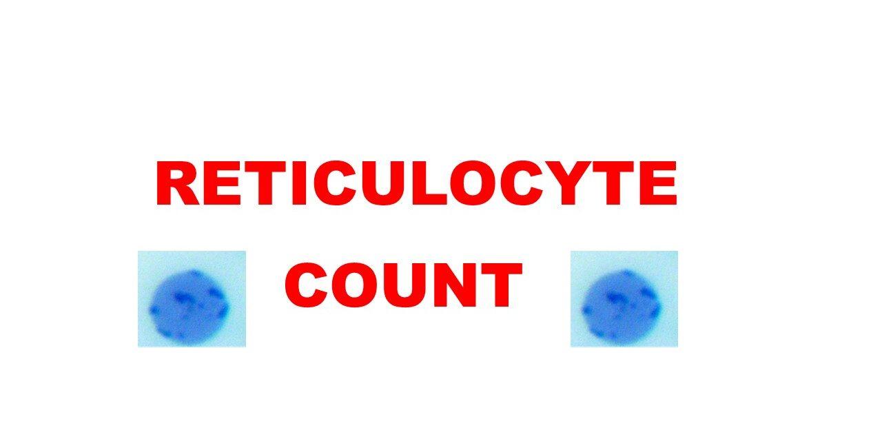
Historically, a retic count required staining blood with a supravital dye, such as methylene blue, which caused the ribosomal RNA in young red blood cells to clump into a visible “reticulum” or network. A technician would then manually count these cells against a total of 1,000 red blood cells. This method was fraught with human error and inter-observer variability.
Modern hematology analyzers have digitized this entire workflow. By utilizing automated sample aspiration and specialized reagent mixing, these machines can process hundreds of samples per hour. This transition from manual labor to automated hardware has increased throughput by over 500% in clinical environments, making rapid diagnostics accessible at scale.
The Role of Fluorescent Flow Cytometry
The core technology used in premium analyzers today is fluorescent flow cytometry. In this process, the blood sample is treated with a fluorescent dye that binds specifically to the RNA inside the reticulocytes. The cells are then hydrodynamically focused—pushed through a narrow channel one by one.
As each cell passes through a laser beam, the machine captures light scatter and fluorescence. The intensity of the fluorescence is directly proportional to the amount of RNA present. This high-tech approach allows the analyzer to differentiate between mature red blood cells (which have no RNA) and reticulocytes with extreme accuracy. This is a prime example of how hardware engineering solves biological measurement challenges.
2. The Software Engine: Data Analysis and Cellular Gating
The hardware generates the raw signal, but the software is what defines a retic count in the digital age. Advanced algorithms are required to translate pulses of light into clinical data points.
Laser Scattering and Light Absorption Algorithms
When the laser hits a cell, it scatters in different directions. Frontal light scatter (forward scatter) indicates the cell’s size, while side scatter indicates the cell’s internal complexity or granularity. The analyzer’s software uses these data points to create a three-dimensional “scattergram.”
Software developers in the medical tech space have designed complex “gating” logic that allows the system to ignore debris, platelets, and white blood cells, focusing exclusively on the red blood cell population. This real-time signal processing is what allows for the high precision required in modern pathology.
The Reticulocyte Hemoglobin (Ret-He) Metric
Beyond a simple count, technology has introduced new parameters like the Reticulocyte Hemoglobin Equivalent (Ret-He). This is a software-calculated value that measures the actual hemoglobin content within the newest red blood cells. By utilizing tech-driven refractive index measurements, clinicians can see the immediate effect of iron therapy or the early onset of anemia—data that was impossible to gather using traditional manual methods. This represents a shift from “counting” to “molecular profiling” via software.
3. AI and Machine Learning in Morphological Recognition

The next frontier of the retic count lies in Artificial Intelligence (AI). While flow cytometry is the current standard, digital morphology powered by AI is beginning to redefine how we visualize and categorize blood cells.
Computer Vision and Image Analysis
Newer diagnostic platforms utilize high-resolution digital cameras to take thousands of photos of a blood smear. AI-driven computer vision models, trained on millions of annotated images, can identify reticulocytes based on their shape, texture, and color profile.
Unlike a human, these AI tools do not suffer from fatigue. They can identify subtle patterns in cell morphology—such as the specific distribution of RNA remnants—that might indicate specific rare disorders. This integration of computer vision into hematology is transforming the laboratory into a data-centric hub where software serves as a second pair of expert eyes.
Predictive Analytics for Anemia Classification
The data generated by a retic count is rarely looked at in isolation. AI tools integrated into Laboratory Information Systems (LIS) can now correlate retic counts with other markers like Mean Corpuscular Volume (MCV) and Ferritin levels.
By using predictive modeling, these systems can flag “high-risk” samples and suggest specific diagnostic pathways (such as iron deficiency vs. hemolytic anemia) before a pathologist even looks at the results. This proactive tech stack optimizes the clinical workflow and speeds up the “time-to-diagnosis,” which is critical in acute care settings.
4. Connectivity, Cloud Integration, and Remote Diagnostics
The “what” of a retic count is increasingly defined by where the data goes and how it is stored. In the era of the Internet of Medical Things (IoMT), the retic count is a mobile data point.
Cloud-Based LIS Integration
Modern hematology analyzers are no longer standalone units. They are connected via secure APIs to centralized Cloud-based Laboratory Information Systems. This allows for “real-time” hematology. A blood sample taken in a remote clinic can have its retic count data uploaded, analyzed by an AI in the cloud, and reviewed by a specialist in a different city within minutes. This connectivity is essential for managing global clinical trials and monitoring patient health across sprawling hospital networks.
Point-of-Care (POC) Tech and Remote Monitoring
We are seeing the emergence of “Point-of-Care” devices—handheld or tabletop units that can perform a retic count outside of a traditional lab. These devices use microfluidic “lab-on-a-chip” technology. By shrinking the components of a massive flow cytometer into a small cartridge, tech companies are enabling retic counts to be performed in rural areas, on the battlefield, or even in a patient’s home. The software on these devices is often optimized for low-power processing, yet it maintains the accuracy of much larger machines through sophisticated error-correction code.
5. Why the Tech Matters: The Impact on Precision Medicine
The technological sophistication behind a retic count has profound implications for the future of precision medicine and biotech research.
Reducing Human Error and Enhancing Throughput
In a high-volume diagnostic lab, the margin for error is slim. The automation of the retic count has reduced laboratory error rates by over 90% compared to manual methods. This reliability is the foundation of modern evidence-based medicine. Furthermore, the speed of automated tech allows labs to process thousands of “retic” requests daily, ensuring that patients receive results in hours rather than days.
Real-Time Monitoring in Biotech R&D
For biotech companies developing new treatments for chronic kidney disease or oncology, the retic count is a vital “bio-signal” used to measure a drug’s efficacy. High-tech hematology platforms allow researchers to monitor “erythropoietic response” (the production of red blood cells) in real-time during clinical trials. By leveraging digital dashboards and longitudinal data tracking, researchers can see how a new drug influences bone marrow activity on a day-by-day basis.

Conclusion: The Retic Count as a Digital Asset
When we ask “what is a retic count” in the 21st century, we are asking about a complex intersection of biology and technology. It is a measurement that relies on the precision of laser optics, the intelligence of machine learning algorithms, and the speed of cloud connectivity.
As diagnostic technology continues to advance, the retic count will likely become even more granular, providing insights not just into the quantity of cells, but into the very genetic and molecular health of our blood production systems. For tech enthusiasts and medical professionals alike, the retic count stands as a testament to how digital innovation can unlock the secrets of human physiology, one cell at a time.
aViewFromTheCave is a participant in the Amazon Services LLC Associates Program, an affiliate advertising program designed to provide a means for sites to earn advertising fees by advertising and linking to Amazon.com. Amazon, the Amazon logo, AmazonSupply, and the AmazonSupply logo are trademarks of Amazon.com, Inc. or its affiliates. As an Amazon Associate we earn affiliate commissions from qualifying purchases.